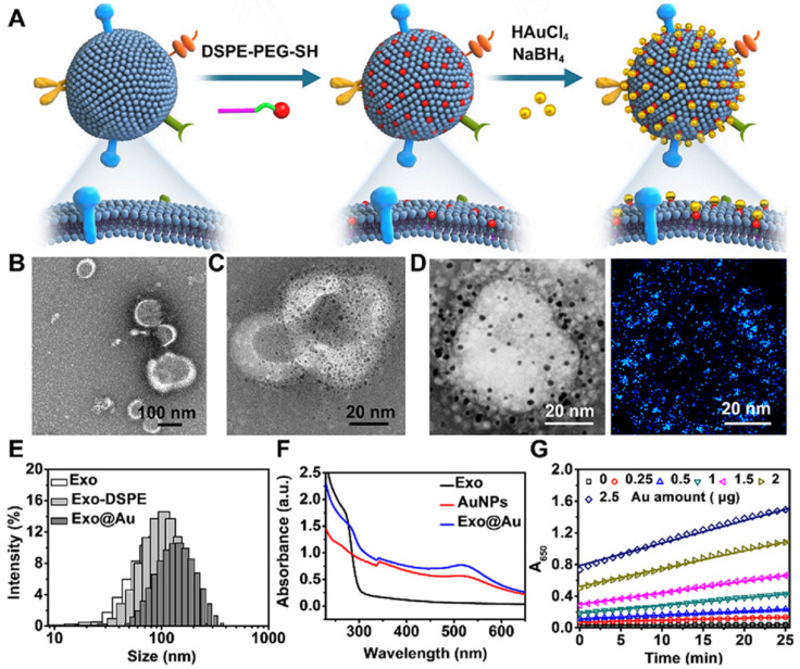
Figure 1.
Schematic of the preparation of the gold nanoparticle-decorated exosomes (Exo@Au) and characterization data for the Exo@Au. (A) Stepwise preparation of the Exo@Au nanozymes. Representative TEM images of (B) the native Exos and (C) the Exo@Au nanozymes. (D) High-resolution TEM and elemental mapping analysis of Exo@Au. Blue dots represent AuNPs. (E) Size distribution of native Exos (Exo, white), DSPE-functionalized Exos (Exo-DSPE, light gray), and Exo@Au (dark gray) as measured by DLS. (F) UV-Vis absorption spectra of native Exos (black line), free AuNPs (red line), and Exo@Au (blue line). (G) Kinetic measurements of the catalytic activity of Exo@Au with different amounts of Au (0, 0.25, 0.5, 1, 1.5, 2, and 2.5 µg) at 37 °C for 25 min.

