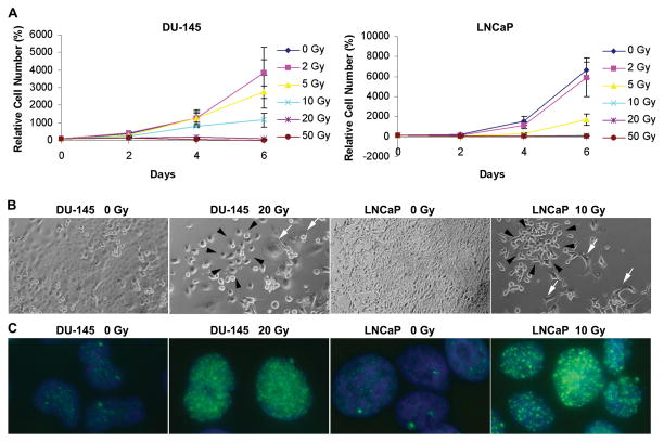
Fig. 1.
Radiation-induced growth inhibition and DNA damage. (A) Viable cell numbers of LNCaP and DU-145 cells following irradiation, normalized to the number of cells plated. (B) Phase-contrast images of irradiated DU-145 and LNCaP cultures. Note repopulating colonies (black arrows) and degenerating cells (white arrows). (Original magnification: x100) (C) Immunocytochemistry of radiation-induced γ-H2AX foci (green), 2 hours post-irradiation. Images from DU-145 and LNCaP cells irradiated with 20 and 10 Gy, respectively. (Original magnification: x 600.)

