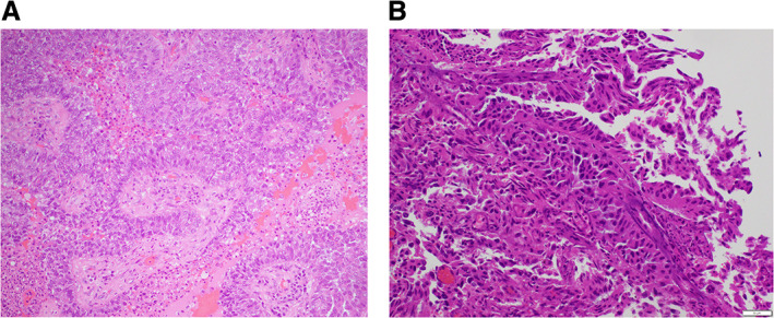
Figure 3.

Photomicrographs of H&E‐stained cross‐sectional images of the temporal brain metastases resected from Mrs. ND 2014 (A) and right vertex lesion from 2019 (B). Image in (B) demonstrates a morphological difference from the original (A) resected brain specimen with poorly differentiated nests and sheets of epithelioid cells with ample eosinophilic cytoplasm and scanty intratumoral lymphocytes, necrosis, and hemorrhage. This image has unavoidable changes from intraoperative heat effect. The original (A) was composed of lager pleomorphic cytoplasm abundant cells with a papillary architecture and prominent fibrovascular cores and an acute on chronic inflammatory infiltrate
