

Abstract
Nudix proteins catalyze the hydrolysis of pyrophosphate bonds in a variety of substrates and are ubiquitous in all domains of life. The genome of an important opportunistic human pathogen, Pseudomonas aeruginosa, encodes multiple Nudix proteins. To determine the role of nine Nudix hydrolases of the P. aeruginosa PAO1161 strain in its fitness, virulence or antibiotic resistance mutants devoid of individual enzymes were constructed and analyzed for growth rate, motility, biofilm formation, pyocyanin production, and susceptibility to oxidative stress and different antibiotics. The potential effect on bacterial virulence was studied using the Caenorhabditis elegans–P. aeruginosa infection model. Of the nine mutants tested, five had an altered phenotype in comparison with the wild‐type strain. The ΔPA3470, ΔPA3754, and ΔPA4400 mutants showed increased pyocyanin production, were more resistant to the β‐lactam antibiotic piperacillin, and were more sensitive to killing by H2O2. In addition, ΔPA4400 and ΔPA5176 had impaired swarming motility and were less virulent for C. elegans. The ΔPA4841 had an increased sensitivity to oxidative stress. These changes were reversed by providing the respective nudix gene in trans indicating that the observed phenotype alterations were indeed due to the lack of the particular Nudix protein.
Keywords: motility, Nudix mutants, oxidative stress, Pseudomonas aeruginosa, pyocyanin, virulence
Nudix proteins catalyze the hydrolysis of pyrophosphate bonds in a variety of substrates and are ubiquitous in all domains of life. The genome of an important opportunistic human pathogen, Pseudomonas aeruginosa, encodes multiple Nudix proteins. Most of them are important for different cellular activities.

1. INTRODUCTION
Pseudomonas aeruginosa, a ubiquitous environmental bacterium capable of infecting a wide variety of organisms, has emerged as a leading source of nosocomial infections that displays not only an intrinsic resistance to many antibiotics but also a remarkable ability to adapt and develop novel mechanisms of resistance during treatment. Moreover, its pathogenicity is mediated by multiple cell‐associated and excreted factors. Type III secretion, quorum sensing, biofilm formation, and motility are the most studied factors of P. aeruginosa that impact infections (Jimenez et al., 2012). However, since almost half of the proteins encoded by the P. aeruginosa genome have not been assigned a function yet (Winsor et al., 2011), numerous additional determinants of pathogenesis, factors involved in resistance to antibacterial treatments and elements responsible for the high adaptive capacity are likely to exist. Among such putative unrecognized factors are members of the Nudix family which could have a significant impact on these processes. The P. aeruginosa genome encodes fourteen Nudix proteins: PA0336, PA0990, PA1823, PA2625, PA2769, PA3180, PA3470, PA3754, PA3755, PA4400, PA4841, PA4916, PA4971, and PA5176. Only for PA0336, PA1823, and PA4916 have the physiological consequences of their lack been described (Kujawa et al., 2017; Modzelan, Kujawa, Głąbski, Jagura‐Burdzy, & Kraszewska, 2014; Okon et al., 2017).
Genes encoding Nudix hydrolases are present not only in all organisms but also found in viral genomes indicating that they are conserved evolutionarily. Nudix proteins are typically small (16–35 kDa) and have two domains. A highly conserved Nudix motif GX5EX7REUXEEXGU (where U is a hydrophobic amino acid, usually leucine, isoleucine, or valine, and X may be any amino acid) is located in the C‐terminal domain and functions as the catalytic site. This motif is a part of the Nudix fold characteristic for all Nudix family members. Nudix proteins are enzymes catalyzing the hydrolysis of pyrophosphate bonds in a variety of substrates, mainly nucleoside diphosphate derivatives such as (d)NTPs and (r)NTPs (canonical and modified), nucleoside sugars, and coenzymes including NAD, NADH, and CoA (McLennan, 2006). Also, proteins from this family hydrolyze the m7GTP mRNA cap in eukaryotes and the 5′‐triphosphorylated bacterial transcripts (Messing et al., 2009; Song, Bail, & Kiledjian, 2013). Apart from their catalytic activity, some Nudix proteins can act directly as transcription factors (Gao, Wei, Hassan, Li, Deng, & Feng, 2019; Rodionov et al., 2008), and some play important regulatory functions in response to stress and in pathogenesis (Alva‐Pérez, Arellano‐Reynoso, Hernández‐Castro, & Suárez‐Güemes, 2014; Modzelan et al., 2014; Okon et al., 2017; Wagley et al., 2018; Zhang, Zborníková, Rejman, & Gerdes, 2018).
To address the question of the possible involvement of Nudix proteins in pathogenesis and stress response of P. aeruginosa, nine single nudix mutants were constructed and their phenotypic features determined.
2. EXPERIMENTAL PROCEDURES
2.1. Bacterial strains, plasmids, primers, and media
The Escherichia coli and P. aeruginosa strains used in this study are listed in Table 1. Plasmids and primers are listed in Tables A1 and A2, respectively. Primer synthesis and DNA sequencing were performed at the Institute of Biochemistry and Biophysics, PAS. Bacteria were grown in Luria‐Bertani (L‐broth) medium, on L‐agar (L‐broth with 1.5% w/v agar) at 37°C or in minimal media M9 (Sambrook, Fritsch, & Maniatis, 1989), supplemented with thiamine (1 µg/ml). If needed, appropriate antibiotics were added to the media as follows: ampicillin, 100 µg/ml for ApR in E. coli; chloramphenicol, 25 µg/ml for CmR in E. coli and 200 µg/ml in P. aeruginosa; kanamycin sulfate, 50 µg/ml for KmR in E. coli and 500 µg/mlin P. aeruginosa; carbenicillin disodium salt, 300 µg/ml for CbR in P. aeruginosa; and rifampicin, 300 µg/ml in P. aeruginosa.
TABLE 1.
Bacterial strains used in this study
| Strain | Description | Source |
|---|---|---|
| Pseudomonas aeruginosa | ||
| PAO1161 | leu‐,r‐,RifR | Lasocki, Bartosik, Mierzejewska, Thomas, and Jagura‐Burdzy (2007) |
| PAO1161ΔPA0990 | leu,r‐, RifR, ΔPA0990 | This study |
| PAO1161ΔPA2769 | leu‐,r‐, RifR, ΔPA2769 | This study |
| PAO1161ΔPA3180 | leu‐,r‐, RifR, ΔPA3180 | This study |
| PAO1161ΔPA3470 | leu‐,r‐, RifR, ΔPA3470 | This study |
| PAO1161ΔPA3754 | leu‐,r‐, RifR, ΔPA3754 | This study |
| PAO1161ΔPA3755 | leu‐,r‐, RifR, ΔPA3755 | This study |
| PAO1161ΔPA4400 | leu‐,r‐, RifR, ΔPA4400 | This study |
| PAO1161ΔPA4841 | leu‐,r‐, RifR, ΔPA4841 | This study |
| PAO1161ΔPA5176 | leu‐,r‐, RifR, ΔPA5176 | This study |
| Escherichia coli | ||
| S17‐1 | recA pro hsdRhsdMTpRStrR ΩRPT‐Tc::Mu‐Km::TnT | Simon, O'Connell, Labes, and Puhler (1986) |
| XL1‐Blue MRF′ | ∆(mcrA)183 ∆(mcrCB‐hsdSMR‐mrr)173 endA1 supE44 thi‐1 recA1 gyrA96 relA1 lac [F′ proAB | Baba et al. (2006) |
| HB101 | supE44 hsdS20(rB‐mB‐) recA13 ara‐14 proA2 lacY1 galK2 rpsL20 (SmR) xyl‐5 mtl‐1 | Boyer and Roulland‐Dussoix (1969) |
| DH5αλpir | supE44, ΔlacU169 (φ80 lacZΔM15), hsdR17 (rk‐mk+), recA1, endA1, thi1, gyrA, relA, λpirlysogen | Martínez‐García and de Lorenzo (2011) |
| OP50 | Uracil auxotroph | Brenner (1974) |
2.2. Introduction of mutant nudix alleles into Pseudomonas aeruginosa PAO1161
Mutants were obtained with the use of two slightly different methods. ΔPA0990, ΔPA3180, ΔPA3755, ΔPA4841, and ΔPA5176 mutants were obtained as follows: for each gene, upstream and downstream DNA fragments of about 300–500 nucleotides were amplified using chromosomal DNA as a template. These fragments were subsequently ligated to the suicide pAKE600 vector and a gentamicin cassette was ligated in between. The pAKE600 vector encodes a pMBI ori that enables it to replicate in P. aeruginosa (El‐Sayed, Hothersall, & Thomas, 2001). Escherichia coli S17‐1 was transformed with the obtained plasmids (pAKEΔ 0990, pAKEΔ 3180, PAKEΔ 3755, pAKEΔ 4841, or pAKEΔ 5176), and the transformants were conjugated with P. aeruginosa PAO1161 as the recipient strain using the procedure described by Bartosik, Mierzejewska, Thomas, and Jagura‐Burdzy (2009). Following removal of the integrated suicide vector, P. aeruginosa colonies were analyzed by RT‐PCR to determine whether the allele exchange was successful and the transcript of the particular nudix gene was absent (Figure A1).
The ΔPA3470, ΔPA3754, and ΔPA4400 mutants were obtained by the method developed by Martínez‐García and de Lorenzo (2011). For each gene, upstream and downstream DNA fragments of 300–500 nucleotides in length were amplified as before and ligated to the suicide pEMG KmR vector and a gentamicin cassette was ligated in between. The obtained plasmids (pEMGΔ 3470, pEMGΔ 3754, and pEMGΔ 4400) were introduced into E. coli DH5αλpir strain and transformants were conjugated by triparental mating with P. aeruginosa PAO1161 as the recipient strain with the assistance of the helper strain E. coli HB101 bearing the pRK2013 plasmid (de Lorenzo & Timmis, 1994). The pEMG‐derived plasmids unable to replicate in P. aeruginosa integrated into the PAO1161 chromosome. The obtained transformants were selected and the pSW‐I plasmid, carrying the SceI nuclease under the control of 3‐methylbenzoate (3MB)‐inducible promoter, was introduced to them by triparental mating. The excision of the integrated vector was achieved by adding 3MB (15 mM, final concentration) to bacterial cultures. Following the removal of the integrated vector, P. aeruginosa colonies were analyzed for the absence of the respective transcript (Figure A1).
2.3. Introduction of wild‐type allele into mutant strain‐ trans‐complementation
The PA3470, PA3754, PA4400, PA4841, or PA5176 genes were PCR‐amplified from the P. aeruginosa genome and cloned into pQE80L vectors and subsequently subcloned into the pBBR plasmids (Table A1). The plasmid carrying the wild‐type copy of a nudix gene was introduced by conjugation (see above) into the appropriate deletion mutant. Production of the protein was induced by 0.02% arabinose.
2.4. Biofilm production assay
Overnight cultures of P. aeruginosa PAO1161 and mutant strains were diluted 1:100 in fresh L‐broth medium in three replicates, and 100 µl of each diluted culture was transferred into 8 wells on a 96‐well plate (wild‐type P. aeruginosa PAO1161 and one mutant strain on each plate). The plates were incubated statically at 37°C for approximately 20 hr. OD600 was measured with a plate reader. The medium with planktonic bacteria was removed, the wells were washed three times with PBS, 200 µl of 0. 1% crystal violet solution was added to each well and incubated for 30 min at room temperature. The solution was removed, and the wells were washed three times with water and once with PBS. The plates were dried and 100 µl of 96% ethanol was added to dissolve the bound stain. After 10 min of incubation at room temperature, the solution was mixed by pipetting, OD590 was determined, and the OD590/OD600 ratio was calculated.
2.5. Motility assays
The assays were performed according to Rashid and Kornberg (2000). Swimming plates (1% tryptone, 0.5% NaCl, 0.3% agar), swarming plates (0.8% nutrient broth, 0.5% dextrose, 0.5% agar), and twitching plates (1% bactotryptone, 0.5% NaCl, 1.5% agar) were inoculated from fresh overnight cultures on L‐agar plates with a sterile toothpick and observed after incubation at 37°C for 24 hr. Motility tests were repeated at least three times.
2.6. Pyocyanin quantification
Overnight cultures of P. aeruginosa PAO1161 and mutant strains were inoculated 1:100 in 20 ml of L‐broth and at grown in triplicate at 37°C with aeration. After 12 hr of incubation, two 7.5‐ml samples were withdrawn from each culture and extracted with 4.5 ml of chloroform and then 1.5 ml 0.2 M HCl was added to the extract causing the color change. OD520 was determined and the obtained values were converted to pyocyanin content following Essar, Eberly, Hadero, and Crawford (1990). The experiment was repeated at least three times.
2.7. Antibiotic sensitivity tests
Overnight cultures of P. aeruginosa PAO1161 and mutant strains were diluted 1:100 in 15 ml of fresh L‐broth medium and incubated at 37°C to OD600 equal to 0.1. Bacteria were spread evenly on Mueller–Hinton plates (Biomaxima S.A; 17.6 g/L casein hydrolase, 2.0 g/L beef extract, 1.5 g/L starch, 17 g/L agar) to give a homogenous lawn, and then, antibiotic disks (Oxoid) were placed on the center of each plate. Disks with antibiotics from different groups such as cephalosporins: ceftazidime (10 µg) (CAZ); quinolones: ciprofloxacin (5 µg) (CIP); β‐lactams: imipenem (10 µg) (IPM), meropenem (10 µg) (MEM), and piperacillin (100 µg) (PRL); polymyxins: polymyxin B (300 µg) (PB) and colistin (10 µg) (CT); and aminoglycosides: tobramycin (10 µg) (TOB) were used. The plates were incubated at 37°C for 20 hr, and the diameter of growth inhibition was measured. The tests were repeated several times.
2.8. Determination of H2O2‐induced killing
Overnight cultures of P. aeruginosa PAO1161 and mutant strains were diluted 1:100 in 15 ml of fresh L‐broth medium and grown to an OD600 of ~0.6. For each assay, 8 ml of culture was centrifuged and the pellet suspended in 8 ml of sterile 0.8% NaCl. A sample of the cell suspension was diluted and spread on L‐agar plates and H2O2 (Sigma‐Aldrich) was added to the remaining cells immediately to a final concentration of 200 mM. After 15 min of shaking at 37°C, the cells were transferred to the fresh L‐broth medium to obtain appropriate dilutions. To determine survivability, the dilutions were spread on L‐broth agar plates, incubated overnight at 37°C and colonies were counted. The survival rate was calculated as a ratio of the number of colonies formed by cells treated with H2O2 to that of colonies formed by untreated cells (taken as 100%).
2.9. Mutation frequency
To determine the mutation frequency of PAO1161 and mutant strains in response to fosfomycin (Fos) or streptomycin (Str), overnight culture in 10 ml L‐broth was centrifuged and the pellet was suspended in 1ml of L‐broth. Portions (100 μl) from this suspension and successive dilutions were plated onto L‐agar plates as well as onto L‐agar plates containing 128 μg/ml fosfomycin or 300 μg/ml streptomycin. The number of colonies (CFU) was counted after incubation at 37°C for 48 hr, and the ratio between colonies on L‐agar agar and antibiotic plates refers to the mutation frequency. These experiments were reproduced at least three times.
2.10. Nematode handling
Caenorhabditis elegans wild‐type strain N2 (Brenner, 1974) used in this study was obtained from the Caenorhabditis Genetics Center (GCG), University of Minnesota, USA. Worms were grown at 20°C on nematode growth medium (NGM) plates with E. coli HB101 as a food source. Gravid adults were synchronized by hypochloride treatment and eggs hatched in S‐Basal buffer (Brenner, 1974) with vigorous shaking at 20°C. At approximately 18–20 hr, L1 larvae were harvested and grown to the L4 stage at 25°C for use in killing experiments.
2.11. Caenorhabditis elegans killing assay
This method was first described by Tan et al. (1999). Overnight cultures of P. aeruginosa PAO1161 and mutant strains were diluted 1:100 in 15 ml of fresh L‐broth medium, and 120 μl was spread on 35‐mm NGM plates and incubated at 37°C for 24 hr and then for 24 hr at 25°C. Each plate was then seeded with 50 L4‐stage hermaphrodite worms. Plates were incubated at 25°C and scored for live worms every 24 hr. Three or four replicates were carried out for each bacterial strain. Escherichia coli OP50 was used as a negative control. A worm was considered dead when it no longer responded to touch. Any worms that died as a result of getting stuck to the wall of the plate were excluded from the analysis. The experiment was repeated at least three times for each strain.
3. RESULTS
Phenotypic characteristics of ΔPA0990, ΔPA2769, ΔPA3180, ΔPA3470, ΔPA3754, ΔPA3755, ΔPA4400, ΔPA4841, and ΔPA5176 deletion mutants.
3.1. Growth
To examine the biological impact of Nudix hydrolases on P. aeruginosa, we tested the effects of individual chromosomal deletions of the PA0990, PA2769, PA3180, PA3470, PA3754, PA3755, PA4400, PA4841, or PA5176 genes on P. aeruginosa cells. The mutated allele in which the coding sequence was replaced with an antibiotic cassette was cloned into a suicide vector and introduced by mobilization into the P. aeruginosa PAO1161 strain and then incorporated into the chromosome by homologous allele exchange (Bartosik et al., 2009; Martínez‐García & de Lorenzo, 2011). The effect of the insertion was verified by RT‐PCR, and no wild‐type transcripts of the respective genes were detected in any of the mutant strains (Figure A1).
To determine how the lack of the nudix genes affected growth, single bacterial colonies of each mutant were transferred into L‐broth and growth was monitored. No major differences in the growth rate were observed between the mutants and the parental strain in either the exponential or stationary phase. (Figure 1a). Similarly, no growth alterations were found when the rich medium was replaced with the M9 minimal medium. The only mutant that showed slower growth was ΔPA4400 (Figure 1b). Interestingly, the amino acid sequence of the C‐terminal part of PA4400 protein is 47% identical to the consensus sequence of thiamine monophosphate synthase (Figure 1c), suggesting that PA4400 is bifunctional and apart from its nudix function plays a role in thiamine biosynthesis. Indeed, when the M9 medium was supplemented with thiamine no differences in growth were observed between the mutant and the parental strain (Figure 1d).
FIGURE 1.

Effect of nudix mutations on the growth of Pseudomonas aeruginosa. (a) Growth curve of wild‐type P. aeruginosa PAO1161 and ΔPA0990, ΔPA2769, ΔPA3180, ΔPA3470, ΔPA3754, ΔPA3755, ΔPA4400, ΔPA4841, and ΔPA5176 mutants on L‐broth. (b) Growth curve of wild‐type P. aeruginosa PAO1161 and ΔPA0990, ΔPA2769, ΔPA3180, ΔPA3470, ΔPA3754, ΔPA3755, ΔPA4400, ΔPA4841, and ΔPA5176 mutants on M9 minimal medium. (c) Growth curve of wild‐type P. aeruginosa PAO1161 and ΔPA4400 mutant on M9 minimal medium and M9 supplemented with thiamine (1 µg/ml). (d) Alignment of P. aeruginosa PA4400 and Burkholderia vietnamensis thiamine synthase ThiE amino acid sequences. The alignment was performed using NCBI Protein BLAST®. Identical amino acids are in white‐on‐black, and similar ones are on a gray background. Nudix motif is boxed and the fragment used for alignment is underlined in red
3.2. Biofilm formation
Recently, it was shown that the RenU Nudix hydrolase of Mycobacterium smegmatis influences biofilm formation (Wolff et al., 2015). To check whether any of the P. aeruginosa Nudix enzymes tested had a similar effect, biofilm production by the mutants was determined. No significant differences between the mutants and the parental strain were observed indicating that none of these Nudix proteins participated in this process (Figure 2).
FIGURE 2.

Effect of nudix mutations on biofilm formation. Biofilm production by wild‐type Pseudomonas aeruginosa PAO1161 and nudix mutants was determined as in Experimental Procedures. The experiment was repeated at least three times with similar results
3.3. Motility
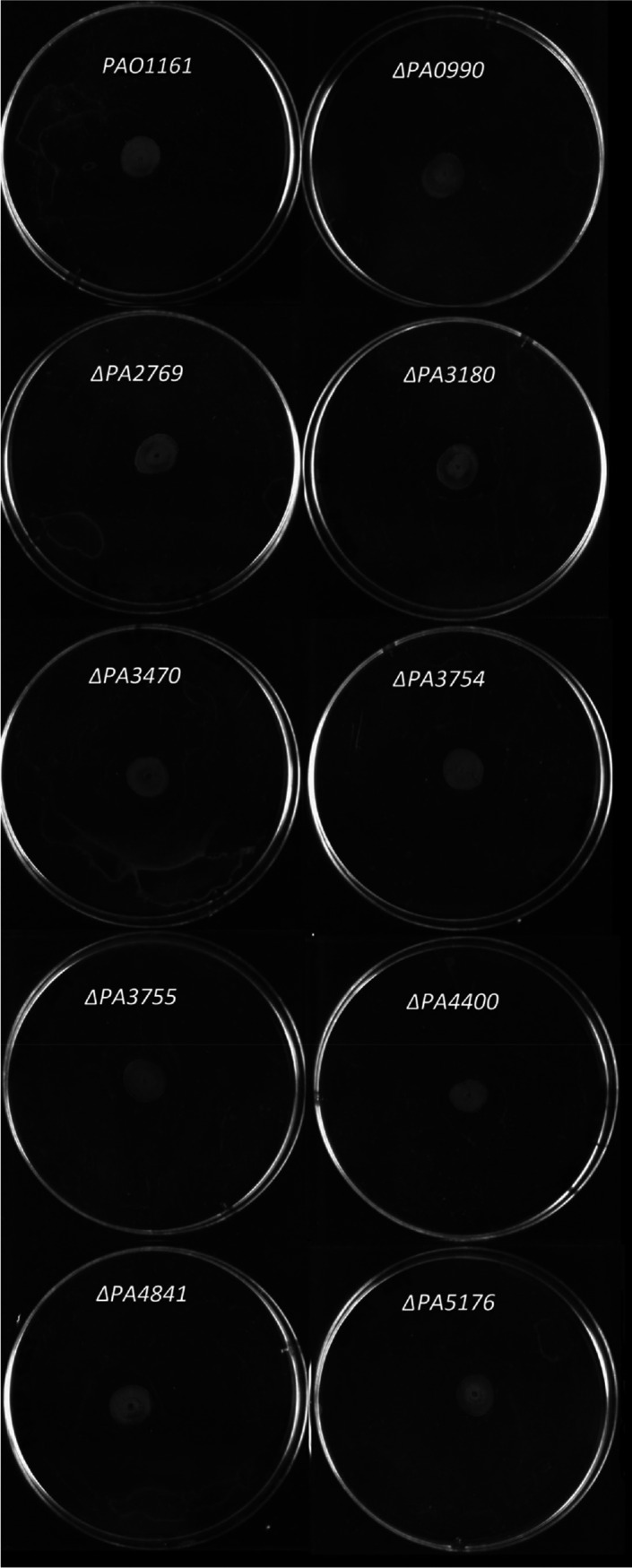
It is well recognized that motility is strongly associated with bacterial adaptation and virulence. To establish whether the Nudix proteins influenced motility, each mutated strain was tested for their swimming, swarming, and twitching ability. While the swimming and twitching motility was not disturbed in the mutants (Figures A2 and A4), the lack of either PA4400 or PA5176 protein severely impaired the type IV pili–flagella–rhamnolipids‐dependent swarming as compared to the wild‐type cells (Figures 3 and A3).
FIGURE 3.

Effect of ΔPA4400 and ΔPA5176 mutation on swarming. Swarming motility of wild‐type Pseudomonas aeruginosa PAO1161 and ΔPA4400 or ΔPA5176 mutants was examined as described in Experimental Procedures. Representative results obtained from three independent experiments are shown
3.4. Pyocyanin production and antibiotic susceptibility
We have earlier shown that Nudix hydrolase PA0336 (RppH) influences pyocyanin production in P. aeruginosa (Kujawa et al., 2017). To check whether any other Nudix protein could be important for this process, the level of pyocyanin was determined in the nudix mutants. Pyocyanin content was increased in the ΔPA3470, ΔPA3754, and ΔPA4400 mutants by 43%, 58%, and 33%, respectively (Figure 4a). Interestingly, the same mutants were also more resistant to piperacillin than was the parental strain or the other nudix mutants. (Figure 5a). Notably, both differences were nullified following trans‐complementation with the respective nudix gene (Figures 4b and 5b). Except for piperacillin, no significant differences in the response to other antibiotics were observed between the strains assayed (Figure A5).
FIGURE 4.

Effect of nudix mutation on pyocyanin production. (a) Production of pyocyanin by wild‐type Pseudomonas aeruginosa PAO1161 and nudix mutants. (b) Production of pyocyanin by nudix mutants transformed either with empty pBBR vector or pBBR carrying wild‐type copy of the respective nudix gene. Pyocyanin level (μg/ml) was determined at the stationary phase of growth. The mean value of at least three independent replicates ± SD is shown. Significant differences were indicated as *p ≤ .05
FIGURE 5.

Effect of nudix mutation on antibiotic susceptibility. (a) Susceptibility to piperacillin of wild‐type Pseudomonas aeruginosa PAO1161 and nudix mutants. (b) Susceptibility to piperacillin of ΔPA3470, ΔPA3754, and ΔPA4400 transformed either with empty pBBR vector or pBBR carrying wild‐type copy of the respective nudix gene. Susceptibility to the antibiotic was determined by disk diffusion assay as described in Experimental Procedures. The mean value of at least three independent replicates ± SD is shown. Significant differences were indicated as *p ≤ .05
3.5. Oxidative stress and mutation frequency
As a consequence of inflammation, bacteria colonizing human airways are exposed to massive oxidative stress caused by the reactive oxygen species released by leukocytes as the first line of defense (Ciofu, Riis, Pressler, Poulsen, & Høiby, 2005). To infect humans effectively P. aeruginosa responds with adaptive and protective strategies against these toxic molecules. To examine whether Nudix hydrolases participate in the response to such stress, the mutated P. aeruginosa strains were exposed to hydrogen peroxide. The ΔPA3470, ΔPA3754, ΔPA4400, and ΔPA4841 mutants were sensitized to killing by H2O2 (Figure 6a), indicating that these Nudix proteins may play a protective role, for example, by hydrolyzing the oxidized dNTPs which appear in the nucleotide pool following oxidative stress. Oxidized dNTPs are highly mutagenic (Pericone et al., 2002). To establish whether indeed these Nudix hydrolyze mutagenic dNTP derivatives mutation frequency was determined for the nudix mutants by measuring the frequency of appearance of antibiotic‐resistant colonies for two types of antibiotics. Apart from ΔPA4400, no mutant tested displayed an increased spontaneous mutation frequency (Table 2), suggesting that except PA4400, the other Nudix enzymes have no antimutator activity. Interestingly, the lack of PA5176 hydrolase increases bacterial resistance to killing by H2O2. Comparing to the parental strain, the ΔPA3470, ΔPA3754, ΔPA4400, and ΔPA4841 mutants complemented with a wild‐type copy of the respective gene were no longer sensitized to killing by H2O2 (Figure 6b).
FIGURE 6.

Effect of nudix mutation on susceptibility to killing by H2O2. (a) Susceptibility to H2O2 of Pseudomonas aeruginosa wild‐type PAO1161 and nudix mutant mutants. (b) Susceptibility to H2O2 of ΔPA3470, ΔPA3754, Δ PA4400, ΔPA4841, or ΔPA5176 transformed either with empty pBBR vector or pBBR carrying wild‐type copy of the gene indicated. To determine survivability, the bacteria were treated with 200 mM H2O2 as described in Experimental Procedures. The mean value of at least three independent replicates ± SD is shown. Significant differences were indicated as *p ≤ .05
TABLE 2.
StrR and FosR mutation frequency of PAO1161 and nudix mutants. Mutation frequency was measured as described in Experimental Procedures
| Strain | StrR | Fold change | FosR | Fold change |
|---|---|---|---|---|
| PAO1161 RifR | 6.26 × 10⁻¹⁰ | ‐ | 2.30 × 10⁻⁶ | ‐ |
| PAO1161 RifR Δ0990 | 6.22 × 10⁻¹⁰ | ‐ | 2.00 × 10⁻⁶ | ‐ |
| PAO1161 RifR Δ2769 | 5.80 × 10⁻¹⁰ | ‐ | 2.52 × 10⁻⁶ | ‐ |
| PAO1161 RifR Δ3180 | 6.20 × 10⁻¹⁰ | ‐ | 1.92 × 10⁻⁶ | ‐ |
| PAO1161 RifR Δ3470 | 5.95 × 10⁻¹⁰ | ‐ | 2.33 × 10⁻⁶ | ‐ |
| PAO1161 RifR Δ3754 | 6.00 × 10⁻¹⁰ | ‐ | 2.30 × 10⁻⁶ | ‐ |
| PAO1161 RifR Δ3755 | 5.50 × 10⁻¹⁰ | ‐ | 1.88 × 10⁻⁶ | ‐ |
| PAO1161 RifR Δ4400 | 2.33 × 10⁻⁷ | 372 | 0.80 × 10⁻⁴ | 35 |
| PAO1161 RifR Δ4841 | 6.18 × 10⁻¹⁰ | ‐ | 2.20 × 10⁻⁶ | ‐ |
| PAO1161 RifR Δ5176 | 5.89 × 10⁻¹⁰ | ‐ | 2.41 × 10⁻⁶ | ‐ |
3.6. Virulence
To determine whether the lack of Nudix hydrolase affected bacterial virulence, we employed C. elegans, a widely used model eukaryotic organism to study various aspects of host‐pathogen interactions (Tan, Mahajan‐Miklos, & Ausubel, 1999). Only the ΔPA4400 and ΔPA5176 mutants displayed an attenuated virulence compared to the wild type, indicating that these Nudix proteins participate in P. aeruginosa pathogenicity (Figure 7a and Appendix Figure A6). As before, the differences in virulence between the mutants and the parental strain were lost when the mutants were trans‐complemented with a wild‐type copy of the PA4400 or PA5176 gene (Figure 7b).
FIGURE 7.

Effect of nudix mutation on virulence. (a) Effect of ΔPA4400 and ΔPA5176 mutation on Pseudomonas aeruginosa virulence. (b) Effect of Δ PA4400 and ΔPA5176 transformed either with empty pBBR vector or pBBR, carrying wild‐type copy of the gene indicated, on P. aeruginosa virulence. Pseudomonas aeruginosa–Caenorhabditis elegans infection model was used to determine the pathogenicity of Pseudomonas strains as described in Experimental Procedures. The experiment was repeated at least three times. Shown are results of the representative experiment presented as the Kaplan–Meier survival curves of C. elegans fed wild‐type PAO1161, nudix mutants or mutants complemented by the wild‐type copy of the PA4400 or PA5176 gene. The significant differences between wild type and mutant were determined by the log‐rank test (**** p < .0001)
4. DISCUSSION
Here, we present characteristics of the ΔPA0990, ΔPA2769, ΔPA3180, ΔPA3470, ΔPA3754, ΔPA3755, ΔPA4400, ΔPA4841, and ΔPA5176 nudix mutants of P. aeruginosa, focusing on pathogenesis and stress‐related features.
The first characterized Nudix family member was the MutT protein from E. coli. A mutT1 mutant strain displayed a significantly higher spontaneous mutation frequency than the wild type (Treffers, Spinelli, & Belser, 1954). Expression, purification, and characterization of the gene product led to the identification of a new enzyme, a nucleoside triphosphatase with a preference for dGTP and its mutagenic oxidized derivative 8‐oxo dGTP (Yanofsky, Cox, & Horn, 1966).
Functional homologues of the E. coli antimutator MutT protein were identified in other bacteria including Mycobacterium tuberculosis, Bacillus subtilis, Bdellovibrio bacteriovorus, Vibrio parahaemolyticus, and Streptococcus oligofermentans (Castellanos‐Juárez et al., 2006; Patil, Sang, Govindan, & Varshney, 2013; Steyert, Messing, Amzel, Gabelli, & Piñeiro, 2008; Wagley et al., 2018; Zhou, Liu, Tong, & Dong, 2012). It was also noticed that the PA4400 gene of P. aeruginosa can complement mutT‐ deficient strain of E. coli (Oliver, Sánchez, & Blázquez, 2002). Inactivation of the P. aeruginosa PA4400 gene increased spontaneous mutation frequency indicating that the encoded protein has an antimutator effect (Sanders, Sudhakara, & Sutton, 2009). In addition to these observations, we found here that the ΔPA4400 mutation stimulates pyocyanin production, severely impairs swarming motility, decreases virulence, and increases resistance to piperacillin. Moreover, we found possible participation of PA4400 protein in thiamine biosynthesis. This hypothesis is under investigation.
A vast majority of Nudix enzymes are not highly specific and exhibit considerable substrate ambiguity, which makes it difficult to assess their biological functions based solely on the substrate preferences established in vitro. Up to now, more than one hundred chemical compounds that are hydrolyzed by Nudix enzymes have been identified (Srouji, Xu, Park, Kirsch, & Brenner, 2017). Despite this, in numerous cases, the identity of the true physiological substrate of many of these hydrolases is uncertain (McLennan, 2013; Nguyen et al., 2016). It appears therefore that the best way to assess the biological role of a Nudix hydrolase is to study the cellular effects of its deficiency.
Of the nine nudix mutants tested, four, ΔPA0990, ΔPA2769, ΔPA3180, and ΔPA3755, did not display any significant phenotypic changes compared to the parental strain. This observation suggests that either these proteins are not essential for the bacteria under the experimental conditions used or that they can be functionally substituted by other Nudix enzymes. Despite the effort, we were unable to construct the ΔPA2625 and ΔPA4971 mutants. Whether it was due to the physiological significance of these genes or to the recombination difficulties in the respective loci remains to be recognized.
Similarly to the ΔPA4400, disabling of the PA3470, PA3754, or PA4841 genes sensitized P. aeruginosa to killing by H2O2, suggesting that the gene products could participate in the repair of the cytotoxic lesions caused by oxidative stress as has been shown for the P. aeruginosa PA4400 protein (Oliver et al., 2002 and Table 2 here). It has also been observed that in addition to their preferred substrates some other non‐MutT Nudix hydrolases often display a residual antimutator activity (Arczewska et al., 2011; Dos Vultos, Blazquez, Rauzier, Matic, & Gicquel, 2006). However, no increase in the mutation frequency was observed in these nudix mutants indicating that the PA3470, PA3754 and PA4841 proteins have no such activity.
It is well known that cell motility and pyocyanin production are among the factors modulating P. aeruginosa virulence (Hall et al., 2016; Kazmierczak, Schniederberend, & Jain, 2015). However, the increased pyocyanin level in ΔPA3470, ΔPA3754, and ΔPA4400 was not accompanied by an increased virulence. Most probably, this was due to the mutant's increased sensitivity to H2O2. It is well recognized that H2O2 production by macrophages is often the host's first line of defense against a pathogen.
Of the mutants tested only ΔPA4400 and ΔPA5176 with severely affected swarming motility also displayed lower virulence as compared to the parental strain, which suggests that in the experimental conditions used swarming motility is the most significant determinant of P. aeruginosa pathogenicity.
Taken together, we have demonstrated that most of the Nudix hydrolases present in P. aeruginosa are important in the response to genotoxic stress and only a few play a role in bacterial pathogenicity.
CONFLICT OF INTEREST
None declared.
AUTHOR CONTRIBUTIONS
Elzbieta Kraszewska: Conceptualization (lead); Funding acquisition (lead); Supervision (lead); Writing – review and editing (lead). Joanna Drabinska: Formal analysis (equal); Investigation (equal); Methodology (equal). Mateusz Ziecina: Data curation (equal); Investigation (equal); Methodology (equal). Marta Modzelan: Investigation (supporting); Methodology (supporting). Grazyna Jagura‐Burdzy: Funding acquisition (supporting); Supervision (supporting); Writing – review and editing (supporting).
ETHICS STATEMENT
None required.
ACKNOWLEDGMENTS
This work was supported by grant UMO‐2014/15/B/NZ6/02562 from the National Science Center (Narodowe Centrum Nauki). Caenorhabditis elegans strain was provided by the CGC, which is funded by the NIH Office of Research Infrastructure Programs (P40 OD010440).
APPENDIX 1.
SUPPLEMENTARY EXPERIMENTAL PROCEDURES
Bacterial transformation
Competent Escherichia coli were prepared by the standard CaCl2 method, and plasmid DNA was introduced by the heat shock method (Sambrook et al., 1989).
DNA manipulations
Purification of plasmids and Pseudomonas genomic DNA was conducted using Plasmid MiniKit and Genomic Mini Kit, respectively (A&A Biotechnology), according to the manufacturer's instructions. To purify DNA from agarose, gel slices were cut out and processed using the Gel‐Out system (A&A Biotechnology) according to the manufacturer's instructions. PCR amplification was performed using high fidelity Pfu DNA polymerase (Promega). Usually, 30 cycles of amplification were performed, at reaction conditions depending on the pairs of primers and the size of the final amplification product. The PCR reactions were conducted on a PTC‐200 EngineCycler (MJ Research) and the PCR products were identified by agarose gel electrophoresis. The primers are listed in Table A2.
RNA isolation and RT‐PCR
RNA was isolated with the use of Total RNA Zol‐OutTM Kit (A&A Biotechnology) and genomic DNA was removed with the use of the Clean‐UP RNA Concentrator kit (A&A Biotechnology), according to the manufacturer's instructions. The RT‐PCR reaction was performed with the use of the QuantiTect® Reverse transcription Kit (Qiagen) according to the manufacturer's instructions. A reaction without the addition of reverse transcriptase was prepared for negative control. RNA isolated from wild‐type P. aeruginosa PAO1161RifR was used as a positive control.
TABLE A1.
Plasmids used in this study
| Name | Relevant feature | Source |
|---|---|---|
| pQE−80L | oriColE1 ApR T5p lacOlacIq His6 tag, expression vector | Qiagen |
| pBBR | araBADp, araC,CmR broad‐host‐range expression vector | Bartosik et al., (2014) |
| pAKE600 | oriMB1 oriRK2 ApR sacB | El‐Sayed et al. (2001) |
| pEMG | KmR, oriR6K, lacZα with two flanking I‐SceI sites | Martínez‐García and de Lorenzo (2011) |
| pRK2013 | ori (ColE1) tra + (RK2) KmR helper plasmid for conjugation | Martínez‐García and de Lorenzo (2011) |
| pSW‐I | ApR, oriRK2, xylS, Pm → I‐sceI (transcriptional fusion of I‐sceI to Pm) | Wong and Mekalanos (2000) |
| pAKEΔ0990 | pAKE600 derivative with 297 upstream bp (incl. first 15 bp of the gene) and 300bp downstream incl. 3 bp of PA0990 gene cloned as EcoRI‐BamHI fragment separated by GmR gene cloned into PstI site | This study |
| pAKEΔ2769 | pAKE600 derivative with 368 bp upstream (incl. first 6 bp of the gene) and 249 bp downstream incl. 7 bp of PA2769 gene cloned as EcoRI‐BamHI fragment separated by GmR gene cloned into PstI site | This study |
| pAKEΔ3180 | pAKE600 derivative with 300 bp upstream (incl. first 15 bp of the gene) and 351 bp downstream incl. last 3 bp of PA3180 gene cloned as EcoRI‐BamHI fragment separated by GmR gene cloned into PstI site | This study |
| pEMGΔ3470 | pEMG derivative with 320 bp upstream (incl. first 15 bp of the gene) and the last 435 bp downstream incl. 7 bp of PA3470 gene cloned as EcoRI‐BamHI fragment separated by GmR gene cloned into PstI site | This study |
| pEMGΔ3754 | pEMG derivative with 446 bp upstream (incl. first 9 bp of the gene.) and the 369 bp downstream incl. 9 bp of PA3754 gene cloned as EcoRI‐BamHI fragment separated by GmR gene cloned into XbaI site | This study |
| pAKEΔ3755 | pAKE600 derivative with 305 bp upstream(incl. 6 first bp of the gene) and 297 bp downstream incl. 3 bp of PA3755 gene cloned as EcoRI‐BamHI fragment separated by GmR gene cloned into PstI site | This study |
| pEMGΔ4400 | pEMG derivative with 302 bp upstream (incl. first 3 bp of the gene) and the 317 bp downstream incl. 9 bp of PA4400 gene cloned as EcoRI‐BamHI fragment separated by GmR gene cloned into XbaI site | This study |
| pEMGΔ4841 | pEMG derivative with 301 bp upstream (first 3 bp of the gene) and 301 bp incl. 12 bp of PA4841 gene cloned as EcoRI‐BamHI fragment separated by GmR gene cloned into KpnI site | This study |
| pAKEΔ5176 | pAKE600 derivative with 327 bp upstream (incl. first 22 bp of the gene) and 300 bp downstream incl. 50 bp of PA5176 gene cloned as EcoRI‐BamHI fragment separated by GmR gene cloned into PstI site | This study |
| pQE3470 | pQE‐80L derivative with PA3470 cloned into BamHI‐PstI site of the vector | This study |
| pQE3754 | pQE‐80L derivative with PA3754 cloned into BamHI‐PstI site of the vector | This study |
| pQE3755 | pQE‐80L derivative with PA3755 cloned into BamHI‐PstI site of the vector | This study |
| pQE4400 | pQE‐80L derivative with PA4400 cloned into BamHI‐PstI site of the vector | This study |
| pQE4841 | pQE‐80L derivative with PA4841 cloned into BamHI‐PstI site of the vector | This study |
| pQE5176 | pQE‐80L derivative with PA5176 cloned into BamHI‐PstI site of the vector | This study |
| pBBR3470 | pBBR derivative with His6‐tag‐PA3470 cloned into BamHI‐PstI site of the vector | This study |
| pBBR3754 | pBBR derivative with His6‐tag‐PA3754 cloned into EcoRI‐SacI site of the vector | This study |
| pBBR3755 | pBBR derivative with His6‐tag‐PA3755 cloned into EcoRI‐SacI site of the vector | This study |
| pBBR4400 | pBBR derivative with His6‐tag‐PA4400 cloned into EcoRI‐SacI site of the vector | This study |
| pBBR4841 | pBBR derivative with His6‐tag‐PA4841 cloned into EcoRI‐SacI site of the vector | This study |
| pBBR5176 | pBBR derivative with His6‐tag‐PA5176 cloned into EcoRI‐SacI site of the vector | This study |
TABLE A2.
Primers used in this study
| Name | Sequence 5′ → 3′ |
Restriction enzyme if used (underlined) |
Usage |
|---|---|---|---|
| RT0990F | CCGTCTTCTGGAATTGATACT | RT‐PCR analysis | |
| RT0990R | CCAGAGCGATCCAGTTCTCG | RT‐PCR analysis | |
| RT2625F | CTGAGCGCCGTCACCGGCAT | RT‐PCR analysis | |
| RT2625R | AGTTCGTCGCGGGTCAGCCA | RT‐PCR analysis | |
| RT2769F | TGATCTTGCGGGACGGCAAG | RT‐PCR analysis | |
| RT2769R | CGCCCCTCGAAGACGTCATT | RT‐PCR analysis | |
| RT3180F | AACCTCGTATCGAATGGCAG | RT‐PCR analysis | |
| RT3180R | ATCGCCAGTATTGTCCTTCG | RT‐PCR analysis | |
| RT3470F | GCACCTCGGCAGCTTCCAG | RT‐PCR analysis | |
| RT3470R | GAGATTGTCCGGCTGCGCTTG | RT‐PCR analysis | |
| RT3754F | GCCTTCATTCCCGACTTCGT | RT‐PCR analysis | |
| RT3754R | TTGACCAACTCCACCACCATGA | RT‐PCR analysis | |
| RT3755F | ATGGAGAACGGCGAGACCCT | RT‐PCR analysis | |
| RT3755R | GGGAATCTCCGCTTCGTCGA | RT‐PCR analysis | |
| RT4400F | AGTGGAGTTCGTCACCCTTT | RT‐PCR analysis | |
| RT4400R | GCCAGAACGCACGGATAC | RT‐PCR analysis | |
| RT4841F | GCCTCCGACGCCGAACTCAT | RT‐PCR analysis | |
| RT4841R | GATAGACCGCCTTGCTCAGGG | RT‐PCR analysis | |
| RT4971F | GCTTCCGTGGCTTCTATCGT | RT‐PCR analysis | |
| RT4971R | GGTTGGCGAGCTTCTGCATC | RT‐PCR analysis | |
| RT5176F | GAGTTGCAACTGCGCTTCAG | RT‐PCR analysis | |
| RT5176R | CTTGGGCAACGACAACTGGT | RT‐PCR analysis | |
| Δ0990‐F1 | CCGGAATTCCTCCTACGGTAAGTC | EcoRI | Mutant construction |
| Δ0990‐R1 | AACTGCAGGCTGACCGATGGCAT | PstI | Mutant construction |
| Δ0990‐F2 | AACTGCAGTAGCCCCCCGACCTC | PstI | Mutant construction |
| Δ0990‐R2 | CGGGATCCCTAGCACGGATGAGC | BamHI | Mutant construction |
| Δ2625‐F1 | CGGAATTCCGAAGACGCTGAGC | EcoRI | Mutant construction |
| Δ2625‐R1 | AACTGCAGCCAGCTCATGGATGTC | PstI | Mutant construction |
| Δ2625‐F2 | AACTGCAGGCCTGATAGAATCCGCG | PstI | Mutant construction |
| Δ2625‐R2 | CGGGATCCAAGTGCTCGAACACG | BamHI | Mutant construction |
| Δ2769‐F1 | CGGAATTCATTACCTCGTGGTGGTCGA | EcoRI | Mutant construction |
| Δ2769‐R1 | AACTGCAGCGGCATCGTCGTACTCCTG | PstI | Mutant construction |
| Δ2769‐F2 | AACTGCAGGCGCTGAGCGACAGGC | PstI | Mutant construction |
| Δ2769‐R2 | CGGGATCCATGTCCTGCTGGGCTG | BamHI | Mutant construction |
| Δ3180‐F1 | CGGAATTCCTGGCCGAGGCTG | EcoRI | Mutant construction |
| Δ3180‐R1 | AACTGCAGAGGGACATCGTGCACC | PstI | Mutant construction |
| Δ3180‐F2 | AACTGCAGTAACGCCGATGACGTTAG | PstI | Mutant construction |
| Δ3180‐R2 | CGGGATCCCTTCAAGCTGTTTCC | BamHI | Mutant construction |
| Δ3470‐F1 | CGGAATTCGTGCTCACCCGCAATC | EcoRI | Mutant construction |
| Δ3470‐R1 | AACTGCAGCAGGTTGTCGGTCATTGC | PstI | Mutant construction |
| Δ3470‐F2 | AACTGCAGCATGTGCGCTGAGCG | PstI | Mutant construction |
| Δ3470‐R2 | CGGGATCCACAACGCCTATATCTTCC | BamHI | Mutant construction |
| Δ3754‐F1 | CGGAATTCAAGGGTATAGAGCTGCAG | EcoRI | Mutant construction |
| Δ3754‐R1 | TATCTAGAGCAGTTCATACTGCCGGG | XbaI | Mutant construction |
| Δ3754‐F2 | TCTCTAGACTGAGTTGACCGACGG | XbaI | Mutant construction |
| Δ3754‐R2 | AGGGATCCGTACTTGCCGATCTTC | BamHI | Mutant construction |
| Δ3755‐F1 | CGGAATTCATGGGTGGACAAACCG | EcoRI | Mutant construction |
| Δ3755‐R1 | AACTGCAGTTTCATGCCAGGTTCTTC | PstI | Mutant construction |
| Δ3755‐F2 | AACTGCAGTGAACGCCAAACACTCG | PstI | Mutant construction |
| Δ3755‐R2 | CGGGATCCTCCAGTAGAAGCCTTC | BamHI | Mutant construction |
| Δ4400‐F1 | CGGAATTCGCGACACGACAGGATA | EcoRI | Mutant construction |
| Δ4400‐R1 | CGTCTAGACACGGATCATCTC | XbaI | Mutant construction |
| Δ4400‐F2 | GCTCTAGAGGCCTTTGACGG | XbaI | Mutant construction |
| Δ4400‐R2 | ATGGATCCGGGCCCGCTGAA | BamHI | Mutant construction |
| Δ4841‐F1 | CGGAATTCCTGGCTGCAGATCGAC | EcoRI | Mutant construction |
| Δ4841 R1 | GGGGTACCCATGGGTCAGCC | KpnI | Mutant construction |
| Δ4841‐F2 | GGGGTACCCTGGACGCCTGAG | KpnI | Mutant construction |
| Δ4841‐R2 | CGGGATCCGCGTCTTTGGGTTGC | BamHI | Mutant construction |
| Δ4971‐F1 | CGGAATTCCTTGAAGAGGCT | EcoRI | Mutant construction |
| Δ4971‐R1 | TATCTAGAGGTTTCGGACATC | XbaI | Mutant construction |
| Δ4971‐F2 | TATCTAGAGCCTGAACCTGCT | XbaI | Mutant construction |
| Δ4971‐R2 | CGGGATCCTCATTCAACTGG | BamHI | Mutant construction |
| Δ5176‐F1 | CGGAATTCAGTACCAGTTGAGCTGGC | EcoRI | Mutant construction |
| Δ5176‐R1 | AACTGCAGGTACGGTGGGTTTCTGAC | PstI | Mutant construction |
| Δ5176‐F2 | AACTGCAGTGTACCTGGTGCGCG | PstI | Mutant construction |
| Δ5176‐R2 | CGGGATCCTGAACTCCTCGCTAC | BamHI | Mutant construction |
| gentxbaF | GCTCTAGAATGTTACGCAGCA | XbaI | Mutant construction |
| gentxbaR | GCTCTAGATTAGGTGGCGGT | XbaI | Mutant construction |
| gentpstF | ATCTGCAGATGTTACGCAGCA | PstI | Mutant construction |
| gentpstR | ATCTGCAGTTAGGTGGCGGT | PstI | Mutant construction |
| gentkpnF | ATGGTACCATGTTACGCAGCA | KpnI | Mutant construction |
| gentkpnR | ATGGTACCTTAGGTGGCGGT | KpnI | Mutant construction |
| QE3470F | TAGGATCCACCGACAACCTGCTG | BamHI | Mutation complementation |
| QE3470R | GCCTGCAGTCAGCGCACATGAT | PstI | Mutation complementation |
| QE3754F | TAGGATCCAACTGCACGCTCGA | BamHI | Mutation complementation |
| QE3754R | GCCTGCAGTCAACTCAGTTTG | PstI | Mutation complementation |
| QE3755F | CGGGATCCAAATTCTGCAGCCTG | BamHI | Mutation complementation |
| QE3755R | ACAAGCTTTCAGTCTTTCTTATAGGAAGCC | HindIII | Mutation complementation |
| QE4400F | CGGGATCCAAACGAGTACATGTCG | BamHI | Mutation complementation |
| QE4400R | ACAAGCTTTCAAAGGCCGCCCG | HindIII | Mutation complementation |
| QE4841F | ATGGATCCATGGCCGCGCCGAT | BamHI | Mutation complementation |
| QE4841R | CGCAAGCTTTCAGGCGTCCAGGT | HindIII | Mutation complementation |
| QE5176F | ATGGATCCCGTCAGAAACCCAC | BamHI | Mutation complementation |
| QE5176R | AGCTGCAGTCATGCCTGGTACTC | PstI | Mutation complementation |
| QEecoF | CCGGAATTCATGAGAGGATCGCATCACCATC | EcoRI | Mutation complementation |
| BBR3470R | GCGAGCTCTCAGCGCACATGATC | SacI | Mutation complementation |
| BBR3754R | GCGAGCTCTCAACTCAGTTTGATG | SacI | Mutation complementation |
| BBR3755R | GCGAGCTCTCAGTCTTTCTTATAGGAA | SacI | Mutation complementation |
| BBR4400R | TTGAGCTCTCAAAGGCCGCCCGG | SacI | Mutation complementation |
| BBR4841F | GCGCTAGCATGAGAGGATCGCA | NheI | Mutation complementation |
| BBR4841R | ATGAGCTCTCAGGCGTCCAGGT | SacI | Mutation complementation |
| BBR5176R | CGAGCTCTCATGCCTGGTACTC | SacI | Mutation complementation |
FIGURE A1.

Expression of nudix genes in Pseudomonas aeruginosa wild‐type PAO1161 and mutant strains analyzed by RT‐PCR. Lane 1: positive control, RT‐PCR productfrom wild‐type P. aeruginosa genomic DNA; Lane 2: negative control; Lane 3 RT‐PCR product from mutant strain genomic DNA
FIGURE A2.

Swimming motility of Pseudomonas aeruginosa PAO1161 wild type and mutant strains, tested as described under Experimental Procedures
FIGURE A3.

Swarming motility of Pseudomonas aeruginosa PAO1161 wild type and mutant strains. Tested as described under Experimental Procedures
FIGURE A4.
Twitching motility of Pseudomonas aeruginosa PAO1161 wild type and mutant strains. Tested as described under Experimental Procedures
FIGURE A5.

Antibiotics susceptibility of wild type Pseudomonas aeruginosa PAO1161 and nudix mutants tested with the use of disc diffusion assay as described in Experimental Procedures. CAZ10—ceftazidime (10 μg), CIP—ciprofloxacin (5 μg) CT—colistin (10 μg), IMP—imipenem (10 μg), MEM—meropenem (10 μg), PB—polymyxin B (300 μg), PRL—piperacillin (100 μg), TOB—tobramycin (10 μg)
FIGURE A6.

Effect of nudix mutations on Pseudomonas aeruginosa virulence. Pseudomonas aeruginosa–Caenorhabditis elegans infection model was used to determine pathogenicity of Pseudomonas strains as described under Experimental Procedures. The experiment was repeated at least three times. Shown are results of the representative experiments presented as the Kaplan–Meier survival curves of C. elegans fed wild type PAO1161 and nudix mutant strains. The significant differences between wild type and mutant were determined by the log‐rank test (****p < .0001)
Drabinska J, Ziecina M, Modzelan M, Jagura‐Burdzy G, Kraszewska E. Individual Nudix hydrolases affect diverse features of Pseudomonas aeruginosa . MicrobiologyOpen. 2020;9:e1052 10.1002/mbo3.1052
DATA AVAILABILITY STATEMENT
All data are provided in full in the results section of this paper.
REFERENCES
- Alva‐Pérez, J. , Arellano‐Reynoso, B. , Hernández‐Castro, R. , & Suárez‐Güemes, F. (2014). The invA gene of Brucella melitensis is involved in intracellular invasion and is required to establish infection in a mouse model. Virulence, 5, 563–574. [DOI] [PMC free article] [PubMed] [Google Scholar]
- Arczewska, K. D. , Baumeier, C. , Kassahun, H. , SenGupta, T. , Bjørås, M. , Kuśmierek, J. T. , & Nilsen, H. (2011). Caenorhabditis elegans NDX‐4 is a MutT‐type enzyme that contributes to genomic stability. DNA Repair, 10, 176–187. 10.1016/j.dnarep.2010.10.009 [DOI] [PubMed] [Google Scholar]
- Baba, T. , Ara, T. , Hasegawa, M. , Takai, Y. , Okumura, Y. , Baba, M. , … Mori, H. (2006). Construction of Escherichia coli K‐12 in‐frame, single‐gene knockout mutants: The Keio collection. Molecular Systems Biology, 2, 2006.0008. [DOI] [PMC free article] [PubMed] [Google Scholar]
- Bartosik, A. A. , Glabski, K. , Jecz, P. , Lasocki, K. , Mikosa, M. , Plochocka, D. , … Jagura‐Burdzy, G. (2014). Dissection of the region of Pseudomonas aeruginosa ParA that is important for dimerization and interactions with its partner ParB. Microbiology, 160, 2406–2420. 10.1099/mic.0.081216-0 [DOI] [PMC free article] [PubMed] [Google Scholar]
- Bartosik, A. A. , Mierzejewska, J. , Thomas, C. M. , & Jagura‐Burdzy, G. (2009). ParB deficiency in Pseudomonas aeruginosa destabilizes the partner protein ParA and affects a variety of physiological parameters. Microbiology, 155, 1080–1092. 10.1099/mic.0.024661-0 [DOI] [PMC free article] [PubMed] [Google Scholar]
- Boyer, H. W. , & Roulland‐Dussoix, D. (1969). A complementation analysis of the restriction and modification of DNA in Escherichia coli . Journal of Molecular Biology, 41, 459–472. 10.1016/0022-2836(69)90288-5 [DOI] [PubMed] [Google Scholar]
- Brenner, S. (1974). The genetics of Caenorhabditis elegans . Genetics, 77, 71–94. [DOI] [PMC free article] [PubMed] [Google Scholar]
- Castellanos‐Juárez, F. X. , Alvarez‐Alvarez, C. , Yasbin, R. E. , Setlow, B. , Setlow, P. , & Pedraza‐Reyes, M. (2006). YtkD and MutT protect vegetative cells but not spores of Bacillus subtilis from oxidative stress. Journal of Bacteriology, 188, 2285–2289. 10.1128/JB.188.6.2285-2289.2006 [DOI] [PMC free article] [PubMed] [Google Scholar]
- Ciofu, O. , Riis, B. , Pressler, T. , Poulsen, H. E. , & Høiby, N. (2005). Occurrence of hypermutable Pseudomonas aeruginosa in cystic fibrosis patients is associated with the oxidative stress caused by chronic lung inflammation. Antimicrobial Agents Chemotherapy, 49, 2276–2282. 10.1128/AAC.49.6.2276-2282.2005 [DOI] [PMC free article] [PubMed] [Google Scholar]
- de Lorenzo, V. , & Timmis, K. N. (1994). Analysis and construction of stable phenotypes in gram‐negative bacteria with Tn5‐ and Tn10‐derived mini transposons. Methods Enzymology, 235, 386–405. [DOI] [PubMed] [Google Scholar]
- Dos Vultos, T. , Blazquez, J. , Rauzier, J. , Matic, I. , & Gicquel, B. (2006). Identification of Nudix hydrolase family members with an antimutator role in Mycobacterium tuberculosis and Mycobacterium smegmatis . Journal of Bacteriology, 188, 3159–3161. 10.1128/JB.188.8.3159-3161.2006 [DOI] [PMC free article] [PubMed] [Google Scholar]
- El‐Sayed, A. K. , Hothersall, J. , & Thomas, C. M. (2001). Quorum‐sensing dependent regulation of biosynthesis of the polyketide antibiotic mupirocin in Pseudomonas fluorescens NCIMB 10586. Microbiology, 147, 2127–2139. [DOI] [PubMed] [Google Scholar]
- Essar, D. W. , Eberly, L. , Hadero, A. , & Crawford, I. P. (1990). Identification and characterization of genes for a second anthranilate synthase in Pseudomonas aeruginosa: Interchangeability of the two anthranilate synthases and evolutionary implications. Journal of Bacteriology, 172, 884–900. 10.1128/JB.172.2.884-900.1990 [DOI] [PMC free article] [PubMed] [Google Scholar]
- Gao, R. , Wei, W. , Hassan, B. H. , Li, J. , Deng, J. , & Feng, Y. (2019). A single regulator NrtR controls bacterial NAD+ homeostasis via its acetylation. eLife, 8, e51603. [DOI] [PMC free article] [PubMed] [Google Scholar]
- Hall, S. , McDermott, C. , Anoopkumar‐Dukie, S. , McFarland, A. , Forbes, A. , Perkins, A. , … Grant, G. D. (2016). Cellular effects of pyocyanin, a secreted virulence factor of Pseudomonas aeruginosa . Toxins, 8, E236. [DOI] [PMC free article] [PubMed] [Google Scholar]
- Jimenez, P. N. , Koch, G. , Thompson, J. A. , Xavier, K. B. , Cool, R. H. , & Quax, W. J. (2012). The multiple signaling systems regulating virulence in Pseudomonas aeruginosa . Microbiolology and Molecular Biolology Review, 76, 46–65. 10.1128/MMBR.05007-11 [DOI] [PMC free article] [PubMed] [Google Scholar]
- Kazmierczak, B. I. , Schniederberend, M. , & Jain, R. (2015). Cross‐regulation of Pseudomonas motility systems: The intimate relationship between flagella, pili and virulence. Current Opinion in Microbiology, 28, 78–82. 10.1016/j.mib.2015.07.017 [DOI] [PMC free article] [PubMed] [Google Scholar]
- Kujawa, M. , Lirski, M. , Ziecina, M. , Drabinska, J. , Modzelan, M. , & Kraszewska, E. (2017). Nudix‐type RNA pyrophosphohydrolase provides homeostasis of virulence factor pyocyanin and functions as a global regulator in Pseudomonas aeruginosa . Molecular Microbiology, 106, 381–394. [DOI] [PubMed] [Google Scholar]
- Lasocki, K. , Bartosik, A. A. , Mierzejewska, J. , Thomas, C. A. , & Jagura‐Burdzy, G. (2007). Deletion of the ParA (Soj) homologue in Pseudomonas aeruginosa causes ParB instability and affects growth rate, chromosome segregation, and motility. Journal of Bacteriology, 189, 5762–5772. 10.1128/JB.00371-07 [DOI] [PMC free article] [PubMed] [Google Scholar]
- Martínez‐García, E. , & de Lorenzo, V. (2011). Engineering multiple genomic deletions in Gramnegative bacteria: Analysis of the multiresistant antibiotic profile of Pseudomonas putida KT2440. Environmental Microbiology, 13, 2702–2716. 10.1111/j.1462-2920.2011.02538.x [DOI] [PubMed] [Google Scholar]
- McLennan, A. G. (2006). The Nudix hydrolase superfamily. Cellular and Molecular Life Sciences, 63, 123–143. 10.1007/s00018-005-5386-7 [DOI] [PMC free article] [PubMed] [Google Scholar]
- McLennan, A. G. (2013). Substrate ambiguity among the Nudix hydrolases: Biologically significant, evolutionary remnant, or both? Cellular and Molecular Life Sciences, 70, 373–385. 10.1007/s00018-012-1210-3 [DOI] [PMC free article] [PubMed] [Google Scholar]
- Messing, S. A. , Gabelli, S. B. , Liu, Q. , Celesnik, H. , Belasco, J. G. , Piñeiro, S. A. , & Amzel, L. M. (2009). Structure and biological function of the RNA pyrophosphohydrolase BdRppH from Bdellovibrio bacteriovorus . Structure, 17, 472–481. 10.1016/j.str.2008.12.022 [DOI] [PMC free article] [PubMed] [Google Scholar]
- Modzelan, M. , Kujawa, M. , Głąbski, K. , Jagura‐Burdzy, G. , & Kraszewska, E. (2014). NudC Nudix hydrolase from Pseudomonas syringae, but not its counterpart from Pseudomonas aeruginosa, is a novel regulator of intracellular redox balance required for growth, motility and biofilm formation. Molecular Microbiology, 93, 867–882. [DOI] [PubMed] [Google Scholar]
- Nguyen, V. N. , Park, A. , Xu, A. , Srouji, J. R. , Brenner, S. E. , & Kirsch, J. F. (2016). Substrate specificity characterization for eight putative Nudix hydrolases. Evaluation of criteria for substrate identification within the Nudix family. Proteins, 84, 1810–1822. 10.1002/prot.25163 [DOI] [PMC free article] [PubMed] [Google Scholar]
- Okon, E. , Dethlefsen, S. , Pelnikevich, A. , Barneveld, A. V. , Munder, A. , & Tümmler, B. (2017). Key role of an ADP ‐ ribose ‐ dependent transcriptional regulator of NAD metabolism for fitness and virulence of Pseudomonas aeruginosa . International Journal of Medical Microbiology, 307, 83–94. 10.1016/j.ijmm.2016.09.007 [DOI] [PubMed] [Google Scholar]
- Oliver, A. , Sánchez, J. M. , & Blázquez, J. (2002). Characterization of the GO system of Pseudomonas aeruginosa . FEMS Microbiology Letters, 217, 31–35. [DOI] [PubMed] [Google Scholar]
- Patil, A. G. , Sang, P. B. , Govindan, A. , & Varshney, U. (2013). Mycobacterium tuberculosis MutT1 (Rv2985) and ADPRase (Rv1700) proteins constitute a two‐stage mechanism of 8‐oxo‐dGTP and 8‐oxo‐GTP detoxification and adenosine to cytidine mutation avoidance. Journal of Biological Chemistry, 288, 11252–11262. [DOI] [PMC free article] [PubMed] [Google Scholar]
- Pericone, C. D. , Bae, D. , Shchepetov, M. , McCool, T. , Jeffrey, N. , & Weise, J. N. (2002). Short‐sequence tandem and nontandem DNA repeats and endogenous hydrogen peroxide production contribute to genetic instability of Streptococcus pneumoniae . Journal of Bacteriology, 184, 4392–4399. 10.1128/JB.184.16.4392-4399.2002 [DOI] [PMC free article] [PubMed] [Google Scholar]
- Rashid, M. H. , & Kornberg, A. (2000). Inorganic polyphosphate is needed for swimming, swarming, and twitching motilities of Pseudomonas aeruginosa . Proceedings of the National Academy of Sciences of the United States of America, 97, 4885–4890. 10.1073/pnas.060030097 [DOI] [PMC free article] [PubMed] [Google Scholar]
- Rodionov, D. A. , De Ingeniis, J. , Mancini, C. , Cimadamore, F. , Zhang, H. , Osterman, A. L. , & Raffaelli, N. (2008). Transcriptional regulation of NAD metabolism in bacteria: NrtR family of Nudix‐related regulators. Nucleic Acids Research, 36, 2047–2059. 10.1093/nar/gkn047 [DOI] [PMC free article] [PubMed] [Google Scholar]
- Sambrook, J. , Fritsch, E. F. , & Maniatis, T. (1989). Molecular cloning: A laboratory manual (2nd ed.) Cold Spring, NY: Cold Spring Harbor Laboratory Press. [Google Scholar]
- Sanders, L. H. , Sudhakara, J. , & Sutton, M. D. (2009). The GO system prevents ROS induced mutagenesis and killing in Pseudomonas aeruginosa . FEMS Microbiology Letters, 294, 89–96. [DOI] [PMC free article] [PubMed] [Google Scholar]
- Simon, R. , O'Connell, M. , Labes, M. , & Puhler, A. (1986). Plasmid vectors for the genetic analysis and manipulation of rhizobia and other gram‐negative bacteria. (1986). Methods in Enzymology, 118, 640–659. [DOI] [PubMed] [Google Scholar]
- Song, M. G. , Bail, S. , & Kiledjian, M. (2013). Multiple Nudix family proteins possess mRNA decapping activity. RNA, 19, 390–399. 10.1261/rna.037309.112 [DOI] [PMC free article] [PubMed] [Google Scholar]
- Srouji, J. R. , Xu, A. , Park, A. , Kirsch, J. F. , & Brenner, S. E. (2017). The evolution of function within the Nudix homology clan. Proteins, 85, 775–811. 10.1002/prot.25223 [DOI] [PMC free article] [PubMed] [Google Scholar]
- Steyert, S. R. , Messing, S. A. , Amzel, L. M. , Gabelli, S. B. , & Piñeiro, S. A. (2008). Identification of Bdellovibrio bacteriovorus HD100 Bd0714 as a Nudix dGTPase. Journal of Bacteriology, 190, 8215–8219. 10.1128/JB.01009-08 [DOI] [PMC free article] [PubMed] [Google Scholar]
- Tan, M. W. , Mahajan‐Miklos, S. , & Ausubel, F. M. (1999). Killing of Caenorhabditis elegans by Pseudomonas aeruginosa used to model mammalian bacterial pathogenesis. Proceedings of the National Academy of Sciences of the United States of America, 96, 715–720. 10.1073/pnas.96.2.715 [DOI] [PMC free article] [PubMed] [Google Scholar]
- Treffers, H. P. , Spinelli, V. , & Belser, N. O. (1954). A factor (or Mutator Gene) influencing mutation rates in Escherichia coli . Proceedings of the National Academy of Sciences of the United States of America, 40, 1064–1071. 10.1073/pnas.40.11.1064 [DOI] [PMC free article] [PubMed] [Google Scholar]
- Wagley, S. , Borne, R. , Harrison, J. , Baker‐Austin, C. , Ottaviani, D. , Leoni, F. , … Titball, R. W. (2018). Galleria mellonella as an infection model to investigate virulence of Vibrio parahaemolyticus . Virulence, 9, 197–207. [DOI] [PMC free article] [PubMed] [Google Scholar]
- Winsor, G. L. , Lam, D. K. , Fleming, L. , Lo, R. , Whiteside, M. D. , Yu, N. Y. , … Brinkman, F. S. (2011). Pseudomonas Genome Database: Improved comparative analysis and population genomics capability for Pseudomonas genomes. Nucleic Acids Research, 39(Database issue), D596–D600. 10.1093/nar/gkq869 [DOI] [PMC free article] [PubMed] [Google Scholar]
- Wolff, K. A. , de la Peña, A. H. , Nguyen, H. T. , Pham, T. H. , Amzel, L. M. , Gabelli, S. B. , & Nguyen, L. (2015). A redox regulatory system critical for mycobacterial survival in macrophages and biofilm development. PLoS Pathogology, 11, e1004839. [DOI] [PMC free article] [PubMed] [Google Scholar]
- Wong, S. M. , & Mekalanos, J. J. (2000). Genetic footprinting with mariner‐based transposition in Pseudomonas aeruginosa . Proceedings of the National Academy of Sciences of the United States of America, 97, 10191–10196. 10.1073/pnas.97.18.10191 [DOI] [PMC free article] [PubMed] [Google Scholar]
- Yanofsky, C. , Cox, E. C. , & Horn, V. (1966). The unusual mutagenic specificity of an E. coli mutator gene. Proceedings of the National Academy of Sciences of the United States of America, 55, 274–281. 10.1073/pnas.55.2.274 [DOI] [PMC free article] [PubMed] [Google Scholar]
- Zhang, Y. , Zborníková, E. , Rejman, D. , & Gerdes, K. (2018). Novel (p)ppGpp binding and metabolizing proteins of Escherichia coli .Mbio, 9, e02188‐17. [DOI] [PMC free article] [PubMed] [Google Scholar]
- Zhou, P. , Liu, L. , Tong, H. , & Dong, X. (2012). Role of operon aaoSo‐mutT in antioxidant defense in Streptococcus oligofermentans . PLoS ONE, 7, e38133 10.1371/journal.pone.0038133 [DOI] [PMC free article] [PubMed] [Google Scholar]
Associated Data
This section collects any data citations, data availability statements, or supplementary materials included in this article.
Data Availability Statement
All data are provided in full in the results section of this paper.