Significance
The emergence of antibiotic resistance poses a critical threat to the efficacy of antibiotic treatments. A resistant bacterial population must originally arise from a single cell that mutates or acquires a resistance gene. This single cell may, by chance, fail to successfully reproduce before it dies, leading to loss of the nascent resistant lineage. Here, we show that antibiotic concentrations that selectively favor resistance are nonetheless sufficient to reduce the chance of outgrowth from a single cell to a very low probability. Our findings suggest that lower antibiotic concentrations than those required to clear a large resistant population may be sufficient to prevent, with high probability, outgrowth of initially rare resistant mutants.
Keywords: antimicrobial resistance, Pseudomonas aeruginosa, inoculum effect, mathematical model, extinction probability
Abstract
A better understanding of how antibiotic exposure impacts the evolution of resistance in bacterial populations is crucial for designing more sustainable treatment strategies. The conventional approach to this question is to measure the range of concentrations over which resistant strain(s) are selectively favored over a sensitive strain. Here, we instead investigate how antibiotic concentration impacts the initial establishment of resistance from single cells, mimicking the clonal expansion of a resistant lineage following mutation or horizontal gene transfer. Using two Pseudomonas aeruginosa strains carrying resistance plasmids, we show that single resistant cells have <5% probability of detectable outgrowth at antibiotic concentrations as low as one-eighth of the resistant strain’s minimum inhibitory concentration (MIC). This low probability of establishment is due to detrimental effects of antibiotics on resistant cells, coupled with the inherently stochastic nature of cell division and death on the single-cell level, which leads to loss of many nascent resistant lineages. Our findings suggest that moderate doses of antibiotics, well below the MIC of resistant strains, may effectively restrict de novo emergence of resistance even though they cannot clear already-large resistant populations.
Antibiotics have had a huge impact on human health by reducing the burden associated with bacterial infections, and the use of antibiotics now underpins many areas of medicine. Unfortunately, antibiotic treatment is also associated with the evolution of resistance (1), resulting in poorer patient outcomes (2). A better understanding of how antibiotic dosing affects resistance evolution could aid the design of more effective treatment strategies that suppress pathogenic bacteria while reducing the risk of emergence of resistance.
Susceptibility of a bacterial strain to a particular antibiotic is typically quantified by the minimum inhibitory concentration (MIC), the lowest antibiotic concentration that prevents growth of this strain in a standardized assay, such as in ref. 3. Here, we will refer to any strain with reduced susceptibility relative to a reference “sensitive” strain simply as “resistant,” as is common in evolutionary microbiology literature (e.g., refs. 4–6), as opposed to defining resistance with respect to clinical breakpoints. Although antibiotic dosing strategies initially focused only on efficacy against sensitive bacteria (7), the past two to three decades have seen development of a large body of work investigating how antibiotic exposure affects emergence of resistance (8, 9). A prominent concept is that preexisting resistant subpopulations will be selectively enriched within a particular range of antibiotic concentrations, an idea first proposed in the 1990s (10–12), then refined by the definition of the “mutant prevention concentration” giving the upper bound of this range (13) and further developed into the “mutant selection window” (MSW) hypothesis (14–16). This hypothesis predicts that outgrowth of resistance occurs at antibiotic concentrations ranging between the MIC of the sensitive strain (which we denote MICS) and the mutant prevention concentration, which is approximated by the MIC of the most resistant single-step mutant (16). The MSW hypothesis has gained support from in vitro and animal model studies, and has been extended to consider time-varying drug concentrations (reviewed in ref. 17).
The MSW is defined by thresholds in absolute fitness (growth rates) of each strain in isolation, i.e., their MIC values. In evolutionary biology, however, selection refers to changes in proportions of genotypes in a population according to their differences in fitness relative to one another. Direct competition experiments have shown that resistant strains can have a competitive fitness advantage over sensitive strains, even at concentrations well below MICS (4, 11, 12, 18, 19). Thus, resistance can be selectively favored over a potentially very wide range of antibiotic concentrations (5), from concentrations considered too low to have any clinical benefit (below MICS), up to concentrations above the MIC of a resistant strain (MICR) that may be too high to achieve in clinical practice, because of physiological constraints on the accumulation of antibiotics in tissues (pharmacokinetics) and/or toxic side effects (20–22).
Selection operates efficiently when both sensitive and resistant populations are large, resulting in an increase in relative frequency of the fitter strain. Correspondingly, selection coefficients are typically measured by competition between large numbers of cells (typically >104 colony-forming units [CFU]) of both resistant and sensitive strains across a gradient of antibiotic concentrations (e.g., ref. 18). However, the de novo emergence of resistant strains should be subject to stochastic processes (23) that are not captured by the aforementioned experiments. First, resistance must stochastically arise in a sensitive cell by mutation, genomic instability (24), or acquisition of a resistance gene through horizontal gene transfer. Next, the single resistant cell thus generated must survive and successfully divide to produce daughter cells that likewise survive, and so on to generate a large number of resistant descendant cells. The latter process, which we will refer to throughout as “establishment” of resistance (23), will be our focus here. Importantly, due to the stochastic nature of cell divisions and deaths on the individual cell level, establishment is not guaranteed, even if the resistant strain has positive expected growth rate and a selective advantage over the sensitive strain (25). In particular, if antibiotics partially inhibit the resistant strain below its MICR, in the sense of increasing the chance that a resistant cell dies or fails to divide (i.e., decreasing absolute fitness), then the risk that a resistant lineage is stochastically lost should increase with antibiotic concentration, despite its selective (i.e., relative fitness) advantage. Despite the substantial body of work addressing the selection of resistance, very little experimental work has addressed the stochastic establishment phase (see, however, refs. 26–28).
We set out to quantify stochastic establishment in vitro in Pseudomonas aeruginosa, an important opportunistic pathogen that evolves resistance at an exceptionally high rate during infections (1, 29). To isolate the establishment phase, we inoculated hundreds of cultures, each with a very small number of resistant cells (on average, ∼1 to 3), and assessed culture growth. We tested two strains carrying nonconjugative plasmids (Rms149 and PAMBL2) that confer resistance to streptomycin and meropenem, respectively, across a range of antibiotic concentrations below the corresponding MICR. By fitting mathematical models to these data, we estimated the probability of establishment, i.e., of detectable culture growth due to clonal expansion from a single resistant cell, as a function of antibiotic concentration. Our key finding is that the establishment probability of resistant cells drastically declines at concentrations well below the MIC of the resistant strain, reaching at one-eighth of MICR in both systems. This result can be reconciled with the standard definition of the MIC through a simple quantitative relationship between inoculum size and probability of population growth (experimentally validated in the streptomycin system). Our results highlight that antibiotic selection pressure is not a sufficient condition for de novo emergence of resistance starting from single cells; it is also crucial to account for demographic stochasticity when evaluating the impact of dosing strategies on emergence of resistance. Our results suggest that lower antibiotic doses than those required to clear large resistant populations (i.e., sub-MICR) may be effective, with high probability, when resistant mutants are initially rare.
Results
Establishment of Resistance Is Inhibited by Sub-MICR Antibiotic Concentrations.
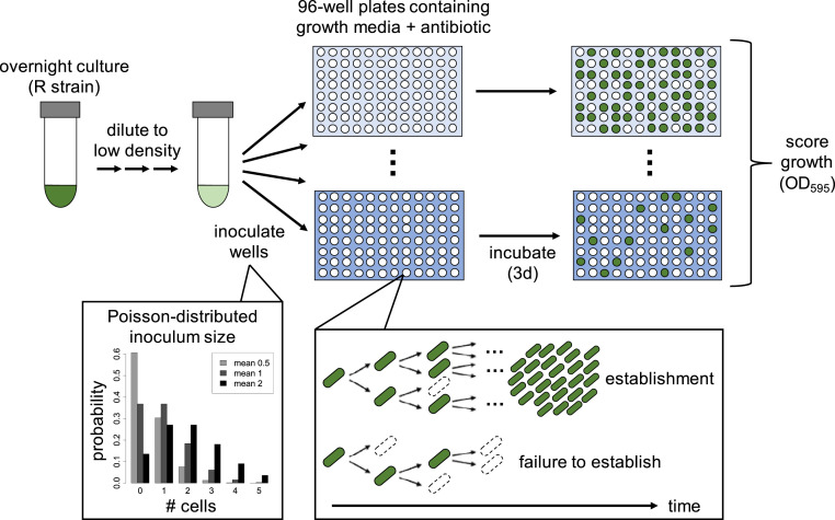
To elucidate the direct impact of antibiotics on resistant cells, we first investigated establishment of a resistant strain in the absence of a sensitive strain. We first focused on the streptomycin-resistant PA01:Rms149 strain. To estimate its probability of establishment, defined as outgrowth of a detectable (i.e., large) population from a single cell, we conducted large-scale “seeding” experiments (see also ref. 28). In this assay (Fig. 1), a highly diluted overnight culture of the resistant strain is inoculated into fresh media in a large number of replicate cultures. The high dilution factors yield average inoculum sizes of <1 to ∼3 cells per culture. Importantly, however, the actual number of cells inoculated into each replicate culture is random, and can be described by a Poisson distribution (SI Appendix, Fig. S1). One implication of this protocol is that many cultures are not inoculated with any cells, while others receive more than one cell; our modeling approach will account for this variation statistically. We inoculated parallel replicate cultures in streptomycin-free media and at a range of streptomycin concentrations below the MIC of the resistant strain, denoted MICR, as measured using standard protocols with larger inoculum size (ref. 3) (SI Appendix, Table S1). We then scored the number of replicate cultures showing growth based on reaching a threshold optical density (OD) of 0.1 within 3 d postinoculation.
Fig. 1.
Design of seeding experiments to estimate establishment probability. An overnight culture of the resistant strain is highly diluted and used to inoculate 96-well plates containing growth media (LB broth) with antibiotic at various concentrations (shades of blue). The number of cells inoculated per well follows a Poisson distribution (examples plotted for mean inoculum size of 0.5, 1, or 2 cells per well). Within these culture wells, stochastic population dynamics imply that each inoculated cell may either produce a large number of descendants (establishment) or produce no/few descendants that ultimately die out (failure to establish). Plates are incubated for 3 d, and optical density is measured to score growth in wells (OD595 > 0.1; dark green). The number of replicate cultures showing growth is used to estimate the per-cell establishment probability at each antibiotic concentration by fitting a mathematical model.
A culture could fail to grow either because the inoculum did not contain any cells, or because every cell in the inoculum failed to give rise to a surviving lineage. To infer the probability that a single cell yields detectable population growth (i.e., the per-cell establishment probability), we fit a mathematical model, accounting for both the random inoculum size and demographic stochasticity, to the observed number of replicate cultures showing growth (Materials and Methods). All probabilities are normalized by the result in streptomycin-free media, which corresponds to scaling inoculum size by the mean number of cells that establish in benign conditions (which we call the “effective” inoculum size). Thus, relative establishment probability equals 1 by definition in streptomycin-free conditions, while we expect with streptomycin treatment; however, values larger than 1 can arise due to sampling error.
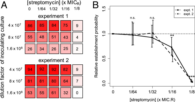
Our seeding experiments revealed that the probability of establishment of a single resistant cell declines with increasing streptomycin concentration (Fig. 2 and SI Appendix, Table S2). While exposure to the lowest tested concentrations of streptomycin (up to 1/32 × MICR) had no detectable impact on establishment, 1/16 × MICR was already sufficient for significant declines, to of 55 to 73% (maximum-likelihood estimates in two independent experiments). At 1/8 × MICR, dropped to just 3 to 5%. These results suggest that a resistant mutant would only rarely establish at antibiotic concentrations that are well below its MICR.
Fig. 2.
Establishment probability of single PA01:Rms149 streptomycin-resistant cells, estimated from seeding experiments. (A) Visual representation of the growth data, indicating the number of replicate cultures (out of 96) that grew in each test condition up to 3 d postinoculation. (B) Estimated relative per-cell establishment probability , scaled by the probability in streptomycin-free medium, as a function of streptomycin concentration, scaled by the standard MIC value of the resistant strain (MICR = 2,048 μg/mL; SI Appendix, Table S1). Results are shown for two separate experiments. Plotted points indicate the maximum-likelihood estimate of , and error bars indicate the 95% confidence interval, using the fitted model selected by the likelihood ratio test (experiment 1: model B′, fixed environmental effect; experiment 2: model C′, the null model [Eq. 1]. Both of these models pool data across three inoculation densities; see SI Appendix, Text, section 10, for details). Significance of the streptomycin effect is determined by fitting a generalized linear model to the population growth data (n.s.: not significant, P > 0.05; **P = 0.01 in experiment 1, P = 2e-7 in experiment 2, and P = 2e-8, pooling both experiments; ***P < 2e-16 in both experiments; see SI Appendix, Text, section 14.1, for full results).
MIC Depends on Inoculum Size.
The frequent failure of the resistant strain to grow in our seeding experiments at concentrations well below its MIC is, at face value, surprising. We hypothesized that these results could be explained by the difference in inoculum size between these assays. Specifically, standard MIC values are assessed from an inoculation density of 5 × 10⁵ CFU/mL (3), which corresponds to an inoculum size of 105 CFU per 200-μL culture on our microtiter plates. In contrast, our seeding experiments used an inoculum size on the order of 1 CFU per culture. MIC for many antibiotics has been observed to increase with higher-than-standard inoculation densities (CFU/ml) (30–32), which corresponds to higher absolute inoculum size (in CFU) for a fixed culture volume. Although less well explored, it has also occasionally been noted that MIC can decrease when lower absolute inoculum sizes are used (33, 34).
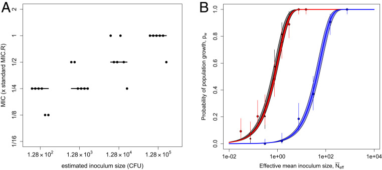
To test the hypothesis that inoculum size influences MIC in the present system, we conducted a modified MIC assay using the PA01:Rms149 strain with inoculum sizes ranging over three orders of magnitude, from ∼102 to 105 CFU per culture (corresponding to inoculation densities of 5 × 10² up to the standard 5 × 10⁵ CFU/mL). We found that MIC indeed increases with inoculum size (Fig. 3A). This pattern arises regardless of whether growth is scored at 20 h, as per the standard MIC assay protocol (3), or up to 3 d postinoculation, as in our seeding experiments, although the number of cultures showing detectable growth, and thus the measured MIC, tends to increase over time (SI Appendix, Fig. S2).
Fig. 3.
Inoculum size effects on MIC and probability of population growth of the resistant PA01:Rms149 strain in streptomycin. (A) MIC as a function of inoculum size. Cultures were inoculated with PA01:Rms149 at four different inoculum sizes. MIC was evaluated as the minimal tested streptomycin concentration that prevented detectable growth up to 3 d postinoculation; a qualitatively similar pattern arose if growth was evaluated at 20 h (SI Appendix, Fig. S2). The y axis is scaled by the MIC of this strain at standard inoculation density (MICR). The points represent six replicates at each inoculum size, with the line segments indicating their median. (B) Null model of the inoculum size effect (Eq. 1) fit to culture growth data. Probability of population growth (pw) is plotted as a function of effective mean inoculum size (, calibrated by the results in streptomycin-free media; SI Appendix, Fig. S4). Black: streptomycin-free; red: streptomycin at 1/16 × MICR; blue: 1/8 × MICR. These results are based on growth in streptomycin up to 5 d postinoculation; see SI Appendix, Text, section 15, for results at 3 d postinoculation. Points indicate the proportion of replicate cultures showing growth, i.e., the maximum-likelihood estimate (MLE) of pw in the full model, with error bars indicating the 95% confidence interval (CI). The solid line shows the best fit of the null model (i.e., Eq. 1 parameterized with the MLE of ), and the shaded area corresponds to the 95% CI. According to the likelihood ratio test, the null model deviance from the full model is not significant at any streptomycin concentration (streptomycin-free: P = 0.55; 1/16 × MICR: P = 0.28; 1/8 × MICR: P = 0.71; see SI Appendix, Text, section 15, for full results).
Since all cultures contained the same volume in the above experiment, this pattern could be due to changes either in absolute inoculum size (i.e., CFU) or in inoculation density (i.e., CFU per unit volume). These two possibilities are not typically distinguished in the literature; however, they lead to distinct interpretations. If demographic stochasticity is the dominant force, we expect absolute numbers to matter, whereas if interactions among cells (e.g., competition or cooperation) affect establishment, cell density per unit volume could be more important. To disentangle these two factors, we repeated the MIC assay covarying inoculation density and culture volume. This experiment confirmed that absolute inoculum size has a strong effect on MIC. In contrast, inoculation density per unit volume does not have a significant effect within the range that we tested, after controlling for absolute cell numbers (SI Appendix, Fig. S3).
Population Growth Can Be Explained by an Independent Chance of Each Cell to Establish.
Taken together, our seeding experiments and MIC assays reveal that the absolute number of cells in the inoculum has a strong effect on whether the culture eventually shows detectable growth. The simplest explanation for this result is that population growth can be attributed to the stochastic outgrowth of one or more lineages, each initiated by a single cell in the inoculum, acting independently. This independence assumption yields a “null model” that mathematically describes the effect of inoculum size on the probability of outgrowth of a detectable population (Materials and Methods and Eq. 1). Here, the probability of establishment of each cell in the inoculum is a scaling parameter, which does not depend on inoculum size. Note that this null model would not hold if interactions among cells substantially influence their chances of successful replication. For example, if cells secrete an enzyme that breaks down an antibiotic extracellularly, then the establishment probability of each cell could increase with inoculum size. On the other hand, if cells compete for limiting resources or secrete toxins, the per-cell establishment probability could decrease with inoculum size.
To formally test the null model, we again conducted seeding experiments with the PA01:Rms149 strain, but now using many different inoculum sizes, spanning approximately three orders of magnitude. We tested two streptomycin concentrations (1/16 and 1/8 × MICR) for which growth often failed from a single cell but succeeded from standard inoculum size in MIC assays. In parallel, we tested growth in streptomycin-free media in order to estimate the effective mean inoculum size (SI Appendix, Fig. S4). This left one free parameter, the per-cell relative establishment probability , to fit at each streptomycin concentration.
We found good agreement between the null model and our experimental data at all tested streptomycin concentrations, consistent with the hypothesis that cells establish independently (main experiment, Fig. 3B, and repeat experiments, SI Appendix, Fig. S5). More precisely, the null model did not show significant deviance from the observed proportion of populations that grew (according to the likelihood ratio test), and thus we accept it as a parsimonious explanation for the data. Furthermore, we obtain estimates of relative establishment probability, , at 1/16 and 1/8 × MICR similar to those from the previous seeding experiments (SI Appendix, Table S2).
To summarize, the probability of culture growth at any given streptomycin concentration depends on inoculum size, according to a simple quantitative relationship. Our experimental data are consistent with a simple stochastic model in which cells behave independently, such that a fixed per-cell establishment probability can explain our growth data across inoculum sizes. That is, cells are not “more susceptible” to streptomycin at lower inoculum sizes, but rather, culture growth is less likely to be observed simply because fewer cells are available to establish, and not all cells succeed. In turn, the minimal concentration of streptomycin required to prevent growth in a given proportion of replicate cultures (i.e., the observed MIC) increases with inoculum size.
Sub-MICR Streptomycin Concentrations Induce Resistant Cell Death and Extend Lag Phase.
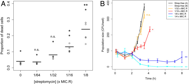
We hypothesized that resistant cells sometimes failed to establish in our seeding experiments because exposure to streptomycin compromised cell division rate and/or viability. As a simple test of this idea, we measured the relative abundance of dead cells in cultures of the resistant strain grown at sub-MICR concentrations of streptomycin. We found that the fraction of dead cells after 7 h of treatment, as determined by propidium iodide staining, increased from an average of 3 to 4% in streptomycin-free conditions to >20% at 1/8 × MICR streptomycin (Fig. 4A and SI Appendix, Fig. S6 and Table S3). Note that this is a conservative measure of cell death, because this assay only detects cells that have compromised membrane permeability, and not, for example, cells that have already lysed. Furthermore, this assay provides only a snapshot in time.
Fig. 4.
Effects of sub-MICR streptomycin treatment on PA01:Rms149 resistant cell dynamics. (A) Proportion of dead cells after 7 h in sub-MICR streptomycin. The proportion of dead cells in streptomycin-treated cultures was estimated using live–dead staining and flow cytometry. Points represent six independent treatment replicates at each concentration, and line segments indicate their mean. Differences from the streptomycin-free control cultures were assessed using a one-way ANOVA followed by a post hoc Dunnett’s test (n.s.: not significant, P > 0.05; *P = 9e-3; **P < 1e-4). Effects identified as significant do not change if we exclude an outlier replicate (shaded-in points) showing consistently elevated dead cell fractions (SI Appendix, Table S3). (B) Viable cell population dynamics in sub-MICR streptomycin. Points with connecting lines indicate the mean number of viable cells across 6 replicate cultures per streptomycin concentration, per sampling time point (or 12 replicates for streptomycin-free controls); the error bars indicate SE. SI Appendix, Fig. S7 shows all individual replicates. Viable cell numbers were estimated by plating undiluted culture samples; plots are truncated when colonies became too dense to count. Significance of each streptomycin concentration compared to the streptomycin-free control was assessed by a post hoc Dunnett’s test (n.s.: not significant, P = 0.87; *P = 4e-4; **P < 1e-4).
To gain further insight into how sub-MICR streptomycin impacts the population dynamics of the resistant strain, we quantified viable cell density over the first few hours after inoculation into streptomycin-containing media. Cultures were inoculated with ∼100 cells in this experiment, to ensure that cell numbers were low enough for demographic stochasticity to be relevant, yet large enough to be detectable using conventional plating methods.
We found that streptomycin treatment has a significant effect on the growth of resistant cultures (ANOVA, main effect: P < 2e-16), and this effect varies over time (ANOVA, interaction term: P < 2e-16; Fig. 4B and SI Appendix, Fig. S7). Following inoculation, cultures exhibited a lag phase of ∼2 h. Control cultures in streptomycin-free media then began to grow exponentially. The lowest tested concentration of streptomycin (1/32 × MICR) had no significant effect on these dynamics (Dunnett’s test: P = 0.87); however, 1/16 × MICR was already sufficient to slow growth (P = 4e-4). Nonetheless, all replicate cultures (n = 48 per concentration) eventually grew, as detected by OD. Meanwhile, higher doses of streptomycin (1/8 × or 1/4 × MICR) had dramatic effects on growth dynamics (P < 1e-4), with cultures exhibiting an extended lag phase of at least 7 to 8 h, in which viable cell density initially declined. After further incubation (up to 3 d), 25% of cultures (15/60) exposed to 1/8 × MICR eventually showed growth, while the remaining 75% (45/60) failed to reach detectable OD. At 1/4 × MICR, no viable cells were detected in most cultures from 4 h on, and only 1/60 cultures reached detectable OD within 3 d.
In summary, sub-MICR streptomycin treatment has the effect of extending the lag phase, before cultures eventually either grow to saturation or die out. Failure to grow can be explained by significantly elevated cell death rates beginning at 1/16 × MICR, which can lead to stochastic loss of initially small populations.
Stochastic Establishment Is Recapitulated for a Clinically Relevant Antibiotic and Resistance Plasmid.
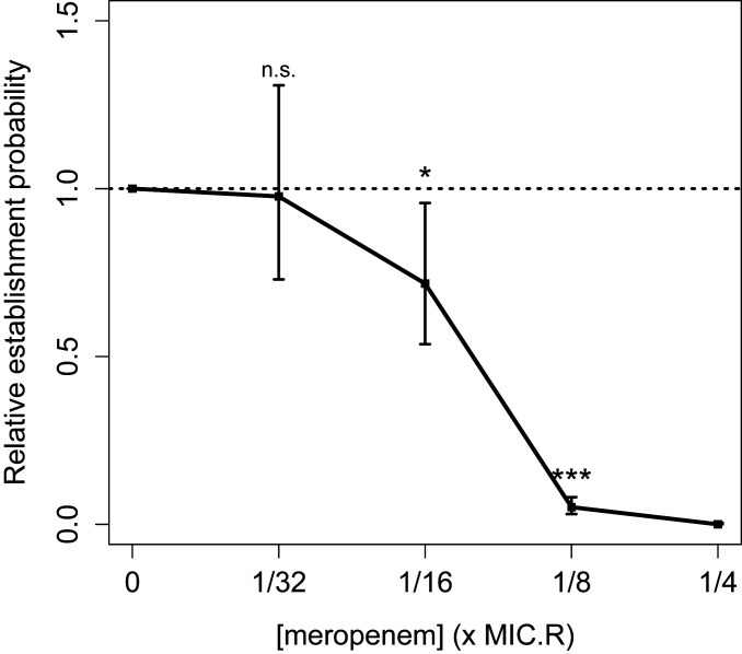
If the frequent failure of resistant cells to establish surviving populations at antibiotic doses well below their MIC is a general phenomenon, it would have important implications for understanding de novo emergence of resistance during antibiotic treatment. To check that our result was not driven by the specific choice of antibiotic or resistance mechanism, we repeated the key seeding experiment using a P. aeruginosa PA01 strain carrying a recently isolated multidrug resistance plasmid, PAMBL2 (35, 36), that confers resistance to meropenem through the blavim-1 carbapenemase. Carbapenems are an important treatment option for serious infections caused by Gram-negative bacterial pathogens (37), and resistance is of current clinical concern (38, 39); carbapenem-resistant P. aeruginosa has been identified as a “critical priority” for new antibiotic development by the World Health Organization (40). In agreement with our previous findings, the establishment probability of PA01:PAMBL2 cells declined at concentrations of meropenem well below this strain’s MICR (SI Appendix, Table S1), reaching ∼5% at 1/8 × MICR, while no establishment was observed at 1/4 × MICR (Fig. 5 and SI Appendix, Table S4). This result demonstrates that the stochastic loss of resistant cells is not unique to our primary model system of PA01:Rms149 in streptomycin.
Fig. 5.
Estimated relative per-cell establishment probability of the PA01:PAMBL2 meropenem-resistant strain as a function of meropenem concentration. Concentration is scaled by the standard MIC of this strain in meropenem (MICR = 512 μg/mL; SI Appendix, Table S1). Plotted points indicate the maximum-likelihood estimate of , and error bars indicate the 95% confidence interval, using the fitted model selected by the likelihood ratio test (model C′, the null model [Eq. 1], which pools data across two tested inoculation densities). Significance of the meropenem effect is determined by fitting a generalized linear model (GLM) to population growth data (n.s.: not significant, P > 0.05; *P = 0.02; ***P < 2e-16; see SI Appendix, Text, section 14.2, for full results). The 1/4 × MICR meropenem result was excluded from the GLM because zero replicates established.
The Sensitive Population Modulates Probability of Establishment of Resistant Cells.
So far, we focused on the direct effects of antibiotics on resistant cells by conducting experiments with monocultures of resistant strains. However, de novo resistance will actually arise within a sensitive population, by mutation or transfer of a mobile genetic element into a sensitive cell. Moreover, antibiotic treatment will only begin in clinical settings once the total pathogen population is large enough to cause symptoms. We therefore asked whether the presence of a large sensitive population affects the establishment of initially rare resistant cells during antibiotic treatment, returning to PA01 (sensitive) and PA01:Rms149 (streptomycin-resistant) as a model system.
We expect the sensitive population and the antibiotic to have interacting effects on establishment of resistance. In particular, at sufficiently low antibiotic concentrations, a sensitive strain is generally expected to outcompete a resistant strain due to the fitness cost associated with resistance (4, 18). We confirmed this expectation in our experimental system using a standard competition assay, where both strains start from large inoculum sizes (SI Appendix, Figs. S8–S10 and Table S5). We found that the sensitive strain is favored up to a minimum selective concentration (MSC) between 1 and 2 μg/mL streptomycin (equivalent to 1/32 to 1/16 × MICS, or 1/2,048 to 1/1,024 × MICR), in agreement with previous results for these strains (41). We hypothesized that competitive suppression by the sensitive strain would reduce absolute fitness of the resistant strain (5) and hence prevent its establishment at streptomycin concentrations below the MSC.
As a simple test of this idea, we modified the seeding experiment to inoculate very few resistant cells into a large sensitive population. Since bacterial densities in clinical infections can vary widely (6, 42), we inoculated the sensitive strain at two different densities: ∼5 × 10⁵ CFU/mL (as in a standard MIC assay; labeled “low”) and 5 × 10⁷ CFU/mL (labeled “high”). The resistant strain was seeded, with mean inoculum size on the order of one cell per culture, immediately thereafter.
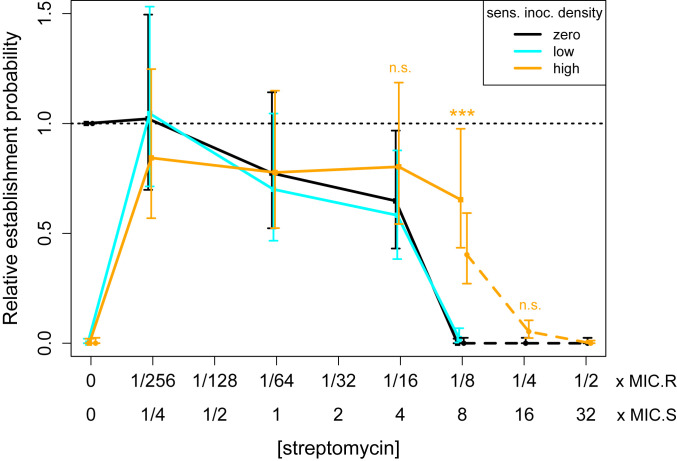
As hypothesized, we found that the presence of the sensitive population (at either density) abolished establishment of resistant cells in the absence of streptomycin (Fig. 6 and SI Appendix, Fig. S11 and Table S6). Meanwhile, at streptomycin concentrations above the MSC (1/256 to 1/8 × MICR, or 1/4 to 8 × MICS), adding the sensitive population at low density had a negligible effect on the probability of establishment of resistant cells. At high density, the sensitive population also had negligible effects on establishment of resistance at streptomycin concentrations up to 1/16 × MICR (4 × MICS). However, at 1/8 × MICR (8 × MICS), the presence of a high-density sensitive population increased the establishment probability from near zero to 65%. To confirm and further probe the extent of this apparent protective effect, we repeated the experiment over a higher range of streptomycin concentrations. The boost in establishment probability was repeatable and highly significant at 1/8 × MICR (Wilcoxon rank-sum test, high- vs. zero or low-density sensitive: P < 5e-8 in both experiments). However, at 1/4 × MICR (16 × MICS), an apparent slight boost in establishment probability was nonsignificant, and by 1/2 × MICR (32 × MICS) the effect was abolished. Thus, a sufficiently dense sensitive population can extend the range of streptomycin concentrations at which the resistant strain is likely to emerge, but does not change the qualitative pattern of stochastic establishment.
Fig. 6.
Impact of a large sensitive population on the establishment probability of a resistant cell. The PA01:Rms149 resistant strain was seeded either alone (black) or into a low-density (cyan) or high-density (orange) sensitive PA01 population, across a range of streptomycin concentrations. Results are shown from two separate experiments, testing different subsets of conditions (experiment 1, data points in squares with solid line; experiment 2, data points in circles with dashed line). Within each experiment, the estimated relative establishment probability per resistant cell in each condition is normalized by the result for the resistant strain alone in streptomycin-free media. Points indicate the maximum-likelihood estimate of , and error bars indicate the 95% confidence interval, using the fitted model selected by the likelihood ratio test (model C′, the null model [Eq. 1] for both experiments). At streptomycin concentrations of particular interest, the number of replicates in which the resistant strain established in the presence of no or low-density sensitive (pooled where applicable) vs. high-density sensitive was compared using a two-sided Wilcoxon rank-sum test, with significance annotated on the plot (1/16 × MICR: experiment 1, P = 0.17; 1/8 × MICR: experiment 1, P < 2.2e-16, and experiment 2, P = 4.8e-8; 1/4 × MICR: experiment 2, P = 0.042, not significant after Bonferroni correction); see SI Appendix, Text, section 16, for further details.
Discussion
In order for resistance to emerge de novo, not only must a resistance gene arise in a bacterial population by mutation or horizontal gene transfer; this first resistant cell must also successfully expand to form a large population. Since any individual cell may fail to replicate, particularly in challenging environmental conditions, the expansion of newly arisen resistant strains is not guaranteed. Our results clearly demonstrate the significance of demographic stochasticity in the de novo emergence of resistance, at antibiotic concentrations well below the resistant strain’s MIC (MICR).
We empirically demonstrated the importance of stochasticity with a simple “seeding experiment” mimicking the growth of clonal resistant lineages founded by single cells. First, to assess the direct impact of antibiotics, we inoculated fresh antibiotic-containing media with approximately one resistant cell per replicate culture and quantified the per-cell probability of establishing a detectable population. Strikingly, this establishment probability dropped off at concentrations well below the MIC of the resistant strain (MICR). For example, the establishment probability of PA01:Rms149 was significantly reduced by streptomycin concentrations as low as 1/16 × MICR and dropped to <5% at 1/8 × MICR (Fig. 2). Resistant cells failed to establish viable populations because of the toxic effects of exposure to sub-MICR concentrations of antibiotics (Fig. 4) coupled with the inherently stochastic nature of individual cell death and division. Importantly, we were able to replicate our key finding of frequent stochastic loss using a different, meropenem-resistant strain (PA01:PAMBL2; Fig. 5). This demonstrated that our results are not limited to a particular model system, but are also relevant to antibiotics used clinically to treat P. aeruginosa infections (37). We expect the qualitative finding of stochastic loss to occur more generally, although the quantitative similarity of results in the two systems tested here is likely to be coincidental.
A natural follow-up question is, then, why some cells establish and others do not. There are several possible mechanistic explanations. First, it is conceivable that the inoculated resistant cells that ultimately established carried additional genetic changes (acquired during growth of the overnight preculture) conferring even higher levels of resistance. In particular, unstable genetic changes such as chromosome inversions (43) and gene amplifications (24) can occur more frequently than point mutations, and amplifications have recently been identified as a major contributor to heteroresistance in bacterial populations (24, 44). However, at the frequencies observed previously (∼10−7 to 10−5 for amplifications conferring at least eightfold increase in MIC; ref. 24), this mechanism is expected to be negligible in our seeding experiments, which inoculated only a few hundred cells in total across replicates. Epigenetic changes have also been suggested as a mechanism for slight decreases in susceptibility occurring at higher frequency (∼1%) (45). Finally, there is a growing recognition that even genetically identical bacteria exhibit phenotypic heterogeneity, which could in particular affect individual cells’ susceptibility to antibiotics (46, 47). For example, those cells that established in our experiments might have been in a particular metabolic state, or had particularly high expression of the resistance gene. Investigating the role of these mechanisms in the establishment of resistant lineages would be an interesting direction for future work.
In clinical settings, antibiotic treatment will typically begin only when the total population of pathogenic bacteria is large enough to cause symptoms. Assuming that the patient was initially infected by a sensitive strain, and given that resistance typically carries a fitness cost in the absence of antibiotics (48, 49), we expect this bacterial population will be predominantly antibiotic sensitive when treatment begins. We therefore next asked how the presence of a large sensitive population would combine with the above effects of antibiotics to shape the establishment of resistant cells, again in the streptomycin model system. As expected, establishment of resistance was abolished in the absence of antibiotics (Fig. 6), presumably due to competitive suppression by the sensitive strain. More interestingly, a sufficiently dense sensitive population (inoculated at ∼5 × 10⁷ CFU/mL here) was able to shift the range of concentrations at which resistance usually established upwards by approximately twofold. We speculate that this apparent protection is due to sensitive cells absorbing antibiotics, thus lowering their concentration in the media (31, 50). A priori, one may have expected outgrowth of resistant cells whenever antibiotic concentration is below MICR, and hence for protection to be irrelevant. However, in the stochastic regime of establishment, any increase in the probability of individual cells surviving and dividing can critically impact the fate of a rare resistant lineage. We emphasize that, although these experiments provide an initial proof of concept, a complete investigation of the interacting effects of sensitive population density, antibiotic dose, and timing remains an important direction for future work. Importantly, however, our main message continues to hold in the more realistic context of resistance emerging within a large sensitive population: Stochastic loss of resistant cells is frequent at antibiotic concentrations well below MICR, despite the selective advantage of resistance in these conditions.
The failure of resistant cells to establish successful lineages at concentrations well below MICR illustrates the limitations of population-based measures in explaining single-cell behavior. To explain this effect rigorously, we quantified the probability of outgrowth of a detectable resistant population at a fixed streptomycin concentration, starting from inoculum sizes spanning three orders of magnitude. We fit these data to a mathematical model relating inoculum size to probability of population growth, under the hypothesis that each cell in the inoculum behaves independently (Eq. 1). This simple stochastic model, parameterized by the per-cell probability of establishment, provides a good explanation for inoculum size-dependent population growth in PA01:Rms149 (Fig. 3B). In the case of cell-to-cell heterogeneity in susceptibility, the establishment probability should be interpreted as an average across cells (SI Appendix, Text, section 10.1), or the proportion of cells that establish at a given antibiotic concentration. Importantly, however, this parameter is independent of inoculum size: that is, individual cells are not “more susceptible” to antibiotic in smaller populations. Instead, the cumulative effect of many cells, each with a small chance of establishment (e.g., <5% at 1/8 × MICR in this system), virtually guarantees population growth from a sufficiently large inoculum size, reconciling our results with the standard definition of the MIC. We thus emphasize that MIC is an emergent property of a population of cells. We also note that the inoculum size effect on MIC that we found here—a purely stochastic phenomenon arising at low absolute numbers (in CFU)—is distinct from the inoculum size effect already widely recognized in the literature, which is seen at high cell density (in CFU/ml) and attributed to various density-dependent mechanisms, such as titration or enzymatic inactivation of antibiotics (31, 32, 41, 51–54). While there are hints of the former absolute-number effect in earlier studies (33, 34), we provide a rigorous explanation in terms of stochastic population dynamics.
This model also provides a simple way to predict whether resistance will emerge during treatment from a preexisting subpopulation of resistant cells, derived from one or more mutations occurring prior to the start of treatment. The mean inoculum size in Eq. 1, representing the number of resistant cells inoculated into antibiotic-containing media in our experiments, can be reinterpreted as the number of resistant cells present in an infection at the start of treatment. Clearly, if the preexisting subpopulation is sufficiently large, the probability of outgrowth during treatment approaches 1 at sub-MICR antibiotic concentrations. However, if resistance must first arise by de novo mutation or horizontal gene transfer, the size of the resistant subpopulation may be small and highly variable, following a form of the Luria–Delbrück distribution (55, 56). Eq. 1 provides a unified way of scaling from single-cell to population behavior across this range of inoculum sizes. We note that an identical relationship arises more generally in models of evolutionary rescue, where adaptation prevents extinction of a population facing severe environmental change (57) (see SI Appendix, Text, section 10.2, for further discussion).
Although the role of demographic stochasticity in the fate of mutant lineages has long been recognized in theoretical population genetics, until very recently it had never been addressed empirically (25). Our study joins a small handful of others that have now experimentally quantified establishment probability from single cells (26, 27, 58, 59), including two (26, 27) investigating establishment of bacterial cells in the presence of antibiotics. These studies used different methods to ours (see SI Appendix, Text, section 10.2, for a more detailed comparison) and did not discuss the implications for emergence of antibiotic resistance. More broadly, the concepts and statistical methods developed here are applicable to a variety of situations where growth depends on success of rare cells and is thus highly stochastic, for instance the establishment of productive infection in a host following pathogen transmission (60), the onset of invasive bacterial infections (61), the outgrowth of bacteria in food products from small initial contaminants (62), or the establishment of metastases from cancerous tumors (63).
In summary, our study highlights the stochastic nature of de novo emergence of antibiotic resistance. In a practical sense, this stochasticity implies that to accurately assess the risk of resistance emerging, we must evaluate not only mutation rates, but also the probability that resistant mutants escape extinction when rare (6), which will depend on antibiotic dosing. Our results underscore the importance of understanding the impact of antibiotic treatment on the single-cell level (see also ref. 45) and caution against the extrapolation of population-level measures. While a positive selection coefficient is a necessary condition for resistance to outcompete an initially prevalent sensitive strain, it does not guarantee establishment when rare; indeed, we showed that single resistant cells are frequently lost at antibiotic concentrations at which they are selectively favored. In cases where MICR lies above the maximum tolerable dose (22), our results have the promising message that lower concentrations may be sufficient to prevent de novo emergence of resistance with high probability. Use of lower antibiotic doses could have further benefits, such as reducing release of antibiotics into the environment (64) and, in the case of antibiotics that have mutagenic effects, perhaps reducing the chance of a resistance mutation arising in the first place (ref. 65; see, however, ref. 66). The possibility of stochastic extinction of small resistant populations opens the door to considering alternative antibiotic dosing strategies, and an important direction for future work will be to evaluate the establishment probability of resistant cells in time-varying antibiotic concentrations reflecting realistic pharmacokinetics (cf. ref. 67).
Materials and Methods
Further details of experimental protocols, data processing, mathematical models, and statistical methods and results are provided in SI Appendix, Text.
Bacterial Strains, Media, and Culture Conditions.
Bacterial strains.
The majority of our experiments, in streptomycin, were conducted with a set of Pseudomonas aeruginosa PA01 strains studied previously (41). The streptomycin-sensitive and -resistant strains are chromosomally isogenic, while resistant strains additionally carry the clinically derived, nonconjugative plasmid Rms149 (68), which is stably maintained in PA01 at approximately two copies per cell (69). Streptomycin resistance is conferred by the aadA5 gene on Rms149, which codes for an enzyme that adenylates streptomycin (70). Both plasmid carriers (resistant) and noncarriers (sensitive) are available with either YFP or DsRed chromosomal fluorescent markers or with no marker (41). The live–dead staining experiment was conducted with the unlabeled resistant strain. All other experiments reported in the main text were conducted with the YFP-labeled resistant strain and, where applicable, the DsRed-labeled sensitive strain. We chose this pairing because YFP provides a stronger signal, facilitating detection of the resistant strain in mixed cultures. Previous work with these strains suggests that the two fluorescent labels have similar fitness effects (41), and we confirmed that the label had no substantive effect on the MIC values of the sensitive strain (SI Appendix, Text, section 2.1). For the seeding experiment in meropenem, we transformed the plasmid PAMBL2 into the same PA01-YFP background (SI Appendix, Text, section 1). This plasmid, isolated in 2007 from a P. aeruginosa clinical isolate (35), confers meropenem resistance through three copies of the blaVIM-1 gene, which codes for a metallo-β-lactamase (35, 36). It is nonconjugative (36) and stably maintained in PA01 at an average of two to three copies per cell (69). MIC values of all relevant strain–antibiotic pairs are reported in SI Appendix, Table S1.
Media and antibiotics.
We cultured bacteria in LB broth containing 5 g/L NaCl (Sigma-Aldrich; product no. L3022). To assess CFU, we plated on LB agar, Vegitone, containing 5 g/L NaCl and 15 g/L agar (Sigma-Aldrich; product no. 19344). Streptomycin was prepared from streptomycin sulfate salt (Sigma-Aldrich; product no. S6501), and meropenem was prepared from meropenem trihydrate (Santa Cruz Biotechnology; product no. SC-485799). Stocks prepared in water were stored according to supplier directions and added to media on the day of experiments. When high antibiotic concentrations were required, stocks were instead prepared directly in LB on the day of experiments to avoid excessive dilution of the media with water. Bacterial cultures were diluted in PBS prepared from tablets (Sigma-Aldrich; product no. P4417). Treatment cultures were set up with 90% media plus 10% inoculating culture by volume; thus, the final concentrations of LB and antibiotics in the treatments are 90% of the prepared media values denoted on plots.
Culture conditions.
All cultures were incubated at 37 °C, shaking at 225 rpm. Overnight cultures were inoculated directly from freezer stocks into 2 mL of LB in 14-mL culture tubes and incubated for ∼16 h. Overnight cultures were then diluted in PBS and used to inoculate treatment plates. Unless otherwise noted, experimental treatments were conducted in 200-μL cultures in flat-bottom 96-well microtiter plates.
Scoring culture growth.
In all experiments, we evaluated culture growth by measuring OD595 using a BioTek Synergy 2 plate reader, at room temperature. Lids on microtiter plates were briefly removed in a nonsterile environment for the reading; comparison to controls mock-inoculated with PBS indicated that contamination was rare (see SI Appendix, Text, for detailed quantification in each experiment). We set a threshold of OD595 > 0.1 to score as growth, whereas background OD in media-only controls was typically below 0.05. Final readings at 3 d postinoculation were used for data analysis unless otherwise noted. By this time, growth had typically stabilized, with OD much higher than the threshold.
MIC Assays.
Standard MIC values for all applicable strain–antibiotic pairs (i.e., resistant Rms149-carrier against streptomycin; resistant PAMBL2-carrier against meropenem; sensitive noncarrier against both antibiotics) were determined under our culture conditions using the broth microdilution method. Overnight cultures were diluted 103-fold and inoculated into antibiotic-containing media at 20 μL/well on 96-well test plates. This dilution factor consistently yielded an inoculation density close to 5 × 10⁵ CFU/mL, in accordance with standard protocol (3); actual density was estimated by plating. Test plates were incubated and scored for growth at ∼20 h [as per standard protocol (3)], 2 d, and 3 d postinoculation. For consistency with growth scoring in seeding experiments, the standard MIC values (MICS and MICR) used to scale antibiotic concentrations on plot axes are based on results at 3 d. Consensus MIC values of all tested strain–antibiotic pairs, at both 20 h and 3 d, are reported in SI Appendix, Table S1, with results of individual replicates reported in SI Appendix, Text, section 2.1. For the YFP-labeled Rms149-carrying resistant strain, an additional MIC assay in streptomycin was conducted, varying inoculum size (Fig. 3A). Here, inoculations were conducted with overnight culture diluted 103-, 104-, 105-, and 106-fold (see SI Appendix, Text, section 2.2, for details).
Seeding Experiments: Resistant Strains in Isolation.
Experimental protocol.
A highly diluted overnight culture of the YFP-labeled resistant strain (Rms149- or PAMBL2-carrier) was inoculated at 20 μL/well into antibiotic-containing media on 96-well test plates. For experiments with PA01:Rms149 screening across many streptomycin concentrations (Fig. 2), we used three dilution factors (4 × 10⁷-, 8 × 10⁷-, and 1.6 × 10⁸-fold), each to inoculate 96 replicate wells at each concentration. To test the null model of the inoculum size effect (Fig. 3B), we screened fewer streptomycin concentrations across a larger number of dilution factors (5 in streptomycin-free conditions and 6 to 10 in each streptomycin concentration), each with 54 replicates. These dilution factors were chosen differently for each streptomycin concentration to capture the range over which the proportion of replicate cultures showing growth increased from near 0 to near 1. For the experiment with PA01:PAMBL2 in meropenem (Fig. 5), we used two dilution factors (5 × 10⁷- and 2 × 10⁸-fold), each with 96 replicates per concentration. In all cases, test plates were incubated and scored for growth after ∼1, 2, and 3 d; for the null model test, incubation and readings were continued up to 5 d to confirm stabilization of growth. See SI Appendix, Text, sections 4 and 5, for further details.
Model fitting.
The number of replicate cultures showing growth by 3 d (or, additionally, by 5 d for the null model test), at each inoculating dilution factor and antibiotic concentration, was used for subsequent model fitting. To estimate single-cell establishment probability and evaluate the null model of the inoculum size effect, likelihood-based methods were used to fit a stochastic model of population growth to these data (see below, Mathematical Model of Establishment). In addition, to evaluate the effect of antibiotic concentration on establishment, generalized linear models were fit to data from the seeding experiments screening across streptomycin (Fig. 2) or meropenem (Fig. 5) concentrations. Using the built-in R function “glm,” growth data were treated as binomial, with inoculating dilution factor and antibiotic concentration taken as explanatory variables, applying the complementary log-log link function (SI Appendix, Text, section 12).
Seeding Experiments: Resistant Strain in Presence of Sensitive Population.
Overnight culture of the DsRed-labeled PA01 sensitive strain was diluted 5-fold to obtain the “high-density” inoculating culture, and (in the first experiment only) further to 500-fold to obtain the “low-density” inoculating culture. Overnight culture of the YFP-labeled PA01:Rms149 resistant strain was diluted up to 5 × 10⁷-fold and 2 × 10⁸-fold. These cultures were inoculated as follows into media at various streptomycin concentrations on 96-well plates. Pure sensitive control cultures (24 replicates per test condition) were inoculated with 10 μL/well of the appropriate diluted culture plus 10 μL/well PBS. “Blank” wells to serve as background fluorescence controls were inoculated with 20 μL/well PBS. Seeding test plates were first inoculated with 10 μL/well of either PBS (for pure resistant control cultures), low-density or high-density sensitive culture. The resistant strain was inoculated at 10 μL/well immediately thereafter (all sensitive and resistant culture inoculations were completed within an hour). Seeding was conducted with 30 to 60 replicates per test condition and resistant dilution factor (see SI Appendix, Text, section 6, for details). All test plates were then incubated as before, with OD595 and fluorescence (excitation: 500 ± 27 nm; emission: 540 ± 25 nm) measured at ∼1, 2, and 3 d postinoculation. Among wells showing growth (OD > 0.1), we considered the YFP-labeled resistant strain to have established if fluorescence exceeded 5 × 10⁵ units, chosen by comparison to pure cultures. In each test condition, the number of replicates in which resistance established was taken as data for model fitting, as in the previous seeding experiments.
Fraction of Dead Cells by Live–Dead Staining.
This experiment used the PA01:Rms149 resistant strain with no fluorescent label, to avoid interfering with the signal from the stains. We inoculated streptomycin treatment cultures (six replicates per concentration) with 103-fold diluted overnight culture, as in the standard MIC assay. After 7 h of treatment, we diluted test cultures 100-fold and stained with thiazole orange and propidium iodide (BD Cell Viability Kit; product no. 349483). In parallel, we diluted and stained media and heat-killed cultures as controls. We sampled 50 μL per diluted culture using flow cytometry (BD Accuri C6 Flow Cytometer with fast fluidics, discarding events with forward scatter FSC-H < 10,000 or side scatter SSC-H < 8,000). The staining and flow cytometry steps were carried out in groups containing one replicate per concentration plus controls, to avoid potentially toxic effects of stain exposure over prolonged times (SI Appendix, Text, section 7). To better discriminate cells from background in the flow cytometry data, we first gated on events according to forward and side scatter before defining clusters of dead (membrane-compromised) and intact cells based on fluorescence; see SI Appendix, Text, section 7 and Fig. S6, for details.
Viable Cell Density Dynamics.
Using the YFP-labeled PA01:Rms149 strain, we tracked the number of viable cells over time in streptomycin-free media (12 replicates per time point) and at 1/32, 1/16, 1/8, and 1/4 × MICR streptomycin (6 replicates per time point). An independent test plate was used for sampling at each time point. Lower (set A) and higher (set B) streptomycin concentrations were split across separate plates and sampled at different times. Cultures were inoculated with 20 μL of 5 × 10⁵-fold diluted overnight culture. At each sampling time, we plated 5 × 4-μL spots of undiluted cultures (10% sampling by volume). The number of viable cells was estimated from total colony count following incubation. Comparison of streptomycin-free controls from both sets (A and B) indicated that the plate set effect was nonsignificant (ANOVA: P = 0.10); thus, controls were pooled for further analysis of the streptomycin effect (see SI Appendix, Text, section 8, for further details).
Mathematical Model of Establishment.
Model.
We denote by pw the probability that a small number of inoculated cells grows into a large population, i.e., that the culture reaches detectable OD as described above. Among a set of n independent replicates, the number of cultures showing growth is thus described by a binomial(n,pw) distribution.
In the “null” model, similar to previous work (57, 60), a simple expression for pw is derived under the assumptions that 1) the number of cells in the inoculum is Poisson-distributed with mean ; 2) each cell, independently, establishes a surviving lineage with probability pc, which depends only on antibiotic concentration x; and 3) culture growth is observed, provided at least one cell establishes a surviving lineage. Then the probability of observing culture growth, as a function of mean inoculum size and antibiotic concentration, can be written as follows (SI Appendix, Text, section 10):
| [1] |
In the second step, we have rewritten the expression in terms of the “effective mean inoculum size,” , which is the mean number of established lineages in the absence of antibiotics; and the “relative establishment probability,” . Although we expect that is close to 1, and play indistinguishable roles in this model, so that in practice we can only estimate their product. This definition of effective inoculum size based on cells that grow in benign conditions is similar to the usual quantification of “viable” cells according to successful formation of a colony; we simply assess growth in liquid rather than on solid medium. Scaling up by the dilution factor applied to the inoculating culture, we have an estimate of bacterial density in this culture, equivalent to the historical “most probable number” method (71, 72). If cells are phenotypically heterogeneous (i.e., vary in their propensity to establish), or if the individual units in the inoculum are actually clumps of cells, then pc should be interpreted as the mean establishment probability among individuals (SI Appendix, Text, section 10.1).
More generally, we need not assume that cells establish independently. If we suppose simply that the number of established lineages is Poisson-distributed with some mean α (which is supported empirically by the distribution of CFU counted in highly diluted cultures; SI Appendix, Fig. S1), we have the following relationship:
| [2] |
where α, and hence pw, have an arbitrary dependence on mean inoculum size and antibiotic concentration. In the statistical “full model,” we estimate a distinct pw (or equivalently α, by the one-to-one mapping in Eq. 2) in each test condition. Relative establishment probability is then generally defined by . Nested models, including the null model above, make additional assumptions about the form of α (see SI Appendix, Text, section 10, for details).
Likelihood-based model fitting and comparisons.
These stochastic models are fit to experimental population growth data using likelihood-based methods (SI Appendix, Text, section 11). Specifically, under each model, we obtain a maximum-likelihood estimate and a 95% confidence interval (determined by the range of parameter values that would not be rejected by a likelihood ratio test at 5% significance level) on the parameter pw, which can be transformed to an estimate for α. In the case of relative establishment probability, , we use a profile likelihood confidence interval accounting for the uncertainty in both numerator (i.e., results at antibiotic concentration x) and denominator (i.e., results in antibiotic-free conditions). The fit of nested models is compared using the likelihood ratio test (LRT) at 5% significance level, i.e., a χ2 test on model deviance with degrees of freedom equal to the difference in number of fitted parameters between the two models.
To test the null model of the inoculum size effect, we neglect any experimental error in preparing overnight culture dilutions, and assume that mean inoculum size is inversely proportional to the applicable dilution factor. Effective mean inoculum size, , is estimated by fitting Eq. 1 to population growth data in antibiotic-free media. Per-cell relative establishment probability then remains as the single free parameter to fit at each tested antibiotic concentration. The goodness of fit of the null model (Eq. 1) is assessed for each test concentration separately, using the LRT to compare it to the fit of the full model (Eq. 2).
All model fitting was implemented in R, version 3.3.1 (The R Foundation for Statistical Computing, 2016).
Data and Code Availability.
Data generated in this study, as well as custom R scripts for likelihood-based model fitting and comparisons, are deposited in Dryad (DOI: 10.5061/dryad.12jm63xtb).
Supplementary Material
Acknowledgments
H.K.A. was supported by an Early Postdoc.Mobility Fellowship (P2EZP3_165188) and an Advanced Postdoc.Mobility fellowship (P300PA_177789) from the Swiss National Science Foundation, and a University Research Fellowship from the Royal Society (URF\R1\191269). R.C.M. was supported by Wellcome Trust Grant 106918/Z/15/Z. We thank Isabel Frost, Natalia Kapel, Lois Ogunlana, Andrei Papkou, and Célia Souque for advice and assistance on experimental protocols.
Footnotes
The authors declare no competing interest.
This article is a PNAS Direct Submission.
Data deposition: The data and custom code reported in this paper have been deposited in the Dryad Digital Repository (DOI: 10.5061/dryad.12jm63xtb).
This article contains supporting information online at https://www.pnas.org/lookup/suppl/doi:10.1073/pnas.1919672117/-/DCSupplemental.
References
- 1.Fish D. N., Piscitelli S. C., Danziger L. H., Development of resistance during antimicrobial therapy: A review of antibiotic classes and patient characteristics in 173 studies. Pharmacotherapy 15, 279–291 (1995). [PubMed] [Google Scholar]
- 2.World Health Organization , Antimicrobial Resistance: Global Report on Surveillance, (World Health Organization, Geneva, 2014). [Google Scholar]
- 3.European Committee for Antimicrobial Susceptibility Testing , Determination of minimum inhibitory concentrations (MICs) of antibacterial agents by broth dilution. Clin. Microbiol. Infect. 9, 1–7 (2003).12691538 [Google Scholar]
- 4.Andersson D. I., Hughes D., Evolution of antibiotic resistance at non-lethal drug concentrations. Drug Resist. Updat. 15, 162–172 (2012). [DOI] [PubMed] [Google Scholar]
- 5.Day T., Huijben S., Read A. F., Is selection relevant in the evolutionary emergence of drug resistance? Trends Microbiol. 23, 126–133 (2015). [DOI] [PMC free article] [PubMed] [Google Scholar]
- 6.Andersson D. I., Improving predictions of the risk of resistance development against new and old antibiotics. Clin. Microbiol. Infect. 21, 894–898 (2015). [DOI] [PubMed] [Google Scholar]
- 7.Drusano G. L., Prevention of resistance: A goal for dose selection for antimicrobial agents. Clin. Infect. Dis. 36 (suppl. 1), S42–S50 (2003). [DOI] [PubMed] [Google Scholar]
- 8.Olofsson S. K., Cars O., Optimizing drug exposure to minimize selection of antibiotic resistance. Clin. Infect. Dis. 45 (suppl. 2), S129–S136 (2007). [DOI] [PubMed] [Google Scholar]
- 9.Drusano G. L., Louie A., MacGowan A., Hope W., Suppression of emergence of resistance in pathogenic bacteria: Keeping our powder dry, part 1. Antimicrob. Agents Chemother. 60, 1183–1193 (2015). [DOI] [PMC free article] [PubMed] [Google Scholar]
- 10.Negri M. C., Morosini M. I., Loza E., Baquero F., In vitro selective antibiotic concentrations of beta-lactams for penicillin-resistant Streptococcus pneumoniae populations. Antimicrob. Agents Chemother. 38, 122–125 (1994). [DOI] [PMC free article] [PubMed] [Google Scholar]
- 11.Baquero F., Negri M. C., Morosini M. I., Blázquez J., The antibiotic selective process: Concentration-specific amplification of low-level resistant populations. Ciba Found. Symp. 207, 93–105, discussion 105–111 (1997). [DOI] [PubMed] [Google Scholar]
- 12.Negri M.-C., Lipsitch M., Blázquez J., Levin B. R., Baquero F., Concentration-dependent selection of small phenotypic differences in TEM beta-lactamase-mediated antibiotic resistance. Antimicrob. Agents Chemother. 44, 2485–2491 (2000). [DOI] [PMC free article] [PubMed] [Google Scholar]
- 13.Dong Y., Zhao X., Domagala J., Drlica K., Effect of fluoroquinolone concentration on selection of resistant mutants of Mycobacterium bovis BCG and Staphylococcus aureus. Antimicrob. Agents Chemother. 43, 1756–1758 (1999). [DOI] [PMC free article] [PubMed] [Google Scholar]
- 14.Zhao X., Drlica K., Restricting the selection of antibiotic-resistant mutants: A general strategy derived from fluoroquinolone studies. Clin. Infect. Dis. 33 (suppl. 3), S147–S156 (2001). [DOI] [PubMed] [Google Scholar]
- 15.Zhao X., Drlica K., Restricting the selection of antibiotic-resistant mutant bacteria: Measurement and potential use of the mutant selection window. J. Infect. Dis. 185, 561–565 (2002). [DOI] [PubMed] [Google Scholar]
- 16.Drlica K., Zhao X., Mutant selection window hypothesis updated. Clin. Infect. Dis. 44, 681–688 (2007). [DOI] [PubMed] [Google Scholar]
- 17.Drlica K., Zhao X., “Mutant selection window hypothesis: A framework for anti-mutant dosing of antimicrobial agents” in National Institute of Allergy and Infectious Disease, NIH Volume 1, Frontiers in Research, Georgiev V. S., Western K., McGowan J. J., Eds. (Humana Press, Totowa, NJ, 2008), pp. 101–106. [Google Scholar]
- 18.Gullberg E. et al., Selection of resistant bacteria at very low antibiotic concentrations. PLoS Pathog. 7, e1002158 (2011). [DOI] [PMC free article] [PubMed] [Google Scholar]
- 19.Liu A. et al., Selective advantage of resistant strains at trace levels of antibiotics: A simple and ultrasensitive color test for detection of antibiotics and genotoxic agents. Antimicrob. Agents Chemother. 55, 1204–1210 (2011). [DOI] [PMC free article] [PubMed] [Google Scholar]
- 20.Kalghatgi S. et al., Bactericidal antibiotics induce mitochondrial dysfunction and oxidative damage in mammalian cells. Sci. Transl. Med. 5, 192ra85 (2013). [DOI] [PMC free article] [PubMed] [Google Scholar]
- 21.Prayle A., Watson A., Fortnum H., Smyth A., Side effects of aminoglycosides on the kidney, ear and balance in cystic fibrosis. Thorax 65, 654–658 (2010). [DOI] [PMC free article] [PubMed] [Google Scholar]
- 22.Drlica K., The mutant selection window and antimicrobial resistance. J. Antimicrob. Chemother. 52, 11–17 (2003). [DOI] [PubMed] [Google Scholar]
- 23.zur Wiesch P. A., Kouyos R., Engelstädter J., Regoes R. R., Bonhoeffer S., Population biological principles of drug-resistance evolution in infectious diseases. Lancet Infect. Dis. 11, 236–247 (2011). [DOI] [PubMed] [Google Scholar]
- 24.Nicoloff H., Hjort K., Levin B. R., Andersson D. I., The high prevalence of antibiotic heteroresistance in pathogenic bacteria is mainly caused by gene amplification. Nat. Microbiol. 4, 504–514 (2019). [DOI] [PubMed] [Google Scholar]
- 25.Patwa Z., Wahl L. M., The fixation probability of beneficial mutations. J. R. Soc. Interface 5, 1279–1289 (2008). [DOI] [PMC free article] [PubMed] [Google Scholar]
- 26.Schenk M. F., Szendro I. G., Krug J., de Visser J. A. G. M., Quantifying the adaptive potential of an antibiotic resistance enzyme. PLoS Genet. 8, e1002783 (2012). [DOI] [PMC free article] [PubMed] [Google Scholar]
- 27.Coates J. et al., Antibiotic-induced population fluctuations and stochastic clearance of bacteria. eLife 7, e32976 (2018). [DOI] [PMC free article] [PubMed] [Google Scholar]
- 28.Levin-Reisman I. et al., Antibiotic tolerance facilitates the evolution of resistance. Science 355, 826–830 (2017). [DOI] [PubMed] [Google Scholar]
- 29.Livermore D. M., Multiple mechanisms of antimicrobial resistance in Pseudomonas aeruginosa: Our worst nightmare? Clin. Infect. Dis. 34, 634–640 (2002). [DOI] [PubMed] [Google Scholar]
- 30.Brook I., Inoculum effect. Rev. Infect. Dis. 11, 361–368 (1989). [DOI] [PubMed] [Google Scholar]
- 31.Udekwu K. I., Parrish N., Ankomah P., Baquero F., Levin B. R., Functional relationship between bacterial cell density and the efficacy of antibiotics. J. Antimicrob. Chemother. 63, 745–757 (2009). [DOI] [PMC free article] [PubMed] [Google Scholar]
- 32.Artemova T., Gerardin Y., Dudley C., Vega N. M., Gore J., Isolated cell behavior drives the evolution of antibiotic resistance. Mol. Syst. Biol. 11, 822 (2015). [DOI] [PMC free article] [PubMed] [Google Scholar]
- 33.Haltalin K. C., Nelson J. D., In vitro susceptibility of Shigellae to sodium sulfadiazine and to eight antibiotics. JAMA 193, 705–710 (1965). [DOI] [PubMed] [Google Scholar]
- 34.McLinn S. E., Nelson J. D., Haltalin K. C., Antimicrobial susceptibility of Hemophilus influenzae. Pediatrics 45, 827–838 (1970). [PubMed] [Google Scholar]
- 35.Tato M., Coque T. M., Baquero F., Cantón R., Dispersal of carbapenemase blaVIM-1 gene associated with different Tn402 variants, mercury transposons, and conjugative plasmids in Enterobacteriaceae and Pseudomonas aeruginosa. Antimicrob. Agents Chemother. 54, 320–327 (2010). [DOI] [PMC free article] [PubMed] [Google Scholar]
- 36.San Millan A. et al., Sequencing of plasmids pAMBL1 and pAMBL2 from Pseudomonas aeruginosa reveals a blaVIM-1 amplification causing high-level carbapenem resistance. J. Antimicrob. Chemother. 70, 3000–3003 (2015). [DOI] [PubMed] [Google Scholar]
- 37.Bassetti M., Vena A., Croxatto A., Righi E., Guery B., How to manage Pseudomonas aeruginosa infections. Drugs Context 7, 212527 (2018). [DOI] [PMC free article] [PubMed] [Google Scholar]
- 38.Fritzenwanker M. et al., Treatment options for carbapenem-resistant gram-negative infections. Dtsch. Arztebl. Int. 115, 345–352 (2018). [DOI] [PMC free article] [PubMed] [Google Scholar]
- 39.Walters M. S. et al., Carbapenem-resistant Pseudomonas aeruginosa at US emerging infections program sites, 2015. Emerg. Infect. Dis. 25, 1281–1288 (2019). [DOI] [PMC free article] [PubMed] [Google Scholar]
- 40.World Health Organization , Prioritization of Pathogens to Guide Discovery, Research and Development of New Antibiotics for Drug-Resistant Bacterial Infections, Including Tuberculosis, (World Health Organization, Geneva, 2017). [Google Scholar]
- 41.Frost I. et al., Cooperation, competition and antibiotic resistance in bacterial colonies. ISME J. 12, 1582–1593 (2018). [DOI] [PMC free article] [PubMed] [Google Scholar]
- 42.Brockhurst M. A. et al., Assessing evolutionary risks of resistance for new antimicrobial therapies. Nat. Ecol. Evol. 3, 515–517 (2019). [DOI] [PubMed] [Google Scholar]
- 43.Guérillot R. et al., Unstable chromosome rearrangements in Staphylococcus aureus cause phenotype switching associated with persistent infections. Proc. Natl. Acad. Sci. U.S.A. 116, 20135–20140 (2019). [DOI] [PMC free article] [PubMed] [Google Scholar]
- 44.Andersson D. I., Nicoloff H., Hjort K., Mechanisms and clinical relevance of bacterial heteroresistance. Nat. Rev. Microbiol. 17, 479–496 (2019). [DOI] [PubMed] [Google Scholar]
- 45.Sorg R. A., Veening J.-W., Microscale insights into pneumococcal antibiotic mutant selection windows. Nat. Commun. 6, 8773 (2015). [DOI] [PMC free article] [PubMed] [Google Scholar]
- 46.El Meouche I., Siu Y., Dunlop M. J., Stochastic expression of a multiple antibiotic resistance activator confers transient resistance in single cells. Sci. Rep. 6, 19538 (2016). [DOI] [PMC free article] [PubMed] [Google Scholar]
- 47.Bergmiller T. et al., Biased partitioning of the multidrug efflux pump AcrAB-TolC underlies long-lived phenotypic heterogeneity. Science 356, 311–315 (2017). [DOI] [PubMed] [Google Scholar]
- 48.Andersson D. I., Hughes D., Antibiotic resistance and its cost: Is it possible to reverse resistance? Nat. Rev. Microbiol. 8, 260–271 (2010). [DOI] [PubMed] [Google Scholar]
- 49.Vogwill T., MacLean R. C., The genetic basis of the fitness costs of antimicrobial resistance: A meta-analysis approach. Evol. Appl. 8, 284–295 (2015). [DOI] [PMC free article] [PubMed] [Google Scholar]
- 50.zur Wiesch P. A., et al. , Classic reaction kinetics can explain complex patterns of antibiotic action. Sci. Transl. Med. 7, 287ra73 (2015). [DOI] [PMC free article] [PubMed] [Google Scholar]
- 51.Yurtsev E. A., Chao H. X., Datta M. S., Artemova T., Gore J., Bacterial cheating drives the population dynamics of cooperative antibiotic resistance plasmids. Mol. Syst. Biol. 9, 683 (2013). [DOI] [PMC free article] [PubMed] [Google Scholar]
- 52.Vega N. M., Gore J., Collective antibiotic resistance: Mechanisms and implications. Curr. Opin. Microbiol. 21, 28–34 (2014). [DOI] [PMC free article] [PubMed] [Google Scholar]
- 53.Sorg R. A. et al., Collective resistance in microbial communities by intracellular antibiotic deactivation. PLoS Biol. 14, e2000631 (2016). [DOI] [PMC free article] [PubMed] [Google Scholar]
- 54.Meredith H. R., Srimani J. K., Lee A. J., Lopatkin A. J., You L., Collective antibiotic tolerance: Mechanisms, dynamics and intervention. Nat. Chem. Biol. 11, 182–188 (2015). [DOI] [PMC free article] [PubMed] [Google Scholar]
- 55.Iwasa Y., Nowak M. A., Michor F., Evolution of resistance during clonal expansion. Genetics 172, 2557–2566 (2006). [DOI] [PMC free article] [PubMed] [Google Scholar]
- 56.Colijn C., Cohen T., Ganesh A., Murray M., Spontaneous emergence of multiple drug resistance in tuberculosis before and during therapy. PLoS One 6, e18327 (2011). [DOI] [PMC free article] [PubMed] [Google Scholar]
- 57.Martin G., Aguilée R., Ramsayer J., Kaltz O., Ronce O., The probability of evolutionary rescue: Towards a quantitative comparison between theory and evolution experiments. Philos. Trans. R. Soc. Lond. B Biol. Sci. 368, 20120088 (2013). [DOI] [PMC free article] [PubMed] [Google Scholar]
- 58.Gifford D. R., de Visser J. A. G. M., Wahl L. M., Model and test in a fungus of the probability that beneficial mutations survive drift. Biol. Lett. 9, 20120310 (2013). [DOI] [PMC free article] [PubMed] [Google Scholar]
- 59.Gifford D. R., MacLean R. C., Evolutionary reversals of antibiotic resistance in experimental populations of Pseudomonas aeruginosa. Evolution 67, 2973–2981 (2013). [DOI] [PubMed] [Google Scholar]
- 60.Druett H. A., Bacterial invasion. Nature 170, 288 (1952). [DOI] [PubMed] [Google Scholar]
- 61.Moxon E. R., Murphy P. A., Haemophilus influenzae bacteremia and meningitis resulting from survival of a single organism. Proc. Natl. Acad. Sci. U.S.A. 75, 1534–1536 (1978). [DOI] [PMC free article] [PubMed] [Google Scholar]
- 62.Koutsoumanis K., A study on the variability in the growth limits of individual cells and its effect on the behavior of microbial populations. Int. J. Food Microbiol. 128, 116–121 (2008). [DOI] [PubMed] [Google Scholar]
- 63.Liotta L. A., Saidel G. M., Kleinerman J., Stochastic model of metastases formation. Biometrics 32, 535–550 (1976). [PubMed] [Google Scholar]
- 64.Dancer S. J., How antibiotics can make us sick: The less obvious adverse effects of antimicrobial chemotherapy. Lancet Infect. Dis. 4, 611–619 (2004). [DOI] [PubMed] [Google Scholar]
- 65.Kohanski M. A., DePristo M. A., Collins J. J., Sublethal antibiotic treatment leads to multidrug resistance via radical-induced mutagenesis. Mol. Cell 37, 311–320 (2010). [DOI] [PMC free article] [PubMed] [Google Scholar]
- 66.Frenoy A., Bonhoeffer S., Death and population dynamics affect mutation rate estimates and evolvability under stress in bacteria. PLoS Biol. 16, e2005056 (2018). [DOI] [PMC free article] [PubMed] [Google Scholar]
- 67.Strukova E. N., Portnoy Y. A., Zinner S. H., Firsov A. A., Predictors of bacterial resistance using in vitro dynamic models: Area under the concentration-time curve related to either the minimum inhibitory or mutant prevention antibiotic concentration. J. Antimicrob. Chemother. 71, 678–684 (2016). [DOI] [PubMed] [Google Scholar]
- 68.Haines A. S., Jones K., Cheung M., Thomas C. M., The IncP-6 plasmid Rms149 consists of a small mobilizable backbone with multiple large insertions. J. Bacteriol. 187, 4728–4738 (2005). [DOI] [PMC free article] [PubMed] [Google Scholar]
- 69.San Millan A., Heilbron K., MacLean R. C., Positive epistasis between co-infecting plasmids promotes plasmid survival in bacterial populations. ISME J. 8, 601–612 (2014). [DOI] [PMC free article] [PubMed] [Google Scholar]
- 70.Sandvang D., Novel streptomycin and spectinomycin resistance gene as a gene cassette within a class 1 integron isolated from Escherichia coli. Antimicrob. Agents Chemother. 43, 3036–3038 (1999). [DOI] [PMC free article] [PubMed] [Google Scholar]
- 71.Cochran W. G., Estimation of bacterial densities by means of the “most probable number.”. Biometrics 6, 105–116 (1950). [PubMed] [Google Scholar]
- 72.Hurley M. A., Roscoe M. E., Automated statistical analysis of microbial enumeration by dilution series. J. Appl. Bacteriol. 55, 159–164 (1983). [Google Scholar]
Associated Data
This section collects any data citations, data availability statements, or supplementary materials included in this article.
Supplementary Materials
Data Availability Statement
Data generated in this study, as well as custom R scripts for likelihood-based model fitting and comparisons, are deposited in Dryad (DOI: 10.5061/dryad.12jm63xtb).


