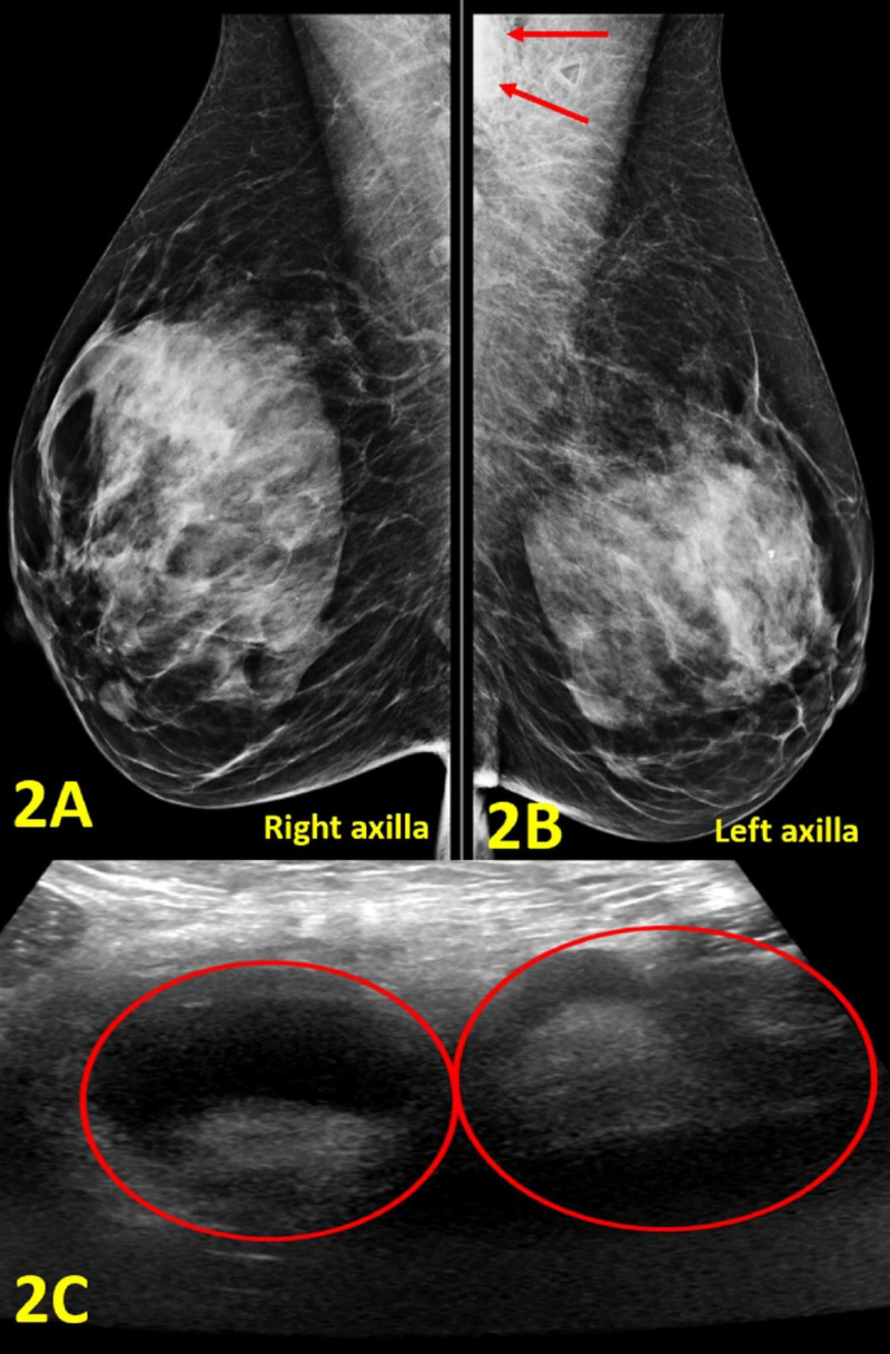
Figure 2. Imaging of Bilateral Breasts and Axillae.
(A) Right axilla diagnostic mammogram demonstrates no abnormal findings. (B) Left axilla diagnostic mammogram shows an enlarged lymph node in the left axilla (outlined by red arrows). (C) Left axilla diagnostic ultrasound shows two adjacent enlarged axillary lymph nodes with cortical thickening measuring 31 mm and 27 mm in diameter, respectively (red circles).

