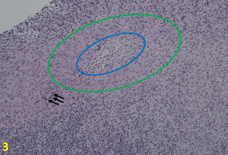
Figure 3. Histologic Findings of Cat Scratch Disease at 10×.
Core needle biopsies show necrotizing lymphadenitis that partially effaces the lymph node architecture. In the necrotic area, there are microabscess (blue oval) with neutrophils and surrounding histiocytes, some of which are palisading (areas between the green and blue circles). Scattered are giant cells (black arrows).

