Hypervariable domains (HVDs) of alphavirus nsP3 proteins recruit host proteins into viral replication complexes. The sets of HVD-binding host factors are specific for each alphavirus, and we have previously identified those specific for EEEV. The results of this study demonstrate that the deletions of the binding sites of the G3BP and FXR protein families in the nsP3 HVD of EEEV make the virus avirulent for mice. Mutations in the nuclear localization signal in EEEV capsid protein have an additional negative effect on viral replication in vivo. Despite the inability to cause a detectable disease, the double HVD and triple HVD/capsid mutants induce high levels of neutralizing antibodies. Single immunization protects mice against infection with the highly pathogenic North American strain of EEEV. High safety, the inability to revert to wild-type phenotype, and high immunogenicity make the designed mutants attractive vaccine candidates for EEEV infection.
KEYWORDS: Eastern equine encephalitis virus, FXR, G3BP, alphavirus, capsid protein, intrinsically disordered, neurovirulence, nsP3, vaccine, viral replication
ABSTRACT
Eastern equine encephalitis virus (EEEV) is the most pathogenic member of the Alphavirus genus in the Togaviridae family. This virus continues to circulate in the New World and has a potential for deliberate use as a bioweapon. Despite the public health threat, to date no attenuated EEEV variants have been applied as live EEEV vaccines. Our previous studies demonstrated the critical function of the hypervariable domain (HVD) in EEEV nsP3 for the assembly of viral replication complexes (vRCs). EEEV HVD contains short linear motifs that recruit host proteins required for vRC formation and function. In this study, we developed a set of EEEV mutants that contained combinations of deletions in nsP3 HVD and clustered mutations in capsid protein, and tested the effects of these modifications on EEEV infection in vivo. These mutations had cumulative negative effects on viral ability to induce meningoencephalitis. The deletions of two critical motifs, which interact with the members of cellular FXR and G3BP protein families, made EEEV cease to be neurovirulent. The additional clustered mutations in capsid protein, which affect its ability to induce transcriptional shutoff, diminished EEEV’s ability to develop viremia. Most notably, despite the inability to induce detectable disease, the designed EEEV mutants remained highly immunogenic and, after a single dose, protected mice against subsequent infection with wild-type (wt) EEEV. Thus, alterations of interactions of EEEV HVD and likely HVDs of other alphaviruses with host factors represent an important direction for development of highly attenuated viruses that can be applied as live vaccines.
IMPORTANCE Hypervariable domains (HVDs) of alphavirus nsP3 proteins recruit host proteins into viral replication complexes. The sets of HVD-binding host factors are specific for each alphavirus, and we have previously identified those specific for EEEV. The results of this study demonstrate that the deletions of the binding sites of the G3BP and FXR protein families in the nsP3 HVD of EEEV make the virus avirulent for mice. Mutations in the nuclear localization signal in EEEV capsid protein have an additional negative effect on viral replication in vivo. Despite the inability to cause a detectable disease, the double HVD and triple HVD/capsid mutants induce high levels of neutralizing antibodies. Single immunization protects mice against infection with the highly pathogenic North American strain of EEEV. High safety, the inability to revert to wild-type phenotype, and high immunogenicity make the designed mutants attractive vaccine candidates for EEEV infection.
INTRODUCTION
The Alphavirus genus in the Togaviridae family contains a wide variety of viral pathogens, which are circulating on all continents (1). In nature, alphaviruses are transmitted mostly by mosquito vectors between vertebrates that serve as amplifying hosts. Some of them represent unquestionable public health threat and cause human diseases of different severities (2). The Old World (OW) representatives are generally less pathogenic and rarely cause life-threatening illnesses. The main symptoms are usually limited to rash and arthritis, but chikungunya virus (CHIKV) infection may be severe, and arthritis symptoms can continue for years (3–5). The New World (NW) alphaviruses, represented by Venezuelan (VEEV), eastern (EEEV), and western (WEEV) equine encephalitis viruses, cause more severe diseases, which are mainly characterized by meningoencephalitis and often result in either lethal outcome or neurological sequelae (6–8). The capacity of NW alphaviruses to be efficiently transmitted by aerosol makes them potential biological warfare agents. EEEV is the most pathogenic among alphaviruses. In the United States it remains relatively rare, with an average of eight EEEV disease cases reported annually during 2003 to 2018. However, in 2019, the CDC received reports of 34 cases of EEEV disease, which resulted in 12 deaths (9, 10). Despite the significant threat to public health, to date, no licensed vaccines or therapeutic means have been developed against the OW and NW alphavirus infections.
The alphavirus genome is represented by a single-stranded genomic RNA (G RNA) of positive polarity that mimics cellular mRNAs in terms of having a Cap structure at the 5′ terminus and a poly(A) tail at the 3′ terminus (1). Upon delivery into the cells, G RNA is translated into viral nonstructural proteins, nsPs, which form replication complexes (vRCs) that mediate G RNA replication and transcription of subgenomic (SG) RNA. The latter RNA is translated into viral structural proteins, which ultimately form infectious viral particles. Alphavirus structural proteins (SPs), particularly the envelope glycoproteins E2 and E1, are greatly less conserved than nsPs because the evolution of nsPs is limited by their enzymatic functions in RNA synthesis. SPs mediate infection of specific tissues and organs, determine sensitivity to type I interferon (IFN), and are critical determinants of viral pathogenesis (11, 12). Accumulation of mutations in SPs appears to adapt alphaviruses to replicate in mosquito and vertebrate species circulating in specific geographical areas.
However, the evolution of SPs does not appear to be the only means of alphavirus adaptation to replication in different hosts. Recently, the nonstructural protein 3 (nsP3) has attracted great attention as an additional major factor responsible for alphavirus evolution and pathogenesis (13–19). This protein contains a long, intrinsically disordered, C-terminal hypervariable domain (HVD) that encodes a variety of linear motifs, which interact with virus-specific host factors (16, 20–22). HVDs function as hubs for the recruitment of host proteins required for alphavirus vRC assembly and function. The G3BP family members, which are expressed in vertebrate cells, together with their mosquito cell-specific homolog Rasputin (Rin1), were shown to be critical HVD-interacting host factors that are specific for all studied OW alphaviruses (16, 23). They drive replication of CHIKV, Ross River (RRV), Semliki Forest (SFV), and Sindbis (SINV) viruses. Separately, replication of the NW representative VEEV critically depends on the members of the cellular FXR protein family (FXR1, FXR2, and FMRP) and not on G3BPs (16, 18). However, EEEV was found to be unique among the OW and NW alphaviruses. Its HVD contains binding motifs for the proteins of both the FXR and G3BP families (19), and in the case of EEEV, members of both families were found to redundantly function in viral RNA replication (19). This virus can replicate essentially with the same efficiency in double knockout (KO) G3bp, triple KO Fxr, and parental cells. The use of a wider range of host factors may be a contributor to the highly pathogenic phenotype of this virus, its greater replication efficiency, and high cytopathogenicity.
Another important determinant of alphavirus pathogenesis, which is relevant to this study, is their ability to induce transcriptional shutoff as a means of inhibiting the development of innate immune response. The nsP2 protein of the OW alphaviruses induces degradation of the catalytic subunit of cellular DNA-dependent RNA polymerase II (24–27). In the case of NW alphaviruses, such as VEEV and EEEV, viral capsid protein binds importin-α/β and exportin CRM1 (28). These large, multimeric complexes block nucleocytoplasmic traffic and ultimately inhibit transcription of cellular RNAs (29). Thus, both OW and NW alphaviruses interfere with activation of the type I IFN response and transcription of interferon-stimulated genes (ISGs) in infected cells (30). To date, the roles of importin-binding nuclear localization signals (NLSs) of VEEV and EEEV, and the effects of capsid-specific modifications on viral replication, have been extensively characterized (31, 32).
In this new study, we have applied the accumulated detailed knowledge of the capsid-specific determinants of EEEV transcription inhibitory functions and the roles of nsP3 HVD-binding host factors in viral vRC assembly. We have designed a set of recombinant EEEV variants that contain extensive irreversible modifications in capsid NLSs and nsP3 HVDs, and experimentally assessed their effects on EEEV pathogenesis in a mouse model. Our data demonstrate that clustered mutations in capsid protein and deletions of FXR- and G3BP-biding sites in nsP3 HVD strongly affect EEEV virulence. Two combinations of mutations made EEEV incapable of inducing detectable disease in mice and, consequently, unable to cause death. The latter two mutants demonstrated no neuroinvasiveness and/or neurovirulence. Importantly, the designed nonpathogenic EEEV mutants remained capable of inducing high levels of neutralizing antibodies (Abs), which protected mice against high doses of the parental North American (NA) strain of EEEV.
RESULTS
Designed capsid and HVD mutants of EEEV are viable and can replicate in various vertebrate cell lines.
In this study, we developed a set of NA EEEV mutants that contained combinations of irreversible mutations in capsid protein and nsP3 HVD (Fig. 1A and B). The infectious cDNAs were designed as follows: (i) EEEV/Cm encoded capsid protein with a mutated NLS; (ii) EEEV/Cm/ΔG contained the above mutations in the capsid gene and its G3BP-binding peptide in nsP3 HVD was deleted; (iii) similarly, EEEV/Cm/ΔF also encoded mutated capsid and nsP3 HVD with a deleted FXR-binding site; (iv) EEEV/Cm/ΔGΔF genome encoded mutated capsid and HVD with deletions of both G3BP- and FXR-binding sites; and (v) EEEV/ΔGΔF genome contained deletions of both G3BP- and FXR-interacting motifs in nsP3 HVD-coding sequence, but the capsid gene remained unmodified. The parental EEEV Florida 93 was used in the study as a highly virulent, positive control. Thus, replication of the designed mutants in vivo and their pathogenesis were expected to be dependent either on mutations in capsid protein or on deletions of the FXR- and G3BP-binding motifs in nsP3 HVD, or on modifications in both nsP3 HVD and capsid protein.
FIG 1.
EEEV variants with mutated capsid and nsP3 HVD are viable. (A) Mutations introduced in the EEEV capsid protein and deletions of G3BP- and FXR-binding motifs in nsP3 HVD. G3BP- and FXR-binding motifs are indicated by red and blue boxes, respectively. Numbers indicate positions of aa in the corresponding proteins. (B) Schematic presentation of the genomes of EEEV variants used in this study. Red and blue boxes indicate positions of G3BP- and FXR-interacting peptides, respectively, in nsP3 HVD. Black box indicates the presence of mutations in capsid protein. (C) Plaques formed by the viruses rescued by transfection of the in vitro-synthesized G RNA on BHK-21 cells. Plaques were stained by crystal violet at 40 h PI.
The in vitro-synthesized G RNAs of the designed mutants and parental EEEV were transfected into BHK-21 cells (see Materials and Methods for details) and viral stocks were harvested at 24 h post RNA transfection. All the mutants were viable. As in the previous studies of alphaviruses that lost the ability to induce transcriptional shutoff, all of them remained capable of developing plaques on BHK-21 cells under agarose cover (Fig. 1C). This strongly simplified the assessment of their titers. Plaques formed by the HVD mutants, particularly by those having both G3BP- and FXR-binding sites in nsP3 HVD deleted (EEEV/Cm/ΔGΔF and EEEV/ΔGΔF), were noticeably smaller. The smaller plaque sizes were expected since these mutants were unable to utilize two families of host factors, which are critically involved in EEEV vRC formation and function.
Our previous studies demonstrated that SINV and VEEV can tolerate extensive modifications in their nsP3 HVDs and replicate in BHK-21 cells, which are commonly used for virus rescue and plaque assay. However, many of these variants were nonviable in other tested vertebrate cell lines. Therefore, we next evaluated the replication efficiency of the designed mutants in a range of the commonly used cell lines, which included Vero, HeLa, Huh7.5, BHK-21, and NIH 3T3 cells. All the designed viruses could replicate in these cells (Fig. 2). However, the mutants that lacked both FXR- and G3BP-binding sites in their HVDs, EEEV/Cm/ΔGΔF and EEEV/ΔGΔF, replicated in HeLa cells to titers that were lower by a few orders of magnitude than those of other variants (Fig. 2). From these results we concluded that the introduced mutations did not make viruses nonviable, and they can be propagated in a variety of vertebrate cell lines.
FIG 2.
The designed EEEV variants with mutated capsid proteins and nsP3 HVDs are capable of replication in commonly used cell lines of vertebrate origin. The indicated cell lines were infected at an MOI of 1 PFU/cell with EEEV and its mutants. At 20 h PI, media were harvested and viral titers were determined by plaque assay on BHK-21 cells. This experiment was reproducibly repeated three times and standard deviations are indicated. Red and blue bars indicate titers of EEEV/ΔGΔF and EEEV/Cm/ΔGΔF, respectively. These mutants demonstrated significant decreases in titers, compared to parental EEEV. Significance of differences was determined by 1-way ANOVA followed by Dunnett’s multiple-comparison test: nonsignificant (n.s.), P > 0.05; *, P < 0.05; **, P < 0.01; ***, P < 0.001, ****, P < 0.0001. Data are represented as means with SD (n = 3).
Modifications introduced into capsid protein and HVD have cumulative negative effects on EEEV virulence.
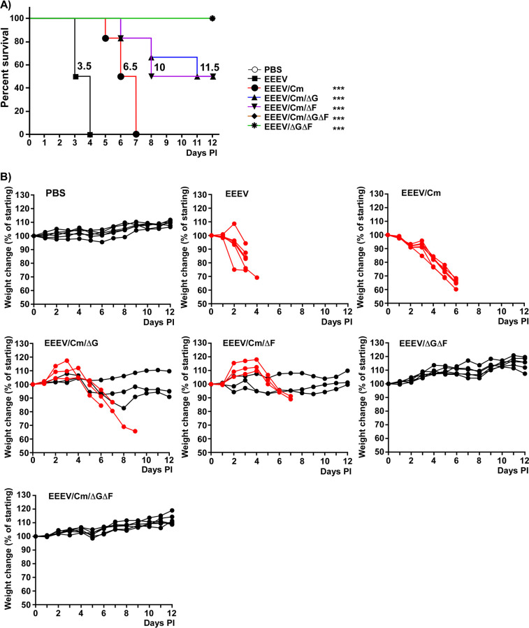
The most critical question concerned the effects of the mutations in capsid protein and nsP3 HVD on EEEV pathogenesis. To address this, 4-to-5-week old CD1 mice were subcutaneously (s.c.) infected with 2 × 10⁵ PFU of parental wild-type (wt) virus and the designed mutants. In this experiment, we intentionally applied very high doses of viruses to definitively identify the most attenuated variants. All the mice infected with wt NA EEEV succumbed to the infection within 4 days postinfection (PI) with the median survival time (MST) of 3.5 days (Fig. 3A). Most of them died before the onset of severe weight loss (Fig. 3B). EEEV/Cm demonstrated statistically significant attenuation characterized by longer MST (6.5 days). However, eventually, all mice were euthanized because of weight loss and development of limb paralysis (Fig. 3B). EEEV/Cm/ΔF and EEEV/Cm/ΔG demonstrated better attenuation that resulted in the survival of some of the infected mice. For both EEEV/Cm/ΔF and EEEV/Cm/ΔG, MST increased to 10 and 11.5 days, respectively. EEEV/ΔGΔF and EEEV/Cm/ΔGΔF were even more attenuated, and none of the infected mice exhibited neurological symptoms or any other signs of the disease, such as weight loss (Fig. 3B), ruffled fur, or limb paralysis, and we recorded complete survival with no morbidity.
FIG 3.
Mutations in capsid protein and nsP3 HVD have negative effects on EEEV virulence in a mouse model. (A) CD1 mice (4-to-5 weeks old) were infected s.c. with the same dose of 2 × 10⁵ PFU of parental EEEV and the designed mutants, or mock-infected (PBS). No death was detected after day 12 PI. Survival analysis was performed in GraphPad software. Numbers on the graphs indicate mean survival times (MSTs). The significance between survivals of mice infected with wt EEEV and mutants was estimated using a log rank test; ***, P < 0.001. (B) Weight changes detected in individual mice infected with the indicated viruses or mock-infected (PBS). Black lines correspond to the mice that survived the infection, and red lines correspond to the mice that succumbed to the infection.
The results of the analyses of the levels of viremia and viral replication in the brain fully correlated with the above data (Fig. 4). Wt EEEV was readily detectable in mouse serum at both day 1 and day 3 PI. The levels of viremia induced by EEEV/Cm, EEEV/Cm/ΔG, EEEV/Cm/ΔF and EEEV/ΔGΔF mutations were lower on day 1 and, for all these variants, viremia fell below the level of detection at day 3 PI. The triple mutant EEEV/Cm/ΔGΔF was present in the serum on day 1 either at the level close to the limit of detection or was undetectable by plaque assay. Like other mutants, EEEV/Cm/ΔGΔF became undetectable in mouse blood by day 3 PI. Taken together, these data suggest the introduced mutations/deletions had negative effects on induction of viremia.
FIG 4.
Mutations in capsid protein and nsP3 HVD affect the development of viremia and brain titers of EEEV. CD1 mice (4-to-5 weeks old) were infected s.c. with 2 × 10⁵ PFU of EEEV and the designed mutants in parallel with the mice used in the analyses presented in Fig. 3. Panels show the levels of viremia and brain titers as means with SD (where applicable) at days 1 and 3 PI. Significance of differences was determined by 2-way ANOVA without correction for multiple comparisons: nonsignificant (n.s.), P > 0.05; *, P < 0.05; **, P < 0.01; ***, P < 0.001, ****, P < 0.0001.
Brain infections (Fig. 4) were in agreement with the abilities of the mutants to induce weight loss and cause death. The parental EEEV accumulated in the brain essentially to the same titers in all the infected mice, which were above 109 PFU/g. At days 1 and 3 PI, titers of the capsid mutant EEEV/Cm were not significantly lower than those of EEEV and, as described above, despite surviving for a longer time, all the mice succumbed to infection by day 7. Deletions of FXR- or G3BP-binding sites significantly reduced brain titers of EEEV/Cm/ΔG and EEEV/Cm/ΔF at day 3 PI. Viral titers also varied by a few orders of magnitude. Taken together with the longer survival, these data suggested that mutants likely became less neurovirulent, but remained neuroinvasive. Since some of the mice survived the infection without profound weight loss and obvious clinical symptoms (Fig. 3A and B), it is reasonable to assume that entry of the mutants to the brain and encephalitis development became more random events than during either wt EEEV or EEEV/Cm infections.
Deletions of both FXR- and G3BP-interacting motifs clearly made EEEV/ΔGΔF even less neuroinvasive and neurovirulent. In this experiment, titers of this double mutant in the brains were close to the limit of detection at both day 1 and day 3 PI. The triple mutant EEEV/Cm/ΔGΔF was not detected in the brains at both times PI. This suggested that it completely lost the ability either to pass into the brain or to replicate there or, most likely, both characteristics.
Double HVD mutants remain highly immunogenic.
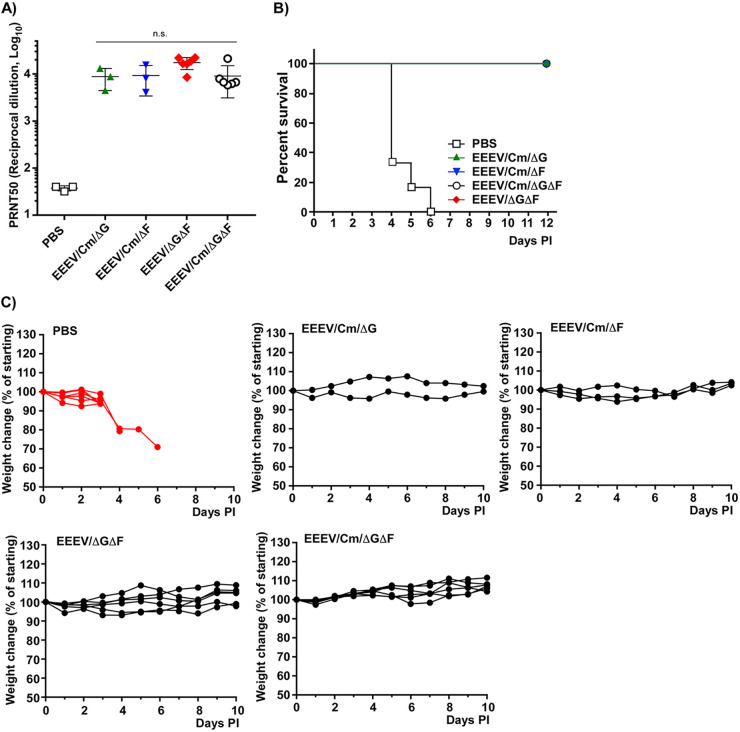
The above data demonstrated cumulative attenuating effects of the mutations in EEEV capsid protein and deletions of the binding motifs of host proteins in EEEV nsP3 HVD on viral infection in vivo. However, the lack of detectable disease in mice could be a result of overattenuation of the mutants, making them poorly immunogenic, if at all. Therefore, we tested the sera from the survived mice for the presence of EEEV-specific neutralizing Abs (see Materials and Methods for details). It was not unexpected to find high levels of EEEV-specific, neutralizing Abs in the sera of mice that had survived the EEEV/Cm/ΔG and EEEV/Cm/ΔF infections (Fig. 5A). To our surprise, EEEV/ΔGΔF and EEEV/Cm/ΔGΔF induced neutralizing Abs to the levels that were not significantly different from those detected in mice that were infected with the above mutants with deleted single FXR- or G3BP-binding sites in nsP3 HVD. Thus, the lack of virulence of EEEV/ΔGΔF and EEEV/Cm/ΔGΔF had no detectable negative effect on the abilities of the double deletion mutants to induce EEEV-specific neutralizing Abs.
FIG 5.
Mice that survived infection with designed EEEV variants were protected against infection with the parental wt EEEV. (A) Serum samples were collected on day 21 PI in the experiment presented in Fig. 3. Titers of neutralizing Abs were evaluated in PRNT50 using the SIN/EEEV chimeric virus (see Materials and Methods for details). Significance of differences was determined by 2-way ANOVA without correction for multiple comparisons: nonsignificant (n.s.), P > 0.05. (B) Mice which survived the primary infection (see Fig. 3 for details) were s.c. challenged with 106 PFU of parental wt EEEV FL-93 to assess protection. Morbidity and mortality were detected only in the PBS group. (C) Weight changes were evaluated in the individual mice until day 10 PI. No deaths were detected in the previously infected groups (black lines).
Next, mice formerly exposed to the mutant viruses were challenged with high doses of the parental EEEV (Fig. 5B). Except for the control group that was previously inoculated with only phosphate-buffered saline (PBS), all the mice that survived the primary infection were resistant to wt EEEV challenge. They did not demonstrate any weight loss (Fig. 5C) or any other symptoms of the disease. Thus, EEEV/ΔGΔF and EEEV/Cm/ΔGΔF induced protective immune response, which correlated with high titers of neutralizing, EEEV-specific Abs.
Lower infectious doses do not reduce the residual virulence of the designed mutants.
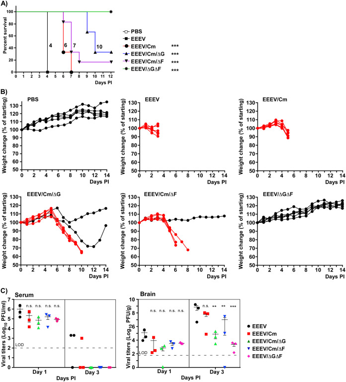
In the above study, mice were infected with very high doses of capsid and HVD mutants of EEEV. In the next round of experiments, we tested whether lowering the inoculation doses could lead to lower mortality and reduced residual neurovirulence of the variants. CD-1 mice were infected with the same doses of 5 × 10³ PFU of EEEV/Cm, EEEV/Cm/ΔG, EEEV/Cm/ΔF, parental EEEV, and the double HVD mutant EEEV/ΔGΔF, which was included in these experiments as a nonvirulent control. The data presented in Fig. 6 were consistent with the results shown in Fig. 3 and 4. EEEV/Cm, EEEV/Cm/ΔG, and EEEV/Cm/ΔF demonstrated statistically significant increases of MST compared to parental EEEV, and partial survival of mice. However, the survival was not better than in the above-described experiments. The levels of viremia were like those in the experiments with high doses of the viruses, and the indicated mutants were readily detectable in the brains. Most of the infected mice developed clear neurological symptoms, such as seizure and partial paralysis, and either succumbed to infections or were euthanized because of severe weight loss.
FIG 6.
Lower inoculation doses of EEEV mutants do not make them less virulent. (A) CD1 mice (4-to-5 weeks old) were infected with the same dose of 5 × 10³ PFU of parental EEEV and the designed mutants, or mock-infected (PBS). No deaths were detected after day 12 PI. Survival analysis was performed in GraphPad software. Numbers on the graphs indicate MSTs. The significance between survivals of mice infected with wt EEEV and mutants was estimated using a log rank test; ***, P < 0.001. (B) Weight changes detected in individual mice infected with the indicated viruses or mock-infected (PBS). Black lines correspond to the mice that survived the infection and red lines correspond to the mice that succumbed to the infection. (C) Levels of viremia and brain titers (means with SD [where applicable]) at days 1 and 3 PI. Panels show the levels of viremia and brain titers as means with SD (where applicable) at days 1 and 3 PI. Significance of differences was determined by 2-way ANOVA without correction for multiple comparisons: nonsignificant (n.s.), P > 0.05; **, P < 0.01; ***, P < 0.001.
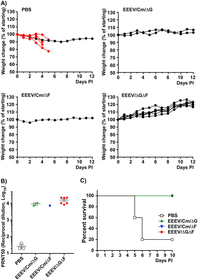
As expected, EEEV/ΔGΔF infections were asymptomatic. Mice demonstrated no weight loss and all of them survived (Fig. 6A and B). As did other survivors, these mice responded to infection with high titers of neutralizing Abs and were fully protected against following infection with parental wt EEEV (Fig. 7A to C). Thus, the use of lower doses did not result in lower residual virulence of EEEV/Cm, EEEV/Cm/ΔG, and EEEV/Cm/ΔF. These mutants retained the ability to randomly pass the blood-brain barrier and remained neurovirulent. However, this experiment additionally confirmed the highly attenuated phenotype of EEEV/ΔGΔF.
FIG 7.
Mice infected with low doses of the mutants are protected against challenge with EEEV. (A) Mice that survived the infection with EEEV mutants or were previously inoculated with PBS (see Fig. 6 for details) were s.c. challenged with 106 PFU of wt EEEV. Weight changes were evaluated in the individual mice until day 12 PI. The black lines indicate mice that survived the challenge and red lines correspond to the mice which succumbed to infection. (B) Titers of neutralizing Abs (PRNT50) in mice at day 21 PI, before challenge with EEEV (means and standard deviations). (C) Survival of mice in the challenge experiment. Morbidity and mortality were detected only in the PBS group. Data were analyzed using GraphPad Prizm software.
DISCUSSION
Alphavirus pathogenesis is a multicomponent event determined by the functioning of viral nonstructural and structural proteins, and nucleotide sequences of the cis-acting elements in viral G and SG RNAs. Mechanistic understanding of their activities in viral replication in vitro and in vivo determines our overall knowledge of viral biology and alphavirus-induced diseases, and ultimately leads to the development of antiviral countermeasures.
Within recent years, alphavirus nsP3 has attracted significant attention as a critical determinant of viral replication in both vertebrate and mosquito cells (16, 17, 19, 23, 33). To date, no enzymatic functions directly involved in the synthesis of viral G and SG RNAs have yet been ascribed to this protein. NsP3 has two conserved, structured domains (the macro domain and alphavirus unique domain [AUD]), and a C-terminal HVD (33). The distinguishing characteristics of alphavirus nsP3 HVDs are as follows: (i) they function as hubs for recruiting virus-specific host factors into large protein complexes during vRC assembly (16, 17, 19–21, 34); (ii) they are intrinsically disordered (22); (iii) they are highly variable even between the closely related viruses that belong to the same serocomplexes (1); and (iv) they contain combinations of short linear motifs that interact with virus-specific sets of host factors. Despite an exceptionally low level of conservation of amino acid (aa) sequence, HVDs of different alphaviruses, including the mosquito-specific Eilat alphavirus, demonstrate some similarities in that (i) they contain motifs that can interact with a variety of the SH3 domain-binding cellular proteins (20, 22, 34) and (ii) most of them have short repeating peptides located at the very C termini (16, 21). Importantly, (iii) nsP3 HVD-binding host factors demonstrate high levels of redundancy in their function in viral replication (17, 19). All members of the families that interact with HVD motifs can support the formation of functional alphavirus vRCs.
The C-terminal HVD repeats interact with the structural components of cellular stress granules, such as G3BP or FXR protein family members. Binding of G3BPs to nsP3 HVD is a prerequisite of replication of the Old World (OW) alphaviruses, such as SFV, CHIKV, and SINV (21). Interaction with the proteins of the FXR (but not G3BP) family is critical for the replication of the New World (NW) alphavirus VEEV (16). However, nsP3 HVD of the most pathogenic member of the NW alphaviruses, EEEV, was found to contain both CHIKV/SINV- and VEEV-specific binding motifs, and demonstrated the ability to interact with both G3BP and FXR proteins (19). These host factors that belong to two different families redundantly supported EEEV replication in vertebrate cells. Consequently, the deletions of individual FXR- and G3BP-interacting motifs in EEEV HVD had no negative effect on viral replication in vitro, but the elimination of both binding sites affected viral growth in cultured vertebrate cells (19) (Fig. 2). Importantly, EEEV variants having both FXR- and G3BP-binding sites deleted remain viable, suggesting that other host factors can still support viral replication, albeit less efficiently than a complete set of HVD-interacting proteins (Fig. 2). The detected cell-specific differences in replication of EEEV HVD mutants suggested that in different cell lines, concentrations of essential host factors may greatly vary. The list of additional EEEV HVD-binding partners includes CD2AP, SNX9, SNX33, and probably some others (19). Their functions in EEEV biology need further investigation.
The in vitro data do not necessarily directly correlate with the results coming from animal experiments. For example, wt VEEV remains encephalitogenic in a mouse model even when the critical motifs in its HVD are mutated and replication in cultured cells is affected (18). Thus, in this new study, we experimentally evaluated the effects of HVD modifications on EEEV virulence. Moreover, since one of the goals of the EEEV-related research is to attenuate this virus and develop new vaccine candidates, we not only focused on deletions in nsP3 HVD but also introduced modifications into viral capsid proteins to test the possibility of additional attenuation. Such mutations make the VEEV and EEEV capsids incapable of inhibiting cellular transcription and interfering with the induction of the innate immune response (29, 32, 35, 36). Thus, we tested the effects of clustered mutations in capsid and deletions in HVD, and their combinations on the ability of EEEV to cause viremia and induce encephalitis in mice. Another critical parameter studied was the ability of the mutants to induce protective levels of neutralizing Abs.
Capsid-specific mutations detectably attenuated EEEV and significantly increased MST, but the designed capsid mutant remained both neuroinvasive and neurovirulent in 100% of infected mice. The deletions of either FXR- or G3BP-binding motifs in the HVD of EEEV/Cm mutant had an incremental and significant negative effect on viral virulence. MSTs became longer than those of parental EEEV and EEEV/Cm mutant, and some mice infected with EEEV/Cm/ΔG or EEEV/Cm/ΔF did not develop neurological symptoms and survived the infections. From this, we concluded that the capsid- and HVD-specific mutations had either synergistic or additive negative effects on the ability of EEEV to induce the disease. However, attenuation remained incomplete, and even the reduction of the infectious doses did not result in better survival of mice and lower virulence of the mutants (Fig. 6). These double mutants appeared to remain capable of randomly passing into the brain, remained neurovirulent, and caused encephalitis.
Elimination of both FXR- and G3BP-binding sites in EEEV nsP3, in contrast, had deleterious effects on viral virulence. Mice infected even with a very high dose of EEEV/ΔGΔF, which encoded wt capsid protein, did not lose weight, did not develop any other symptoms of the disease, and survived the infection. This HVD mutant could still induce a well-detectable viremia at day 1 PI, but was found in the brains at very low levels. Its brain titers were either close to the limit of detection or undetectable. Additional cluster mutations in the capsid protein enhanced attenuation of the EEEV/Cm/ΔGΔF variant compared to EEEV/ΔGΔF. The triple mutant was not present in the brains and poorly detectable in blood of infected mice. Like EEEV/ΔGΔF, the designed EEEV/Cm/ΔGΔF could not cause any detectable disease.
It was reasonable to expect that because of inefficient replication in vivo, EEEV/Cm/ΔGΔF also lost immunogenicity. However, this was not the case. EEEV/Cm/ΔGΔF remained capable of inducing neutralizing Abs as efficiently as EEEV/ΔGΔF, and the developed adaptive immune response protected mice against the following challenge with parental wt EEEV. One of the important characteristics of alphaviruses is that they are highly infectious by aerosol. In this study, we could not answer whether the immune responses developed to EEEV/ΔGΔF and EEEV/Cm/ΔGΔF were sufficient for protecting mice against aerosol infection. However, titers of neutralizing Abs were very high and, based on the previous studies (37), may likely be protective against EEEV delivered by the aerosol route.
The high titers of neutralizing Abs induced by the triple EEEV/Cm/ΔGΔF mutant in the absence of high viremia and any detectable disease correlate with our prior findings. Previously, we detected similar efficient induction of neutralizing Abs by VEE/CHIKV recombinant viruses (38–40). These chimeras were deficient in inhibition of cellular transcription and unable to develop viremia in mice, but induced high levels of CHIKV-specific neutralizing Abs. As suggested previously for VEEV replicons, VEE/CHIKV and EEEV/Cm/ΔGΔF could become more efficient inducers of the innate immune response to viral infection, and the released type I IFN may function as an adjuvant in the development of adaptive immunity (41).
Live attenuated vaccines (LAVs) remain the most efficient means of inducing long-term protective immunity against viral pathogens (42–45). LAVs, but not other viral vaccines, fully mimic natural viral infections in terms of inducing balanced combinations of neutralizing Abs and T cell responses. Among other requirements, safety, efficacy, and stability of the attenuated phenotype remain the major characteristics during LAV development. Serial blind passaging of wt viruses in cultured cells, chicken embryos, and suckling mice was a relatively standard approach in LAV design. In the case of alphaviruses, application of vaccine design resulted in the development of attenuated VEEV TC-83 and CHIKV 181/25 strains (44, 46). However, in both cases, the attenuated phenotypes relied only on two nt substitutions in the viral genomes (47, 48) and, thus, for both mutants, high possibilities of reversions to wt sequence remained an issue. Moreover, both vaccine candidates induced adverse reactions in immunized individuals (49, 50). Despite representing a significant public health threat, to date no licensed LAVs have been developed for EEEV.
Within the recent 2 decades, the advances in molecular virology and application of reverse genetics approaches have opened new opportunities for the development of safe and efficient vaccine candidates for alphaviruses, including EEEV. SIN/EEEV chimeric virus has exhibited a highly attenuated phenotype in mice and nonhuman primates (51, 52). Recombinant Eilat virus encoding EEEV structural proteins (EIL/EEEV chimera), which is incapable of replication in vertebrate cells, was proposed as a fundamentally new type of viral vaccine. EIL/EEEV viral particles fully mimic EEEV virions, do not require any inactivation by formaldehyde or β-propiolactone that can modify particle antigenic structure, and thus are more immunogenic than standard inactivated vaccines (53). An EEEV variant with structural genes cloned under the control of an internal ribosome entry site (IRES) derived from encephalomyocarditis virus (EMCV) was also attenuated and protected mice against following infection with wt EEEV (54). Furthermore, the accumulating new knowledge about alphavirus-host interaction and mechanisms of viral replication led to the identification of the key elements in alphavirus G RNA and encoded proteins that could be modified to cause viral attenuation. In the case of EEEV, they include: (i) the 5′ untranslated region (5′UTR) of G RNA (55, 56); (ii) supra nuclear export and nuclear localization signals (supraNES and NLS, respectively) in capsid protein (28); (iii) E2-specific aa, which mediate the interaction of the glycoprotein with heparan sulfate (57); and (iv) miR-142-3p microRNA-binding sites in the 3′UTR of EEEV G and SG RNAs (58). Mutations and deletions introduced into these sites additively affect EEEV virulence in mice (37).
Our new study was based on the observation that EEEV nsP3 HVD is a key player in recruiting host factors that function in viral vRC assembly and RNA synthesis. It interacts with multiple host proteins, which redundantly mediate RNA replication in different tissues, where the concentration of each factor can vary. Thus, EEEV HVD is a critical new target for modifications aimed at EEEV attenuation. Deletions of FXR- and G3BP-binding sites affected the efficiency of viral replication, both in vivo and in vitro. Mutant viruses, which were incapable of interacting with FXR and G3BP proteins, became no longer neurovirulent and did not induce symptomatic disease in a mouse model. Inactivation of the NLS in the capsid protein additionally affected viral replication in vivo. Importantly, despite the loss of virulence, the developed HVD mutants of EEEV remained highly immunogenic and, at a single dose, protected mice against following infection with wt virus.
MATERIALS AND METHODS
Cell cultures.
Vero E6, HeLa, and NIH 3T3 cells were received from the American Type Culture collection (ATCC, Manassas, VA). Huh7.5 cells were obtained from Charles M. Rice (Rockefeller University, NY, NY) and BHK-21 cells were kindly provided by Paul Olivo (Washington University, St. Louis, MO). BHK-21, Vero, HeLa, and NIH 3T3 cells were maintained in alpha minimum essential medium (αMEM) supplemented with 10% fetal bovine serum (FBS), glutamine, and vitamins. Huh7.5 cells were maintained in Dulbecco’s modified Eagle’s medium (DMEM) supplemented with 10% FBS and glutamine.
Plasmids.
The infectious cDNA clone of North American (NA) EEEV Florida 93 (FL93) was kindly provided by Scott Weaver (University of Texas Medical Branch, Galveston, TX). The original T7 promoter in this plasmid was replaced by the SP6 promoter, and the resulting plasmid construct was used for modification of the capsid gene and nsP3 HVD. The modifications were made by standard PCR-based mutagenesis. The introduced mutations/deletions and the genomes of all the designed mutants are schematically presented in Fig. 1A. In the names of the constructs, Cm indicates the presence of mutations in the NLS-coding sequence of the capsid gene, whereas ΔG and ΔF indicate deletions of the G3BP- and FXR-binding motifs in nsP3 HVD, respectively. Plasmid containing infectious cDNA of recombinant SIN/EEEV virus has been described elsewhere (51). The genome of this chimeric virus encodes SINV-specific nsPs and its cis-acting RNA elements, and structural genes are derived from the North American EEEV (51). SIN/EEEV was used for assessing the levels of EEEV-specific neutralizing Abs in the plaque reduction neutralization test (PRNT50). The nucleotide sequences of the plasmids and details of cloning procedures can be made available upon request.
In vitro RNA transcription and transfections.
Plasmids containing viral cDNAs were purified by ultracentrifugation in CsCl gradients and linearized using the unique restriction sites located downstream of the poly(A) sequence in the 3′UTR of the viral genome. The in vitro transcription reactions were performed in the presence of a cap analog (New England BioLabs) using SP6 RNA polymerase according to the manufacturer’s protocol (Invitrogen). No additional purification of the in vitro-synthesized RNA was done, and 12 μl of the reaction mixtures was directly used for the transfection of BHK-21 cells. EEEV genomes were transfected using TransIT-mRNA transfection reagent according to the manufacturer’s instructions (Mirus). Viral stocks were harvested 24 h posttransfection. All the procedures of the in vitro RNA synthesis, virus rescue, and the following analysis of EEEV replication were performed in biosafety level 3 (BSL3) conditions of SEBLAB of the University of Alabama at Birmingham. The SIN/EEEV viral genome was synthesized in BSL2 conditions, and the virus was rescued by electroporation of the in vitro-synthesized RNA into BHK-21 cells in the previously described conditions (59). Infectious titers of the viruses were assessed by plaque assay on BHK-21 cells (59).
Viral infections in vitro.
For all the cell lines used, cells were seeded into 6-well Costar plates at the concentration of 5 × 10⁵ cells/well. After 4 h of incubation at 37°C, they were infected in PBS supplemented with 1% FBS with recombinant viruses at an MOI of 1 PFU/cell. Media were harvested 20 h PI, and viral titers were determined by plaque assay on BHK-21 cells.
Animal experiments.
CD1 mice (4-to-5-week-old females) were obtained from the Charles River Laboratory. They were s.c. infected with the designed EEEV mutants and parental wt EEEV in 100 μl of PBS supplemented with 1% mouse serum. Mice were monitored twice a day for clinical signs of disease, such as weight loss, change in behavior, seizures, and paralysis. At days 1 and 3 PI, animals were sacrificed and blood and brain samples were collected for assessment of viremia and viral replication in the brains. Titers were determined by plaque assay on BHK-21 cells. At day 21 PI, blood samples were taken for measuring the titers of neutralizing Abs, and mice were challenged with 106 PFU of wt EEEV FL93. Mice were monitored twice a day for clinical signs of disease and weight loss for the next 2 weeks. These experiments were performed in the animal biosafety level 3 (ABSL3) facility of SEBLAB of the University of Alabama at Birmingham according to IACUC-approved protocols.
Evaluation of viremia and viral titers in the brains.
Sera were serially diluted in PBS supplemented with 1% FBS. Semiconfluent BHK-21 cells in 6-well plates were infected for 1 h at 37°C. The cells were overlaid with 0.5% agarose supplemented with DMEM and 3% FBS and incubated for 48 h at 37°C. They were then fixed and stained with crystal violet.
Brain samples were homogenized in PBS and, after low-speed centrifugation, supernatants were used for measuring the titers by plaque assay on BHK-21 cells as described above.
Titers of neutralizing antibodies (PRNT50).
Serum samples were incubated at 50°C for 1 h to inactivate complement and then serially (2-fold) diluted in PBS supplemented with 1% FBS and 250 PFU/ml of SIN/EEEV. Samples were incubated with shaking at 37°C for 2 h, and 0.2-ml aliquots were then applied to monolayers of BHK-21 cells in 6-well Costar plates. After incubation at 37°C for 1 h, the cell monolayers were overlaid with 0.5% agarose supplemented with DMEM and 3% FBS. After 2 days of incubation at 37°C, plaques were stained with crystal violet. The nonlinear curves were used to get best-fit values (slope and intercept) for each sample, and these values were used to calculate dilution with a 50% reduction using GraphPad Prism software.
Statistical analyses.
All the statistical analyses were performed using GraphPad Prism 8.0 software. See figure legends for details.
ACKNOWLEDGMENTS
We thank Scott Weaver for providing the infectious cDNA clone of EEEV Florida 93.
This study was supported by Public Health Service grants R01AI133159 and R01AI118867 to E.I.F. and R01AI095449 to I.F.
REFERENCES
- 1.Strauss JH, Strauss EG. 1994. The alphaviruses: gene expression, replication, evolution. Microbiol Rev 58:491–562. doi: 10.1128/MMBR.58.3.491-562.1994. [DOI] [PMC free article] [PubMed] [Google Scholar]
- 2.Griffin DE. 2013. Alphaviruses, p 1023–1068. Knipe DM, Howley PM (ed), Fields virology, 6th ed Lippincott Williams & Wilkins, Philadelphia, PA. [Google Scholar]
- 3.Weaver SC, Lecuit M. 2015. Chikungunya virus infections. N Engl J Med 373:94–95. doi: 10.1056/NEJMc1505501. [DOI] [PubMed] [Google Scholar]
- 4.Weaver SC, Lecuit M. 2015. Chikungunya virus and the global spread of a mosquito-borne disease. N Engl J Med 372:1231–1239. doi: 10.1056/NEJMra1406035. [DOI] [PubMed] [Google Scholar]
- 5.Weaver SC, Forrester NL. 2015. Chikungunya: evolutionary history and recent epidemic spread. Antiviral Res 120:32–39. doi: 10.1016/j.antiviral.2015.04.016. [DOI] [PubMed] [Google Scholar]
- 6.Weaver SC. 2001. Venezuelan equine encephalitis, p 539–548. Service MW. (ed), The encyclopedia of arthropod-transmitted infections. CAB International, Wallingford, UK. [Google Scholar]
- 7.Weaver SC. 2001. Eastern equine encephalitis, p 151–159. Service MW. (ed), The encyclopedia of arthropod-transmitted infections. CAB International, Wallingford, UK. [Google Scholar]
- 8.Reisen WK. 2001. Western equine encephalitis, p 558–563. Service MW. (ed), The encyclopedia of arthropod-transmitted infections. CAB International, Wallingford, UK. [Google Scholar]
- 9.Lindsey NP, Martin SW, Staples JE, Fischer M. 2020. Notes from the field: multistate outbreak of eastern equine encephalitis virus—United States, 2019. MMWR Morb Mortal Wkly Rep 69:50–51. doi: 10.15585/mmwr.mm6902a4. [DOI] [PMC free article] [PubMed] [Google Scholar]
- 10.Morens DM, Folkers GK, Fauci AS. 2019. Eastern equine encephalitis virus—another emergent arbovirus in the United States. N Engl J Med 381:1989–1992. doi: 10.1056/NEJMp1914328. [DOI] [PubMed] [Google Scholar]
- 11.Brault AC, Powers AM, Chavez CL, Lopez RN, Cachon MF, Gutierrez LF, Kang W, Tesh RB, Shope RE, Weaver SC. 1999. Genetic and antigenic diversity among eastern equine encephalitis viruses from North, Central, and South America. Am J Trop Med Hyg 61:579–586. doi: 10.4269/ajtmh.1999.61.579. [DOI] [PubMed] [Google Scholar]
- 12.Aguilar PV, Adams AP, Wang E, Kang W, Carrara AS, Anishchenko M, Frolov I, Weaver SC. 2008. Structural and nonstructural protein genome regions of eastern equine encephalitis virus are determinants of interferon sensitivity and murine virulence. J Virol 82:4920–4930. doi: 10.1128/JVI.02514-07. [DOI] [PMC free article] [PubMed] [Google Scholar]
- 13.Foy NJ, Akhrymuk M, Akhrymuk I, Atasheva S, Bopda-Waffo A, Frolov I, Frolova EI. 2013. Hypervariable domains of nsP3 proteins of New World and Old World alphaviruses mediate formation of distinct, virus-specific protein complexes. J Virol 87:1997–2010. doi: 10.1128/JVI.02853-12. [DOI] [PMC free article] [PubMed] [Google Scholar]
- 14.Foy NJ, Akhrymuk M, Shustov AV, Frolova EI, Frolov I. 2013. Hypervariable domain of nonstructural protein nsP3 of Venezuelan equine encephalitis virus determines cell-specific mode of virus replication. J Virol 87:7569–7584. doi: 10.1128/JVI.00720-13. [DOI] [PMC free article] [PubMed] [Google Scholar]
- 15.Scholte FE, Tas A, Albulescu IC, Zusinaite E, Merits A, Snijder EJ, van Hemert MJ. 2015. Stress granule components G3BP1 and G3BP2 play a proviral role early in Chikungunya virus replication. J Virol 89:4457–4469. doi: 10.1128/JVI.03612-14. [DOI] [PMC free article] [PubMed] [Google Scholar]
- 16.Kim DY, Reynaud JM, Rasalouskaya A, Akhrymuk I, Mobley JA, Frolov I, Frolova EI. 2016. New World and Old World alphaviruses have evolved to exploit different components of stress granules, FXR and G3BP proteins, for assembly of viral replication complexes. PLoS Pathog 12:e1005810. doi: 10.1371/journal.ppat.1005810. [DOI] [PMC free article] [PubMed] [Google Scholar]
- 17.Meshram CD, Agback P, Shiliaev N, Urakova N, Mobley JA, Agback T, Frolova EI, Frolov I. 2018. Multiple host factors interact with the hypervariable domain of chikungunya virus nsP3 and determine viral replication in cell-specific mode. J Virol 92:e00838-18. doi: 10.1128/JVI.00838-18. [DOI] [PMC free article] [PubMed] [Google Scholar]
- 18.Meshram CD, Phillips AT, Lukash T, Shiliaev N, Frolova EI, Frolov I. 2019. Mutations in hypervariable domain of Venezuelan equine encephalitis virus nsP3 protein differentially affect viral replication. J Virol 94:e01841-19. doi: 10.1128/JVI.01841-19. [DOI] [PMC free article] [PubMed] [Google Scholar]
- 19.Frolov I, Kim DY, Akhrymuk M, Mobley JA, Frolova EI. 2017. Hypervariable domain of eastern equine encephalitis virus nsP3 redundantly utilizes multiple cellular proteins for replication complex assembly. J Virol 91:e00371-17. doi: 10.1128/JVI.00371-17. [DOI] [PMC free article] [PubMed] [Google Scholar]
- 20.Mutso M, Morro AM, Smedberg C, Kasvandik S, Aquilimeba M, Teppor M, Tarve L, Lulla A, Lulla V, Saul S, Thaa B, McInerney GM, Merits A, Varjak M. 2018. Mutation of CD2AP and SH3KBP1 binding motif in alphavirus nsP3 hypervariable domain results in attenuated virus. Viruses 10:226. doi: 10.3390/v10050226. [DOI] [PMC free article] [PubMed] [Google Scholar]
- 21.Panas MD, Ahola T, McInerney GM. 2014. The C-terminal repeat domains of nsP3 from the Old World alphaviruses bind directly to G3BP. J Virol 88:5888–5893. doi: 10.1128/JVI.00439-14. [DOI] [PMC free article] [PubMed] [Google Scholar]
- 22.Agback P, Dominguez F, Pustovalova Y, Lukash T, Shiliaev N, Orekhov VY, Frolov I, Agback T, Frolova EI. 2019. Structural characterization and biological function of bivalent binding of CD2AP to intrinsically disordered domain of chikungunya virus nsP3 protein. Virology 537:130–142. doi: 10.1016/j.virol.2019.08.022. [DOI] [PMC free article] [PubMed] [Google Scholar]
- 23.Fros JJ, Geertsema C, Zouache K, Baggen J, Domeradzka N, van Leeuwen DM, Flipse J, Vlak JM, Failloux AB, Pijlman GP. 2015. Mosquito Rasputin interacts with chikungunya virus nsP3 and determines the infection rate in Aedes albopictus. Parasit Vectors 8:464. doi: 10.1186/s13071-015-1070-4. [DOI] [PMC free article] [PubMed] [Google Scholar]
- 24.Akhrymuk I, Kulemzin SV, Frolova EI. 2012. Evasion of the innate immune response: the Old World alphavirus nsP2 protein induces rapid degradation of Rpb1, a catalytic subunit of RNA polymerase II. J Virol 86:7180–7191. doi: 10.1128/JVI.00541-12. [DOI] [PMC free article] [PubMed] [Google Scholar]
- 25.Garmashova N, Gorchakov R, Frolova E, Frolov I. 2006. Sindbis virus nonstructural protein nsP2 is cytotoxic and inhibits cellular transcription. J Virol 80:5686–5696. doi: 10.1128/JVI.02739-05. [DOI] [PMC free article] [PubMed] [Google Scholar]
- 26.Akhrymuk I, Frolov I, Frolova EI. 2018. Sindbis virus infection causes cell death by nsP2-induced transcriptional shutoff or by nsP3-dependent translational shutoff. J Virol 92:e01388-18. doi: 10.1128/JVI.01388-18. [DOI] [PMC free article] [PubMed] [Google Scholar]
- 27.Akhrymuk I, Lukash T, Frolov I, Frolova EI. 2018. Novel mutations in nsP2 abolish chikungunya virus-induced transcriptional shutoff and make the virus less cytopathic without affecting its replication rates. J Virol 93:e02062-18. doi: 10.1128/JVI.02062-18. [DOI] [PMC free article] [PubMed] [Google Scholar]
- 28.Atasheva S, Fish A, Fornerod M, Frolova EI. 2010. Venezuelan equine encephalitis virus capsid protein forms a tetrameric complex with CRM1 and importin alpha/beta that obstructs nuclear pore complex function. J Virol 84:4158–4171. doi: 10.1128/JVI.02554-09. [DOI] [PMC free article] [PubMed] [Google Scholar]
- 29.Aguilar PV, Weaver SC, Basler CF. 2007. Capsid protein of eastern equine encephalitis virus inhibits host cell gene expression. J Virol 81:3866–3876. doi: 10.1128/JVI.02075-06. [DOI] [PMC free article] [PubMed] [Google Scholar]
- 30.Garmashova N, Gorchakov R, Volkova E, Paessler S, Frolova E, Frolov I. 2007. The Old World and New World alphaviruses use different virus-specific proteins for induction of transcriptional shutoff. J Virol 81:2472–2484. doi: 10.1128/JVI.02073-06. [DOI] [PMC free article] [PubMed] [Google Scholar]
- 31.Atasheva S, Kim DY, Frolova EI, Frolov I. 2015. Venezuelan equine encephalitis virus variants lacking transcription inhibitory functions demonstrate highly attenuated phenotype. J Virol 89:71–82. doi: 10.1128/JVI.02252-14. [DOI] [PMC free article] [PubMed] [Google Scholar]
- 32.Aguilar PV, Leung LW, Wang E, Weaver SC, Basler CF. 2008. A five-amino-acid deletion of the eastern equine encephalitis virus capsid protein attenuates replication in mammalian systems but not in mosquito cells. J Virol 82:6972–6983. doi: 10.1128/JVI.01283-07. [DOI] [PMC free article] [PubMed] [Google Scholar]
- 33.Frolov I, Frolova EI. 2019. Molecular virology of chikungunya virus. Curr Top Microbiol Immunol doi: 10.1007/82_2018_146. [DOI] [PubMed] [Google Scholar]
- 34.Neuvonen M, Kazlauskas A, Martikainen M, Hinkkanen A, Ahola T, Saksela K. 2011. SH3 domain-mediated recruitment of host cell amphiphysins by alphavirus nsP3 promotes viral RNA replication. PLoS Pathog 7:e1002383. doi: 10.1371/journal.ppat.1002383. [DOI] [PMC free article] [PubMed] [Google Scholar]
- 35.Garmashova N, Atasheva S, Kang W, Weaver SC, Frolova E, Frolov I. 2007. Analysis of Venezuelan equine encephalitis virus capsid protein function in the inhibition of cellular transcription. J Virol 81:13552–13565. doi: 10.1128/JVI.01576-07. [DOI] [PMC free article] [PubMed] [Google Scholar]
- 36.Atasheva S, Krendelchtchikova V, Liopo A, Frolova E, Frolov I. 2010. Interplay of acute and persistent infections caused by Venezuelan equine encephalitis virus encoding mutated capsid protein. J Virol 84:10004–10015. doi: 10.1128/JVI.01151-10. [DOI] [PMC free article] [PubMed] [Google Scholar]
- 37.Trobaugh DW, Sun C, Dunn MD, Reed DS, Klimstra WB. 2019. Rational design of a live-attenuated eastern equine encephalitis virus vaccine through informed mutation of virulence determinants. PLoS Pathog 15:e1007584. doi: 10.1371/journal.ppat.1007584. [DOI] [PMC free article] [PubMed] [Google Scholar]
- 38.Wang E, Volkova E, Adams AP, Forrester N, Xiao SY, Frolov I, Weaver SC. 2008. Chimeric alphavirus vaccine candidates for chikungunya. Vaccine 26:5030–5039. doi: 10.1016/j.vaccine.2008.07.054. [DOI] [PMC free article] [PubMed] [Google Scholar]
- 39.Wang E, Kim DY, Weaver SC, Frolov I. 2011. Chimeric Chikungunya viruses are nonpathogenic in highly sensitive mouse models but efficiently induce a protective immune response. J Virol 85:9249–9252. doi: 10.1128/JVI.00844-11. [DOI] [PMC free article] [PubMed] [Google Scholar]
- 40.Kim DY, Atasheva S, Foy NJ, Wang E, Frolova EI, Weaver S, Frolov I. 2011. Design of chimeric alphaviruses with a programmed, attenuated, cell type-restricted phenotype. J Virol 85:4363–4376. doi: 10.1128/JVI.00065-11. [DOI] [PMC free article] [PubMed] [Google Scholar]
- 41.Konopka JL, Thompson JM, Whitmore AC, Webb DL, Johnston RE. 2009. Acute infection with Venezuelan equine encephalitis virus replicon particles catalyzes a systemic antiviral state and protects from lethal virus challenge. J Virol 83:12432–12442. doi: 10.1128/JVI.00564-09. [DOI] [PMC free article] [PubMed] [Google Scholar]
- 42.Lee T, Komiya T, Watanabe K, Aizawa C, Hashimoto H. 1995. Immune response in mice infected with the attenuated Japanese encephalitis vaccine strain SA14-14–2. Acta Virol 39:161–164. [PubMed] [Google Scholar]
- 43.Alevizatos AC, McKinney RW, Feigin RD. 1967. Live, attenuated Venezuelan equine encephalomyelitis virus vaccine. I. Clinical effects in man. Am J Trop Med Hyg 16:762–768. doi: 10.4269/ajtmh.1967.16.762. [DOI] [PubMed] [Google Scholar]
- 44.Levitt NH, Ramsburg HH, Hasty SE, Repik PM, Cole FE Jr, Lupton HW. 1986. Development of an attenuated strain of chikungunya virus for use in vaccine production. Vaccine 4:157–162. doi: 10.1016/0264-410X(86)90003-4. [DOI] [PubMed] [Google Scholar]
- 45.Sabin AB. 1965. Oral poliovirus vaccine. History of its development and prospects for eradication of poliomyelitis. JAMA 194:872–876. doi: 10.1001/jama.194.8.872. [DOI] [PubMed] [Google Scholar]
- 46.Berge TO, Banks IS, Tigertt WD. 1961. Attenuation of Venezuelan equine encephalomyelitis virus by in vitro cultivation in guinea pig heart cells. Am J Hyg 73:209–218. doi: 10.1093/oxfordjournals.aje.a120178. [DOI] [Google Scholar]
- 47.Gorchakov R, Wang E, Leal G, Forrester NL, Plante K, Rossi SL, Partidos CD, Adams AP, Seymour RL, Weger J, Borland EM, Sherman MB, Powers AM, Osorio JE, Weaver SC. 2012. Attenuation of Chikungunya virus vaccine strain 181/clone 25 is determined by two amino acid substitutions in the E2 envelope glycoprotein. J Virol 86:6084–6096. doi: 10.1128/JVI.06449-11. [DOI] [PMC free article] [PubMed] [Google Scholar]
- 48.Kinney RM, Chang GJ, Tsuchiya KR, Sneider JM, Roehrig JT, Woodward TM, Trent DW. 1993. Attenuation of Venezuelan equine encephalitis virus strain TC-83 is encoded by the 5′-noncoding region and the E2 envelope glycoprotein. J Virol 67:1269–1277. doi: 10.1128/JVI.67.3.1269-1277.1993. [DOI] [PMC free article] [PubMed] [Google Scholar]
- 49.Edelman R, Tacket CO, Wasserman SS, Bodison SA, Perry JG, Mangiafico JA. 2000. Phase II safety and immunogenicity study of live chikungunya virus vaccine TSI-GSD-218. Am J Trop Med Hyg 62:681–685. doi: 10.4269/ajtmh.2000.62.681. [DOI] [PubMed] [Google Scholar]
- 50.Powers AM. 2018. Vaccine and therapeutic options to Control chikungunya virus. Clin Microbiol Rev 13:e00104-16. doi: 10.1128/CMR.00104-16. [DOI] [PMC free article] [PubMed] [Google Scholar]
- 51.Wang E, Petrakova O, Adams AP, Aguilar PV, Kang W, Paessler S, Volk SM, Frolov I, Weaver SC. 2007. Chimeric Sindbis/eastern equine encephalitis vaccine candidates are highly attenuated and immunogenic in mice. Vaccine 25:7573–7581. doi: 10.1016/j.vaccine.2007.07.061. [DOI] [PMC free article] [PubMed] [Google Scholar]
- 52.Roy CJ, Adams AP, Wang E, Leal G, Seymour RL, Sivasubramani SK, Mega W, Frolov I, Didier PJ, Weaver SC. 2013. A chimeric Sindbis-based vaccine protects cynomolgus macaques against a lethal aerosol challenge of eastern equine encephalitis virus. Vaccine 31:1464–1470. doi: 10.1016/j.vaccine.2013.01.014. [DOI] [PMC free article] [PubMed] [Google Scholar]
- 53.Erasmus JH, Seymour RL, Kaelber JT, Kim DY, Leal G, Sherman MB, Frolov I, Chiu W, Weaver SC, Nasar F. 2018. Novel insect-specific Eilat virus-based chimeric vaccine candidates provide durable, mono- and multivalent, single-dose protection against lethal alphavirus challenge. J Virol 92:e01274-17. doi: 10.1128/JVI.01274-17. [DOI] [PMC free article] [PubMed] [Google Scholar]
- 54.Pandya J, Gorchakov R, Wang E, Leal G, Weaver SC. 2012. A vaccine candidate for eastern equine encephalitis virus based on IRES-mediated attenuation. Vaccine 30:1276–1282. doi: 10.1016/j.vaccine.2011.12.121. [DOI] [PMC free article] [PubMed] [Google Scholar]
- 55.Hyde JL, Gardner CL, Kimura T, White JP, Liu G, Trobaugh DW, Huang C, Tonelli M, Paessler S, Takeda K, Klimstra WB, Amarasinghe GK, Diamond MS. 2014. A viral RNA structural element alters host recognition of nonself RNA. Science 343:783–787. doi: 10.1126/science.1248465. [DOI] [PMC free article] [PubMed] [Google Scholar]
- 56.Reynaud JM, Kim DY, Atasheva S, Rasalouskaya A, White JP, Diamond MS, Weaver SC, Frolova EI, Frolov I. 2015. IFIT1 differentially interferes with translation and replication of alphavirus genomes and promotes induction of type I interferon. PLoS Pathog 11:e1004863. doi: 10.1371/journal.ppat.1004863. [DOI] [PMC free article] [PubMed] [Google Scholar]
- 57.Gardner CL, Choi-Nurvitadhi J, Sun C, Bayer A, Hritz J, Ryman KD, Klimstra WB. 2013. Natural variation in the heparan sulfate binding domain of the eastern equine encephalitis virus E2 glycoprotein alters interactions with cell surfaces and virulence in mice. J Virol 87:8582–8590. doi: 10.1128/JVI.00937-13. [DOI] [PMC free article] [PubMed] [Google Scholar]
- 58.Trobaugh DW, Gardner CL, Sun C, Haddow AD, Wang E, Chapnik E, Mildner A, Weaver SC, Ryman KD, Klimstra WB. 2014. RNA viruses can hijack vertebrate microRNAs to suppress innate immunity. Nature 506:245–248. doi: 10.1038/nature12869. [DOI] [PMC free article] [PubMed] [Google Scholar]
- 59.Gorchakov R, Hardy R, Rice CM, Frolov I. 2004. Selection of functional 5′ cis-acting elements promoting efficient sindbis virus genome replication. J Virol 78:61–75. doi: 10.1128/jvi.78.1.61-75.2004. [DOI] [PMC free article] [PubMed] [Google Scholar]


