Wellcome Open Research logoLink to Wellcome Open Research
. 2020 Aug 13;5:74. Originally published 2020 Apr 21. [Version 2] doi: 10.12688/wellcomeopenres.15845.2

Silent myelin-weighted magnetic resonance imaging

Tobias C Wood 1,a, Nikou L Damestani 1, Andrew J Lawrence 2, Emil Ljungberg 1, Gareth J Barker 1, Ana Beatriz Solana 3, Florian Wiesinger 1,3, Steven CR Williams 1
PMCID: PMC7431975  PMID: 32832700

Version Changes

Revised. Amendments from Version 1

The manuscript has been updated in response to the reviewer’s helpful and insightful comments. The most important changes are that the figures have been redesigned and the emphasis on the head-orientation study reduced. The MR images have been updated to use a consistent set of slices, Figures 3 & 4 have been merged into a single figure, and the average within-subject CoV has been added. Figure 1 (the number of spokes) and Figure 6 (colour scheme) have been updated for clarity. We hope that these new figures are clearer and more intuitive than the previous figures. The language used to refer to the head orientation study has been clarified to refer to results as “highly statistically significant” rather than “strong”. A reviewer provided a plausible explanation for the negative values of ihMTR in CSF, namely the use of Fermi pulses in the preparation module, and this limitation has been discussed. A table with the mean ihMTR and inverse ihMTR values has been added. The discussion has been expanded to better set the context of the paper within existing literature, with better comparisons between our results and previous papers. We think the resulting paper is much improved and thank the reviewers again for their valued input.

Abstract

Background: Inhomogeneous Magnetization Transfer (ihMT) is an emerging, uniquely myelin-specific magnetic resonance imaging (MRI) contrast. Current ihMT acquisitions utilise fast Gradient Echo sequences which are among the most acoustically noisy MRI sequences, reducing patient comfort during acquisition. We sought to address this by modifying a near silent MRI sequence to include ihMT contrast.

Methods: A Magnetization Transfer preparation module was incorporated into a radial Zero Echo-Time sequence. Repeatability of the ihMT ratio and inverse ihMT ratio were assessed in a cohort of healthy subjects. We also investigated how head orientation affects ihMT across subjects, as a previous study in a single subject suggests this as a potential confound.

Results: We demonstrated that ihMT ratios comparable to existing, acoustically loud, implementations could be obtained with the silent sequence. We observed a small but significant effect of head orientation on inverse ihMTR.

Conclusions: Silent ihMT imaging is a comparable alternative to conventional, noisy, alternatives. For all future ihMT studies we recommend careful positioning of the subject within the scanner.

Keywords: ihMT, Silent MRI, Myelin, ZTE, RUFIS

Introduction

Myelin is a critical part of a healthy nervous system and hence visualising it in vivo is of great use to clinicians. Fortuitously, myelin displays multiple physical properties that give rise to contrast in Magnetic Resonance (MR) images. Tissue containing myelinated axons has lower longitudinal and transverse relaxation times 1, 2, lower susceptibility 3, increased Magnetization Transfer (MT) effects 4, and reduced diffusion 5, compared to non-myelinated tissue. However, while all of these MR parameters are sensitive to myelination, they are not specific, as other biological processes demonstrate the same effects 6. An additional MR-relevant property of myelin is that it is semi-crystalline in nature, being formed of closely packed proteins and lipids 7. This regular structure can maintain dipolar order, and recent work has shown that this can be exploited to produce inhomogeneous Magnetization Transfer (ihMT) contrast 810. Although other substances and tissues such as muscle can exhibit ihMT 11, 12, it is possible to tune the acquisition parameters specifically to the properties of myelin 13. This, along with the fact that there are no other candidate substances that can exhibit ihMT within the central nervous system, suggests that ihMT has the potential to produce genuinely myelin-specific contrast 8.

Previous work has shown that within a single subject the ihMT effect exhibits a dependence on the orientation of axons to the magnetic field 10, 14, 15. This is attributable to the preferential alignment of myelin sheaths with the axons, leading to a non-uniform distribution of orientations of the lipids and proteins in the sheath 16, 17. As the orientation of WM to the magnetic field will also depend on the orientation of the head, this positioning of the patient within the scanner may influence ihMT metrics. To our knowledge, this effect of bulk orientation across subjects has not been directly investigated.

Recent ihMT imaging methods utilise fast gradient-echo sequences to acquire full-brain images in a reasonable time frame 18, 19. Such sequences are among the loudest MR sequences as they utilise a short Repetition Time (TR) and require rapid switching of high amplitude field gradients 20. Acoustic noise is a leading cause of discomfort for subjects during an MR examination 21, 22, and is of particular concern in paediatric and fetal MRI 2326.

It is possible to make 3D gradient echo acquisitions almost silent by swapping from the standard Cartesian to a radial zero echo-time (ZTE) acquisition scheme 2729, but due to the fixed (near zero) echo-time and RF amplifier limits it can be difficult to achieve strong tissue contrasts in such sequences 30, 31. In the current work we incorporate an MT preparation module into a radial ZTE sequence without compromising the acoustic noise level and show that myelin-weighted contrast can be achieved at the expense of only a small increase in scan time. The primary aim was to measure the repeatability of semi-quantitative ihMT and inverse ihMT ratios 14. As a secondary aim we hence investigated the effect of head orientation across subjects on the ihMT effect.

Methods

MR sequence

The Rotating Ultra-Fast Imaging Sequence (RUFIS) was originally introduced to image flowing liquids 27. It is essentially a gradient echo sequence, where each TR consists of a single RF pulse followed by a readout. The principal difference, illustrated in Figure 1, is that the readout gradient is held constant during the TR, including during the excitation pulse, and hence each readout consists of a ‘spoke’ that starts in the center of k-space and moves towards one edge. This is in contrast to a standard Cartesian sequence where the imaging gradients acquire a line from one side of k-space to the other.

Figure 1.

Figure 1.

A - Sequence diagram for a RUFIS segment. The gradients are first ramped to a constant amplitude, and short hard RF pulses are used to avoid slab profile effects from the constant gradient magnitude. For clarity, only four spokes are illustrated in the segment, our acquisition had 32. B - The MT preparation block consists of multiple saturation pulses. To generate the ihMT effect the sign of the saturation frequency is alternated, for standard MT preparation all pulses would have the same sign. C - The overall sequence consists of RUFIS segments separated by MT preparation blocks. At the start of each image a saturation module nulls all signal, and dummy segments where no data is acquired are played until the steady-state magnetization is reached.

The innovations introduced in order to be robust to flow effects have numerous serendipitous effects, chiefly massively reduced acoustic noise levels compared to conventional MR imaging schemes 29. As shown in Figure 1A, the imaging gradients have constant magnitude and only their orientation is varied during the sequence in order to appropriately sample k-space. At the end of each TR the gradient direction is changed by a small step on each gradient channel. As there are no rapid or large gradient changes, which are the major source of acoustic noise, RUFIS acquisitions are extremely quiet.

Because Radio Frequency (RF) excitation occurs in the presence of the imaging gradients in RUFIS, high bandwidth excitation pulses are required to mitigate slab profile effects. In practice, very short hard pulses (on the order of 10µs) are required, which limits the range of available flip-angles to a few degrees due to RF amplifier and transmit coil limitations 30. This restricts a naïve RUFIS implementation to Proton-Density (PD) weighting 31. To circumvent this limitation, the sequence can be segmented, with preparation pulses played in between segments; this approach has previously been utilised for T2 32 and diffusion prepared RUFIS imaging 33.

MT is a common technique for generating contrast in tissues that have high fractions of non-aqueous hydrogen protons 4, 34. Such tissues include White Matter (WM) in the brain, and so MT has seen wide application in WM diseases 35, 36. The most common acquisition method is a Cartesian gradient echo sequence with an off-resonance saturation pulse added to every TR 37. It is not feasible to play a saturation pulse in every TR within RUFIS due to the constant presence of the imaging gradient. If a saturation pulse was played while this gradient was present, the effective frequency offset of the pulse would differ across the field of view. Instead, we added a train of saturation pulses, shown in Figure 1B as a preparation module before each segment.

The approach of prepared segmented MT imaging has already been demonstrated with a Cartesian readout 38, and can be tuned to produce an increased sensitivity to myelin by varying the width and spacing of the saturation pulses 12. Cartesian readouts can choose their k-space view order to preferentially weight the center of k−space to the MT effect. Because the center of k-space is sampled in every repetition in RUFIS, such view re-ordering is not possible. T1 recovery during a segment is hence a significant issue in RUFIS, as it will dilute any weighting from a preparation module. We therefore used a short segment length to minimise T1 recovery. This results in playing the preparation pulses more frequently, which lengthens scan time. However, MT preparation requires very little dead time compared to either T1 or T2 preparation, and so the overall increase in scan time is minimal.

To minimise any contamination from transient signals at the start of scanning or when switching between different MT preparation schemes, we adopted two complementary measures. The first was the addition of a saturation module played once at the start of each volume. This consisted of a single adiabatic 90 degree pulse and spoiler to effectively null all longitudinal magnetization. Following this module, 48 dummy segments were played, where the no data was acquired, to allow the signal in brain parenchyma to approach a steady-state. The 48 dummy segments lasted for 3.3 seconds, which is longer than the approximate T1 of parenchyma (around 1s) at 3T. The full sequence schematic is shown in Figure 1C.

Imaging study

We recruited 6 male and 6 female subjects (age range 25 to 54 years) through local advertisement within our research establishment (King’s College London). Subjects gave written informed consent in accordance with ethics approved by the King’s College London REC (approval number 04/Q0706/72), and standard MRI exclusion criteria were applied. Each subject had two imaging sessions, spaced approximately a week apart, in a 3 Tesla scanner using a 12-channel head coil (Discovery MR750, GE Healthcare). Each session consisted of two ihMT scans, for a total of four ihMT scans per subject. In addition to the ihMT data, to provide an anatomical reference, in the first imaging session a standard T1-weighted image was acquired using the ADNI-GO protocol 39. No special instructions were given to subjects as to their head positions in order to acquire a representative sample of orientations with respect to the main magnetic field.

The ihMT scans consisted of five images (see below). All volumes were acquired with the following parameters: 22cm field of view, 1.5mm isotropic voxel size, readout bandwidth ±25kHz, TR 1.764ms, spokes-per-segment 32. We used a 2° hard pulse for excitation. This was lengthened from the manufacturer default of 8µs to 24µs to lower the B1 amplitude and hence minimise any saturation of the bound pool from the excitation pulses. A train of 10 Fermi saturation pulses was played between each segment with pulse-width of 500µs and a 500µs gap in between. The pulses had a root-mean-square B1 of 8.75µT and an offset frequency of 7kHz. This corresponds to a root-mean-square B1 of 6.2µT over the course of the preparation module. The total RUFIS segment time and preparation time (including ramps and switching time) were 68.6ms and 10.8ms respectively. We did not add an explicit spoiling gradient after the pulse train, instead relying on the initial gradient ramp of the acquisition segment to spoil spuriously generated transverse magnetization from the MT pulses.

To isolate the ihMT contrast images acquired under both single-sided and dual-sided irradiation are required. The five volumes were acquired with the following scheme saturation scheme: +/-, -/+, none, +, -, where + or - refers to the sign of the saturation offset frequency. We refer to the volumes with only positive or negative saturation frequency as MT-weighted and those with dual-sided saturation as enhanced MT (eMT) weighted. Scan time per ihMT volume was 65 seconds, and 5 minutes 41 seconds for all five volumes. The acoustic noise was measured with an MR-compatible microphone (Casella CEL-63X, IDEAL Industries) located in the scanner bore.

Previous work has shown that ihMT ratio (ihMTR) is sensitive to confounds from B1 (RF transmit inhomogeneity) and T1-weighting, but this can be potentially mitigated through the use of an inverse ihMTR 14, and such inverse metrics have also been used to compensate for T1 effects in Chemical Exchange Saturation Transfer experiments 40. To calculate this we additionally acquired a volume with T1-weighting. Because it is difficult to achieve high flip-angles with the short block pulses in RUFIS 30, we opted to replace the ihMT preparation train with a single on-resonance 10ms 25° pulse. The on-resonance preparation pulse generates a large amount of unwanted transverse magnetization compared to the off-resonance MT pulses, and so we added a 10 cycles-per-voxel spoiler gradient which increased the preparation module time to 11.3ms. The spoiler gradient ramp time was lengthened to reduce acoustic noise to the level of the rest of the RUFIS sequence.

The RUFIS images were reconstructed using the manufacturer’s proprietary Orchestra toolbox (GE Healthcare). The reconstruction consisted of nearest-neighbour gridding on a twice-oversampled grid 41, k-space center filling 42, 43, and Total Generalised Variation (TGV) regularization 33, 44. The TGV regularisation parameter was set to λ = 0.01 with a maximum of 64 iterations.

Analysis

We first motion corrected the MT-weighted images with mcFLIRT 45. The MTR, eMTR, ihMTR, inverse ihMTR and MT-asymmetry were calculated using an open-source C++ program added to QUIT 46. MT-asymmetry is a measure of whether the absorption rate differs for positive or negative irradiation frequencies 47. The parameters were defined to match 14:

MTR=1S++S2S0(1)
eMTR=1S+/+S/+2S0(2)
MTasym=S+SS0(3)
ihMTR=2(eMTRMTR)(4)
ihMTRinv=2ST1(1S+/+1S/+1S+1S)(5)

where S +, S , S +/− and S −/+ refer to the signal from the saturation schemes defined above, and S T1 is the signal from the T1-prepared image.

We calculated an affine transform from the eMTR image to each subject’s standard T1-weighted image using ANTs 48. The eMTR image was selected because the contrast is broadly similar to the T1-weighted. We then constructed a study template from all subject’s T1-weighted images, and non-linearly registered the resulting image to the MNI atlas 49, 50. Analysis then proceeded in two complementary directions: first, for illustrative purposes, we resampled each subject’s MT metrics in MNI space, and second, for a quantitative region of interest (ROI) analysis we resampled the JHU WM atlas into the native space of each scanning session. To minimise the number of resampling operations all transforms between the MNI space and the MT-weighted native space were concatenated before application. Ten bilateral white matter tract ROIs were selected 51.

Mean average value within the ROI was calculated for each ROI at each of the 4 scans (2 repeats at 2 sessions) for MTR, eMTR, ihMTR and inverse ihMTR. Intra-class correlation coefficients (ICC) were calculated over the 4 measurements using the regularised mixed-effects method of 52. Specifically, two-way random ICC(2,1) values and 95% confidence intervals (bootstrap percentile method; 1000 resamples) were extracted from a random effects model with random effects of subject and scan. Regularisation of variance components was achieved via a weakly informative gamma prior (shape parameter 2, rate parameter 0.5) 52. Calculations were performed in R version 3.6.2 using the blme package version 1.0.4 53. ICC values were classified as poor if they were less than 0.5, moderate if between 0.5 and 0.75, good between 0.75 and 0.9, and excellent above 0.9 54.

Finally, we investigated how head orientation affects ihMTR. As a proxy for how each subject’s head was aligned with the main magnetic field, we calculated the angle between the Z-direction (head-foot axis) of the MT scan space and the MNI atlas. This was found by first concatenating the three affine transforms (MT- to T1-weighted, T1-weighted to study template, and study template to atlas), applying the concatenated transform to the vector Z = (0, 0, 1) and then calculating the dot product between the result and Z.

We only examined the effect of orientation on ihMTRinv, as this is expected to be robust against B1- and T1-effects 14, which may also have a spatial or angular dependence 55. To probe the impact of head orientation in the presence of potential between-participant differences, we calculated linear mixed effects models using blme as above. These models included mean ihMTRinv values from all 10 ROIs, main effects and interactions of head angle & ROI and both a random and fixed effect for each participant, with subject allowed to interact with ROI. Of prime interest was the average linear effect of head angle within-subject and whether this varied over the ROIs. Statistical significance was tested with ANOVA adjusted by the Kenwood-Roger procedure, with p<0.05 considered significant.

Results

Images

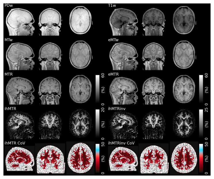
Figure 2 shows the acquired raw images and calculated MT ratios from a single subject. The acoustic noise was measured as 72 dB, compared to a 69 dB background level, which is similar to our previous work where comparable Cartesian sequences were approximately 30 dB louder 30.

Figure 2. Example raw weighted images (PD, T1, MT and eMT), ratio images (MTR, eMTR, ihMTR and ihMTRinv) and within-subject CoV for one subject.

Figure 2.

The inverse ihMTR shows a subtly improved contrast between white and grey matter compared to ihMTR, particularly in the cerebellum and putamen (note the different color scale).

The eMT-weighted image in particular demonstrates good grey matter (GM)/WM contrast, while the MTR image exhibits little GM/WM contrast. The ihMTR, which is the difference between eMTR and MTR (with a scaling factor of two), hence also shows very good GM/WM contrast. It is very close to zero outside the brain, in contrast to both MTR and eMTR which are high in tissues outside the brain. The inverse ihMTR exhibits improved GM/WM contrast compared to ihMTR in the deep GM structures, such as the putamen, the cerebellum and reduced B1 inhomogeneity effects throughout WM.

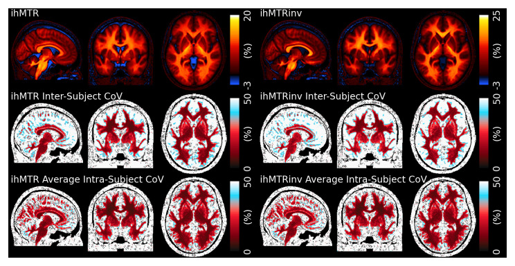
Figure 3 shows the mean, between-subject and average within-subject Coefficient of Variation (CoV) for ihMTR and inverse ihMTR in MNI space, while Table 1 summarises the average ihMTR and inverse ihMTR for the ROIs we examined. The ihMTR and inverse ihMTR values were, respectively, about 12% and 15% in WM, with values in tracts oriented parallel to the main magnetic field slightly higher as expected 15. Values were lower in GM, and we observed a small negative ihMTR in cerebral spinal fluid (CSF) and the eyeballs.

Figure 3. Top - ihMTR and ihMTRinv values averaged across all scans in MNI space.

Figure 3.

An asymmetric color scale has been used to highlight the small negative of ihMTR in cerebral spinal fluid. Heightened values can be observed in WM tracts oritented parallel to B0. Middle – Between-subject CoV calculated across all scans and subjects. Bottom – Average within-subject CoV. The within-subject CoV is lower than the between-subject in the cortex.

Table 1. The mean and standard deviation of ihMTR and inverse ihMTR in the 10 selected ROIs, calculated across all subjects.

ROI ihMTR
Mean
(%)
ihMTR SD
(%)
Inverse ihMTR
Mean (%)
Inverse ihMTR SD
(%)
Genu of CC 10.81 0.16 15.20 0.38
Body of CC 11.79 0.17 14.70 0.44
Splenium of CC 12.52 0.18 15.78 0.52
Corticospinal
tract
15.18 0.08 18.52 0.58
Cerebral
peduncle
13.89 0.17 15.14 0.48
Internal Capsule 12.67 0.22 15.47 0.53
Corona Radiata 12.67 0.16 16.54 0.40
Thalamic
Radiation
12.17 0.19 15.67 0.48
Cingl. Cingulate
Gyrus
10.82 0.15 13.30 0.40
Cingl.
Hippocampus
10.49 0.41 10.49 0.76

The between-subject CoV was approximately 10% in WM, but approaches 50% in GM and reached over 100% in CSF. The average within-subject CoV was lower, at around 8% in WM and 30% in GM. The average value of inverse ihMTR is higher, at approximately 20%. The contrast in parenchyma is broadly similar to ihMTR, but there are subtle differences in deep GM, frontal WM and the brain stem and values in CSF are close to zero instead of negative. Both the between-subject and average within-subject CoV in WM is slightly higher than for ihMTR.

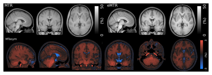
Figure 4 shows the mean MTR, eMTR and MT-asymmetry. The MTR image shows only limited contrast between WM and GM despite the high levels of saturation power, while the eMTR image shows the expected improved contrast. In contrast to ihMTR, non-zero MTR and eMTR can be observed in tissues outside the brain. We found a small consistently positive value of MT asymmetry in cerebral WM, which was increased in cerebellar WM and the major ascending arteries. MT asymmetry was negative in CSF and the eyeballs.

Figure 4. Mean MTR, eMTR and MT-Asymmetry in atlas space.

Figure 4.

Elevated levels of MT asymmetry can be observed in the ascending arteries and cerebellum.

Reliability

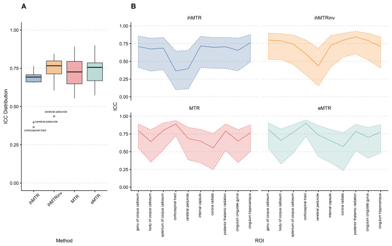
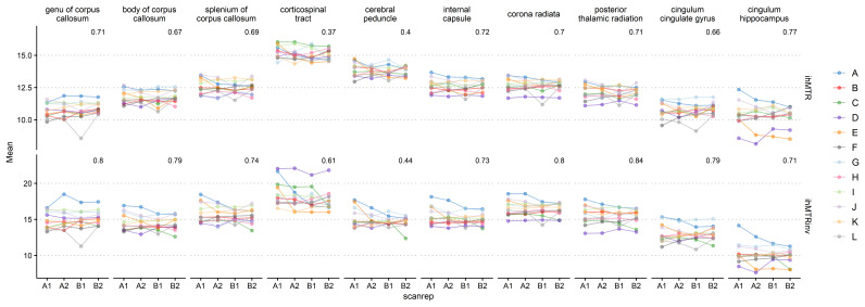
Figure 5 shows the obtained ICC values in the atlas ROIs. ICC values were moderate or good for all measures, except for the cerebral peduncles in both ihMTR and inverse ihMTR and corticospinal tract in ihMTR only, where the ICC values were poor. ICC values were slightly higher for inverse ihMTR compared to ihMTR. The cerebral peduncle and corticospinal tract ROIs commonly gave results atypical of the remaining ROIs. Figure 6 shows the mean values of ihMTR and inverse ihMTR for all ROIs across all four scans. Most ROIs show good reliability and repeatability 56, but there are several obvious outliers, for instance subject D in the corticospinal tract for inverse ihMTR, and subjects D and E for ihMTR in the cingulum hippocampus.

Figure 5.

Figure 5.

A - Intra-class correlation coefficient (ICC) distribution (Tukey Boxplot). B - ICC (solid line) and 95% bootstrap confidence intervals (dashed lines and shaded area), displaying a profile over the regions of interest (ROIs).

Figure 6. Subject-level variability in ihMTR and inverse ihMTR for the 10 atlas regions of interest.

Figure 6.

Head orientation

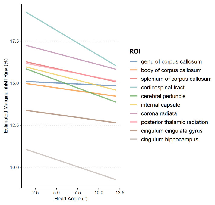
The observed median head orientation angle was 7° (lower quartile 3.2°, upper quartile 10.3°). Subject D showed particularly elevated values around 25° (excluding subject D the maximum observed angle was 12°). Closer examination revealed that subject D had a fairly small head and in both sessions was scanned with their head tilted back within the coil (data not shown). Our analysis revealed a significant main effect of head angle ( F = 27.7, p = 2.50 × 10 −7) on ihMTRinv, such that with increased rotation angle inverse ihMTR values were lower, illustrated in Figure 7. There was no significant interaction between angle and ROI ( F = 1.47, p = 0.15) indicating effects were relatively homogeneous over the ROIs. Because subject D (angle = 25°) could be interpreted as an outlier, we repeated the analysis with subject D excluded and results were comparable (main effect of angle: F = 37.24, p = 3.00 × 10 −9; angle ×ROI interaction: F = 1.32, p = 0.23).

Figure 7. Estimated effects of participant head angle× region of interest (ROI) for inverse ihMTR.

Figure 7.

As the angle of the head increases, the value of inverse ihMTR tends to decrease.

Discussion

We have demonstrated full-brain 3D myelin-weighted ihMT images acquired with a silent and fast imaging sequence. The MT preparation module increases scan-time by a minimal amount and does not compromise the silent nature of RUFIS acquisition. ihMT has shown potential for assessing myelination in multiple sclerosis 57, 58, and the use of a silent sequence will extend this potential to noise intolerant patient cohorts, for instance non-sedated infants.

The single-sided saturation MT-weighted and MTR images showed fairly flat contrast between WM and GM. This is to be expected, as the 7 kHz frequency offset chosen is optimal for the ihMT effect, whereas a smaller offset, for example 2 kHz, would likely generate larger MT contrast. Although the eMTR image exhibits good WM/GM contrast, it also shows significant signal in other tissues outside the brain, such as muscle, cartilage and blood.

Combining the eMT and MT-weighted images into an ihMT ratio increases the specificity of the sequence to myelin, as evidenced by an ihMTR close to zero outside the brain. Our ihMTR values, at around 12% in WM, are similar to previous literature using a comparable preparation module with a Cartesian readout 13, 38, but lower than recent papers using a low-duty cycle preparation module 13, 18. Our protocol was adapted from that presented in 38, which had comparable levels of power deposition during the preparation module, and the principal difference is our acquisition module acquires a larger number (32) of center-out readout spokes with a low flip-angle instead of a small number of Cartesian readouts with a higher flip-angle.

To mitigate against T1 recovery during the readout segment, which repeatedly samples the center of k-space, we minimised the number of acquired spokes to 32. The resulting segment time of less than 70 ms is much shorter than typical T1 times in parenchyma (approximately 1 s at 3T). Increasing the number of spokes per segment would lead to a reduction in scan-time, at a cost of increased T1 recovery and potentially reduced ihMTR.

A particular drawback of radial ZTE sequences compared to Cartesian is constrained SNR. We observed some residual T1-weighted contrast in the PD-weighted reference image. Reducing the excitation flip-angle below 2° to further reduce the T1-weighting would incur a linear reduction in SNR (from the small flip-angle approximation sin αα), which would likely yield unacceptable image quality. We used Total Generalized Variation regularization in our reconstruction to primarily to improve image quality, whereas for Cartesian sequences such methods are generally used with parallel imaging to speed up acquisitions 18, 38, 44. Although non-cartesian parallel imaging methods exist 59, to our knowledge none has been specifically tailored to 3D radial acquisitions. Despite this limitation, we acquired 1.5 mm isotropic maps in 6 minutes, which is competitive with a recent cartesian ihMT acquisition with an MP-RAGE type readout which acquired 2.4 mm isotropic maps in the same time 18.

We observed a small negative ihMTR in CSF and the vitreous humour of the eyeball. The likely cause of this is a small, unwanted, difference in direct saturation effects between our single-sided and dual-sided irradiation preparation modules. Changing the shape of the preparation pulses to one with better controlled sidebands, for example Hann or Gaussian, would likely remove this effect 38. Use of the inverse ihMTR appeared to mitigate T1 and B1+ effects and led to improved contrast between WM and GM, and more consistent contrast within WM, for instance the internal capsule and genu of the corpus callosum have different ihMTR values but similar inverse ihMTR values. To fully determine whether the inverse ihMTR reduced B1+ contributions would require the acquisition of additional B1+ maps, which was beyond the scope of the current work.

We observed a large MT asymmetry in the carotid arteries and an elevated value in the cerebellum compared to the cerebrum. Blood is known to exhibit an MT effect, due to a high concentration of protein 6062, and this is also known to be asymmetric 63. PET and Dynamic Contrast Enhanced MRI measurements indicate that the cerebellum has increased vascularity and cerebral blood volume compared to the cerebrum 64, 65. Hence this result appears to be consistent with previous literature.

The CoV maps in Figure 3 showed high values in cortical GM, indicating poor reliability of ihMTR metrics in the cortex. This is partly to be expected due to the very small absolute values of ihMTR in both GM. However, the average within-subject CoV was lower than the between-subject CoV, indicating that partial volume effects and registration quality affected the between-subject figure. These issues are not unique to ihMTR but affect all quantitative MRI measures 66, 67.

The repeatability of both the ihMTR and inverse ihMTR, as defined by the ICC scores, fell on the boundary of the moderate (0.5-0.75) and good (0.75-0.9) categories with the exception of the cerebral peduncles and corticospinal tract which had notably worse scores. The ICC scores of inverse ihMTR were slightly superior to ihMTR. Our ICC values are lower than a repeatability study of a steady-state Cartesian ihMT study 51. However, the values are not directly comparable as that study used a 2.4×2.4×3.2 mm voxel size compared to our 1.5 mm isotropic voxel size, with a similar overall scan time. Our lower ICC values can hence at least be partly attributed to the smaller voxel size and correspondingly lower SNR.

As shown in Figure 6 most subjects had consistent measures across all four scans but others, notably subject A, showed high variability across sessions. This variation can at least in part be attributed to the orientation of the head with the main magnetic field. We found that the observed ihMTR decreased in all ROIs as the observed rotation angle of the head increased. Our method for quantifying the rotation angle is imperfect, as it cannot distinguish positive and negative rotations and the choice of the MNI template as “zero” is arbitrary. We also did not control for the average angle of each ROI, variations in tract orientation within an ROI, the effect of hemisphere, or the effect of inter-volume motion during the ihMT scan. Despite these limitations, we showed a small but highly statistically significant effect of angle on inverse ihMTR, and so conclude that head orientation is a potential confound in ihMT studies. Recent work has incorporated prospective motion correction into a cartesian ihMT sequence 18, and such approaches could be of benefit in this radial implementation to minimise both inter- and intra-volume motion artefacts in problematic patient cohorts (e.g. infants).

Conclusion

We have demonstrated that MT-weighting can generate significant additional GM/WM contrast in silent ZTE images with minimal extension of scan time. We have shown that the derived semi-quantitative MT ratios have good repeatability, and that the inverse ihMTR has advantages over the ihMTR. However, the ihMT effect depends on the orientation of the subject’s head within the bore, and hence we recommend that careful attention is paid to participant’s positioning in future work.

Data availability

Underlying data

Figshare: Silent Myelin-Weighted MR Imaging, https://doi.org/10.6084/m9.figshare.12090645 68.

This project contains the following data:

  • Atlas images in MNI space format

  • ROI summary statistics in Comma Separated Value format

Data are available under the terms of the Creative Commons Attribution 4.0 International license (CC-BY 4.0).

Funding Statement

TCW received funding from the Wellcome/EPSRC Centre for Medical Engineering (Award Number WT 203148/Z/16/Z). ND, AJL, EL & SCRW thank the NIHR Maudsley Biomedical Research Centre at South London Maudsley Foundation Trust and King's College London for their support and funding this study. EL also thanks GE Healthcare for joint funding of his PhD studentship.

The funders had no role in study design, data collection and analysis, decision to publish, or preparation of the manuscript.

[version 2; peer review: 3 approved, 1 approved with reservations]

References

  • 1. Bock NA, Hashim E, Janik R, et al. : Optimizing T 1-weighted imaging of cortical myelin content at 3.0 T. Neuroimage. 2013;65:1–12. 10.1016/j.neuroimage.2012.09.051 [DOI] [PubMed] [Google Scholar]
  • 2. Liu H, Rubino C, Dvorak AV, et al. : Myelin Water Atlas: A Template for Myelin Distribution in the Brain. J Neuroimaging. 2019;29(6):699–706. 10.1111/jon.12657 [DOI] [PubMed] [Google Scholar]
  • 3. Lee J, Shmueli K, Kang BT, et al. : The contribution of myelin to magnetic susceptibility-weighted contrasts in high-field MRI of the brain. Neuroimage. 2012;59(4):3967–3975. 10.1016/j.neuroimage.2011.10.076 [DOI] [PMC free article] [PubMed] [Google Scholar]
  • 4. Wolff SD, Balaban RS: Magnetization transfer contrast (MTC) and tissue water proton relaxation in vivo. Magn Reson Med. 1989;10(1):135–144. 10.1002/mrm.1910100113 [DOI] [PubMed] [Google Scholar]
  • 5. De Santis S, Drakesmith M, Bells S, et al. : Why diffusion tensor MRI does well only some of the time: variance and covariance of white matter tissue microstructure attributes in the living human brain. Neuroimage. 2014;89(100):35–44. 10.1016/j.neuroimage.2013.12.003 [DOI] [PMC free article] [PubMed] [Google Scholar]
  • 6. Wood TC, Simmons C, Hurley SA, et al. : Whole-brain ex-vivo quantitative MRI of the cuprizone mouse model. PeerJ. 2016;4:e2632. 10.7717/peerj.2632 [DOI] [PMC free article] [PubMed] [Google Scholar]
  • 7. Boullerne AI: The history of myelin. Exp Neurol. 2016;283(Pt B):431–445. 10.1016/j.expneurol.2016.06.005 [DOI] [PMC free article] [PubMed] [Google Scholar]
  • 8. Varma G, Duhamel G, de Bazelaire C, et al. : Magnetization transfer from inhomogeneously broadened lines: A potential marker for myelin. Magn Reson Med. 2015;73(2):614–622. 10.1002/mrm.25174 [DOI] [PMC free article] [PubMed] [Google Scholar]
  • 9. Girard OM, Prevost VH, Varma G, et al. : Magnetization transfer from inhomogeneously broadened lines (ihMT): Experimental optimization of saturation parameters for human brain imaging at 1.5 Tesla. Magn Reson Med. 2015;73(6):2111–2121. 10.1002/mrm.25330 [DOI] [PubMed] [Google Scholar]
  • 10. Manning AP, Chang KL, MacKay AL, et al. : The physical mechanism of “inhomogeneous” magnetization transfer MRI. J Magn Reson. 2017;274(Supplement C):125–136. 10.1016/j.jmr.2016.11.013 [DOI] [PubMed] [Google Scholar]
  • 11. Swanson SD, Malyarenko DI, Fabiilli ML, et al. : Molecular, dynamic, and structural origin of inhomogeneous magnetization transfer in lipid membranes. Magn Reson Med. 2017;77(3):1318–1328. 10.1002/mrm.26210 [DOI] [PubMed] [Google Scholar]
  • 12. Duhamel G, Prevost VH, Cayre M, et al. : Validating the sensitivity of inhomogeneous magnetization transfer (ihMT) MRI to myelin with fluorescence microscopy. Neuroimage. 2019;199:289–303. 10.1016/j.neuroimage.2019.05.061 [DOI] [PubMed] [Google Scholar]
  • 13. Varma G, Girard OM, Mchinda S, et al. : Low duty-cycle pulsed irradiation reduces magnetization transfer and increases the inhomogeneous magnetization transfer effect. J Magn Reson. 2018;296:60–71. 10.1016/j.jmr.2018.08.004 [DOI] [PubMed] [Google Scholar]
  • 14. Geeraert BL, Lebel RM, Mah AC, et al. : A comparison of inhomogeneous magnetization transfer, myelin volume fraction, and diffusion tensor imaging measures in healthy children. Neuroimage. 2018;182:343–350. 10.1016/j.neuroimage.2017.09.019 [DOI] [PubMed] [Google Scholar]
  • 15. Ercan E, Varma G, Mädler B, et al. : Microstructural correlates of 3D steady-state inhomogeneous magnetization transfer (ihMT) in the human brain white matter assessed by myelin water imaging and diffusion tensor imaging. Magn Reson Med. 2018;80(6):2402–2414. 10.1002/mrm.27211 [DOI] [PubMed] [Google Scholar]
  • 16. Pampel A, Müller DK, Anwander A, et al. : Orientation dependence of magnetization transfer parameters in human white matter. NeuroImage. 2015;114:136–146. 10.1016/j.neuroimage.2015.03.068 [DOI] [PubMed] [Google Scholar]
  • 17. Girard OM, Prevost VH, Mchinda M, et al. : 472 - Anisotropy of inhomogeneous Magnetization Transfer (ihMT) in White Matter.2017; ISMRM 25th Annual Meeting. Reference Source [Google Scholar]
  • 18. Varma G, Munsch F, Burns B, et al. : Three-dimensional inhomogeneous magnetization transfer with rapid gradient-echo (3D ihMTRAGE) imaging. Magn Reson Med. 2020. 10.1002/mrm.28324 [DOI] [PMC free article] [PubMed] [Google Scholar]
  • 19. Malik SJ, Teixeira RPAG, West DJ, et al. : Steady-state imaging with inhomogeneous magnetization transfer contrast using multiband radiofrequency pulses. Magn Reson Med. 2020;83(3):935–949. 10.1002/mrm.27984 [DOI] [PMC free article] [PubMed] [Google Scholar]
  • 20. Jin C, Li H, Li X, et al. : Temporary Hearing Threshold Shift in Healthy Volunteers with Hearing Protection Caused by Acoustic Noise Exposure during 3-T Multisequence MR Neuroimaging. Radiology. 2018;286(2):602–608. 10.1148/radiol.2017161622 [DOI] [PubMed] [Google Scholar]
  • 21. McNulty JP, McNulty S: Acoustic noise in magnetic resonance imaging: An ongoing issue. Radiography. 2009;15(4):320–326. 10.1016/j.radi.2009.01.001 [DOI] [Google Scholar]
  • 22. van Minde D, Klaming L, Weda H: Pinpointing Moments of High Anxiety During an MRI Examination. Int J Behav Med. 2014;21(3):487–95. 10.1007/s12529-013-9339-5 [DOI] [PubMed] [Google Scholar]
  • 23. Hughes EJ, Winchman T, Padormo F, et al. : A dedicated neonatal brain imaging system. Magn Reson Med. 2017;78(2):794–804. 10.1002/mrm.26462 [DOI] [PMC free article] [PubMed] [Google Scholar]
  • 24. Raschle N, Zuk J, Ortiz-Mantilla S, et al. : Pediatric neuroimaging in early childhood and infancy: challenges and practical guidelines. Ann N Y Acad Sci. 2012;1252(1):43–50. 10.1111/j.1749-6632.2012.06457.x [DOI] [PMC free article] [PubMed] [Google Scholar]
  • 25. Hutter J, Price AN, Cordero-Grande L, et al. : Quiet echo planar imaging for functional and diffusion MRI. Magn Reson Med. 2018;79(3):1447–1459. 10.1002/mrm.26810 [DOI] [PMC free article] [PubMed] [Google Scholar]
  • 26. Edwards AD, Arthurs OJ: Paediatric MRI under sedation: is it necessary? What is the evidence for the alternatives? Pediatr Radiol. 2011;41(11):1353–1364. 10.1007/s00247-011-2147-7 [DOI] [PubMed] [Google Scholar]
  • 27. Madio DP, Lowe IJ: Ultra-fast imaging using low flip angles and FIDs. Magn Reson Med. 1995;34(4):525–529. 10.1002/mrm.1910340407 [DOI] [PubMed] [Google Scholar]
  • 28. Weiger M, Pruessmann KP: MRI with Zero Echo Time.In Robin K. Harris, editor, Encyclopedia of Magnetic Resonance.John Wiley & Sons, Ltd Chichester, UK.2012. 10.5167/uzh-73614 [DOI] [Google Scholar]
  • 29. Alibek S, Vogel M, Sun W, et al. : Acoustic noise reduction in MRI using Silent Scan: an initial experience. Diagn Interv Radiol. 2014;20(4):360–363. 10.5152/dir.2014.13458 [DOI] [PMC free article] [PubMed] [Google Scholar]
  • 30. Ljungberg E, Wood T, Solana AB, et al. : Silent T 1 mapping using the variable flip angle method with B 1 correction. Magn Reson Med. 2020;84(2):813–824. 10.1002/mrm.28178 [DOI] [PubMed] [Google Scholar]
  • 31. Wiesinger F, Sacolick LI, Menini A, et al. : Zero TE MR bone imaging in the head. Magn Reson Med. 2016;75(1):107–114. 10.1002/mrm.25545 [DOI] [PubMed] [Google Scholar]
  • 32. Solana AB, Menini A, Sacolick LI, et al. : Quiet and distortion-free, whole brain BOLD fMRI using T 2-prepared RUFIS. Magn Reson Med. 2016;75(4):1402–1412. 10.1002/mrm.25658 [DOI] [PubMed] [Google Scholar]
  • 33. Yuan J, Hu Y, Menini A, et al. : Near-silent distortionless DWI using magnetization-prepared RUFIS. Magn Reson Med. 2020;84(1):170–181. 10.1002/mrm.28106 [DOI] [PMC free article] [PubMed] [Google Scholar]
  • 34. Henkelman RM, Stanisz GJ, Graham SJ: Magnetization transfer in MRI: a review. NMR Biomed. 2001;14(2):57–64. 10.1002/nbm.683 [DOI] [PubMed] [Google Scholar]
  • 35. Tozer D, Ramani A, Barker GJ, et al. : Quantitative magnetization transfer mapping of bound protons in multiple sclerosis. Magn Reson Med. 2003;50(1):83–91. 10.1002/mrm.10514 [DOI] [PubMed] [Google Scholar]
  • 36. Dortch RD, Moore J, Li K, et al. : Quantitative magnetization transfer imaging of human brain at 7 T. Neuroimage. 2013;64:640–649. 10.1016/j.neuroimage.2012.08.047 [DOI] [PMC free article] [PubMed] [Google Scholar]
  • 37. Portnoy S, Stanisz GJ: Modeling pulsed magnetization transfer. Magn Reson Med. 2007;58(1):144–155. 10.1002/mrm.21244 [DOI] [PubMed] [Google Scholar]
  • 38. Mchinda S, Varma G, Prevost VH, et al. : Whole brain inhomogeneous magnetization transfer (ihMT) imaging: Sensitivity enhancement within a steady-state gradient echo sequence. Magn Reson Med. 2018;79(5):2607–2619. 10.1002/mrm.26907 [DOI] [PubMed] [Google Scholar]
  • 39. Leung KK, Malone IM, Ourselin S, et al. : Effects of changing from non-accelerated to accelerated MRI for follow-up in brain atrophy measurement. Neuroimage. 2015;107:46–53. 10.1016/j.neuroimage.2014.11.049 [DOI] [PMC free article] [PubMed] [Google Scholar]
  • 40. Zaiss M, Xu J, Goerke S, et al. : Inverse Z-spectrum analysis for spillover-, MT-, and T 1 -corrected steady-state pulsed CEST-MRI--application to pH-weighted MRI of acute stroke. NMR Biomed. 2014;27(3):240–252. 10.1002/nbm.3054 [DOI] [PMC free article] [PubMed] [Google Scholar]
  • 41. Oesterle C, Markl M, Strecker R, et al. : Spiral reconstruction by regridding to a large rectilinear matrix: a practical solution for routine systems. J Magn Reson Imaging. 1999;10(1):84–92. [DOI] [PubMed] [Google Scholar]
  • 42. Wu Y, Dai G, Ackerman JL, et al. : Water- and fat-suppressed proton projection MRI (WASPI) of rat femur bone. Magn Reson Med. 2007;57(3):554–567. 10.1002/mrm.21174 [DOI] [PubMed] [Google Scholar]
  • 43. Froidevaux R, Weiger M, Brunner DO, et al. : Filling the dead-time gap in zero echo time MRI: Principles compared. Magn Reson Med. 2018;79(4):2036–2045. 10.1002/mrm.26875 [DOI] [PubMed] [Google Scholar]
  • 44. Knoll F, Bredies K, Pock T, et al. : Second order total generalized variation (TGV) for MRI. Magn Reson Med. 2011;65(2):480–491. 10.1002/mrm.22595 [DOI] [PMC free article] [PubMed] [Google Scholar]
  • 45. Jenkinson M, Bannister P, Brady M, et al. : Improved optimization for the robust and accurate linear registration and motion correction of brain images. Neuroimage. 2002;17(2):825–841. 10.1016/s1053-8119(02)91132-8 [DOI] [PubMed] [Google Scholar]
  • 46. Wood TC: QUIT: QUantitative Imaging Tools. J Open Source Softw. 2018;3(26):656 10.21105/joss.00656 [DOI] [Google Scholar]
  • 47. Hua J, Jones CK, Blakeley J, et al. : Quantitative description of the asymmetry in magnetization transfer effects around the water resonance in the human brain. Magn Reson Med. 2007;58(4):786–793. 10.1002/mrm.21387 [DOI] [PMC free article] [PubMed] [Google Scholar]
  • 48. Avants BB, Tustison NJ, Song G, et al. : A reproducible evaluation of ANTs similarity metric performance in brain image registration. Neuroimage. 2011;54(3):2033–2044. 10.1016/j.neuroimage.2010.09.025 [DOI] [PMC free article] [PubMed] [Google Scholar]
  • 49. Avants BB, Yushkevich P, Pluta J, et al. : The optimal template effect in hippocampus studies of diseased populations. Neuroimage. 2010;49(3):2457–2466. 10.1016/j.neuroimage.2009.09.062 [DOI] [PMC free article] [PubMed] [Google Scholar]
  • 50. Tustison NJ, Avants BB, Cook PA, et al. : N4ITK: improved N3 bias correction. IEEE Trans Med Imaging. 2010;29(6):1310–1320. 10.1109/TMI.2010.2046908 [DOI] [PMC free article] [PubMed] [Google Scholar]
  • 51. Zhang L, Chen T, Tian H, et al. : Reproducibility of inhomogeneous magnetization transfer (ihMT): A test-retest, multi-site study. Magn Reson Imaging. 2019;57:243–249. 10.1016/j.mri.2018.11.010 [DOI] [PubMed] [Google Scholar]
  • 52. Chen G, Taylor PA, Haller SP, et al. : Intraclass correlation: Improved modeling approaches and applications for neuroimaging. Hum Brain Mapp. 2018;39(3):1187–1206. 10.1002/hbm.23909 [DOI] [PMC free article] [PubMed] [Google Scholar]
  • 53. Chung Y, Rabe-Hesketh S, Dorie V, et al. : A nondegenerate penalized likelihood estimator for variance parameters in multilevel models. Psychometrika. 2013;78(4):685–709. 10.1007/s11336-013-9328-2 [DOI] [PubMed] [Google Scholar]
  • 54. Koo TK, Li MY: A Guideline of Selecting and Reporting Intraclass Correlation Coefficients for Reliability Research. J Chiropr Med. 2016;15(2):155–163. 10.1016/j.jcm.2016.02.012 [DOI] [PMC free article] [PubMed] [Google Scholar]
  • 55. Schyboll F, Jaekel U, Weber B, et al. : The impact of fibre orientation on T 1-relaxation and apparent tissue water content in white matter. MAGMA. 2018;31(4):501–510. 10.1007/s10334-018-0678-8 [DOI] [PubMed] [Google Scholar]
  • 56. Vavasour IM, Clark CM, Li DK, et al. : Reproducibility and reliability of MR measurements in white matter: clinical implications. Neuroimage. 2006;32(2):637–642. 10.1016/j.neuroimage.2006.03.036 [DOI] [PubMed] [Google Scholar]
  • 57. Van Obberghen E, Mchinda S, le Troter A, et al. : Evaluation of the Sensitivity of Inhomogeneous Magnetization Transfer (ihMT) MRI for Multiple Sclerosis. AJNR Am J Neuroradiol. 2018;39(4):634–641. 10.3174/ajnr.A5563 [DOI] [PMC free article] [PubMed] [Google Scholar]
  • 58. Zhang L, Wen B, Chen T, et al. : A comparison study of inhomogeneous magnetization transfer (ihMT) and magnetization transfer (MT) in multiple sclerosis based on whole brain acquisition at 3.0 T. Magn Reson Imaging. 2020;70:43–49. 10.1016/j.mri.2020.03.010 [DOI] [PubMed] [Google Scholar]
  • 59. Luo T, Noll DC, Fessler JA, et al. : A GRAPPA algorithm for arbitrary 2D/3D non-Cartesian sampling trajectories with rapid calibration. Magn Reson Med. 2019;82(3):1101–1112. 10.1002/mrm.27801 [DOI] [PMC free article] [PubMed] [Google Scholar]
  • 60. Morrison C, Henkelman RM: A model for magnetization transfer in tissues. Magn Reson Med. 1995;33(4):475–482. 10.1002/mrm.1910330404 [DOI] [PubMed] [Google Scholar]
  • 61. Spees WM, Yablonskiy DA, Oswood MC, et al. : Water proton MR properties of human blood at 1.5 Tesla: magnetic susceptibility, T 1, T 2, T* 2, and non-Lorentzian signal behavior. Magn Reson Med. 2001;45(4):533–542. 10.1002/mrm.1072 [DOI] [PubMed] [Google Scholar]
  • 62. Kim T, Hendrich K, Kim SG: Functional MRI with magnetization transfer effects: Determination of BOLD and arterial blood volume changes. Magn Reson Med. 2008;60(6):1518–1523. 10.1002/mrm.21766 [DOI] [PMC free article] [PubMed] [Google Scholar]
  • 63. Song AW, Wolff SD, Balaban RS, et al. : The effect of off-resonance radiofrequency pulse saturation on fMRI contrast. NMR Biomed. 1997;10(4–5):208–215. [DOI] [PubMed] [Google Scholar]
  • 64. Turkheimer FE, Edison P, Pavese N, et al. : Reference and Target Region Modeling of [11C]-(R)-PK11195 Brain Studies. J Nucl Med. 2007;48(1):158–67. [PubMed] [Google Scholar]
  • 65. Østergaard L, Weisskoff RM, Chesler DA, et al. : High resolution measurement of cerebral blood flow using intravascular tracer bolus passages. Part I: Mathematical approach and statistical analysis. Magn Reson Med. 1996;36(5):715–725. 10.1002/mrm.1910360510 [DOI] [PubMed] [Google Scholar]
  • 66. Piredda GF, Hilbert T, Granziera C, et al. : Quantitative brain relaxation atlases for personalized detection and characterization of brain pathology. Magn Reson Med. 2020;83(1):337–351. 10.1002/mrm.27927 [DOI] [PubMed] [Google Scholar]
  • 67. Bonnier G, Fischi-Gomez E, Roche A, et al. : Personalized pathology maps to quantify diffuse and focal brain damage. Neuroimage Clin. 2019;21:101607. 10.1016/j.nicl.2018.11.017 [DOI] [PMC free article] [PubMed] [Google Scholar]
  • 68. Tobias W: Silent Myelin-Weighted MR Imaging. figshare.Dataset.2020. 10.6084/m9.figshare.12090645.v1 [DOI] [Google Scholar]
Wellcome Open Res. 2020 Oct 16. doi: 10.21956/wellcomeopenres.17734.r39968

Reviewer response for version 2

Gunther Helms 1

A module of 10 off-resonance Fermi pulses was integrated into a silent 3D radial ZTE sequence (RUFIS) to derive different metrics for magnetization transfer (MT) and inhomogeneous MT (ihMT) of the brain at 3T. It is demonstrated that MT impose additional GM/WM contrast onto the proton density-weighted ZTE images. From these, metrics of inhomogeneous MT (ihMT) are derived, which have been shown to correlate to myelin. Obviously, the presented methodology is especially suited to study myelination in children.  

The sequence implementation is thoroughly motivated. A careful evaluation of 12 healthy adults was conducted to define sequence-specific semi-quantitative values for MT and ihMT.  Repeatability and susceptibility to angulation is described both for individuals and on a group level. The presented documentation may be regarded exemplary for implementation of ihMT into the context of an imaging sequence.

Is the rationale for developing the new method (or application) clearly explained?

Yes

Is the description of the method technically sound?

Yes

Are the conclusions about the method and its performance adequately supported by the findings presented in the article?

Yes

If any results are presented, are all the source data underlying the results available to ensure full reproducibility?

Yes

Are sufficient details provided to allow replication of the method development and its use by others?

Yes

Reviewer Expertise:

Quantitative MRI, especially MT.

I confirm that I have read this submission and believe that I have an appropriate level of expertise to confirm that it is of an acceptable scientific standard.

Wellcome Open Res. 2020 Aug 20. doi: 10.21956/wellcomeopenres.17734.r39965

Reviewer response for version 2

Richard Dortch 1

The authors have addressed all of my critiques. I have no new comments.

Is the rationale for developing the new method (or application) clearly explained?

Partly

Is the description of the method technically sound?

Yes

Are the conclusions about the method and its performance adequately supported by the findings presented in the article?

Partly

If any results are presented, are all the source data underlying the results available to ensure full reproducibility?

Yes

Are sufficient details provided to allow replication of the method development and its use by others?

Yes

Reviewer Expertise:

Imaging science, quantitative MRI, myelin imaging.

I confirm that I have read this submission and believe that I have an appropriate level of expertise to confirm that it is of an acceptable scientific standard.

Wellcome Open Res. 2020 Aug 17. doi: 10.21956/wellcomeopenres.17734.r39967

Reviewer response for version 2

Douglas Dean 1,2,3

The authors have addressed my previous comments and the manuscript is much improved. I have no further comments.

Is the rationale for developing the new method (or application) clearly explained?

Yes

Is the description of the method technically sound?

Partly

Are the conclusions about the method and its performance adequately supported by the findings presented in the article?

Yes

If any results are presented, are all the source data underlying the results available to ensure full reproducibility?

Yes

Are sufficient details provided to allow replication of the method development and its use by others?

Partly

Reviewer Expertise:

quantitative Magnetic Resonance imaging, white matter imaging.

I confirm that I have read this submission and believe that I have an appropriate level of expertise to confirm that it is of an acceptable scientific standard.

Wellcome Open Res. 2020 Jun 4. doi: 10.21956/wellcomeopenres.17381.r38686

Reviewer response for version 1

Gunther Helms 1

An MT-module of 8 off-resonance Fermi pulses was integrated into a 3D silent zero TE radial scan (RUFIS) to derive different metrics for magnetization transfer (MT) and inhomogeneous MT (ihMT). Since is MT is tissue specific, it is demonstrated that MT-weighting can be used to generate additional GM/WM contrast in silent ZTE images, which per se show poor tissue contrast due to low flip angle.

The paper is well motivated, carefully conducted, and presented clearly.

The reviewer has three points of concern:

  1. Since MT is not specific for myelin and the higher specificity of ihMT for myelin is not fully established yet, a more conservative descriptive title is recommended, e.g. Inhomogeneous MT (ihMT) in silent ZTE MRI.

  2. Via the saturation of the bound pool/dipolar reservoir, the imposed saturation by MT will depend on spatial B 1 + inhomogenieties as acknowledged in the paper. Since the 3D images/maps are presented only by sagittal views, it is impossible for the reader to appreciate the residual spatial inhomogeity of the metrics. Transversal images and maps of (ih)MT metrics should also be presented. Since the paper specifically address the influence of orientation relative to B0, it would be good to compare this effect in size to the influence of the local deviation from the nominal B 1 + (varying between 80% and 120% across the brain at 3T). Variation in B 1 + can be mimicked by manipulating the transmit gain. 

  3. To mitigate these spatial effects, the authors calculate an inverse ihMT metric using a reference ZTE image, where T1-weighting has been imposed in the preparation module. However, this metric is not identical to the one in Ref 20, which is based on a simplified signal equation that approximates a sequence where single MT pulses and SPGR readouts are interleaved (Helms et al. 2008 1). To complicate matters, Ref 20 did not show theoretically that their metric actually reduces B 1 +. Qualitative arguments become clear only after lengthy reconstruction, as the inverse MTR (reference of the same flip angle) is proportional to the MTsat. Since the framework of Helms et al. is not applicable to interleaved ZTE , the authors should mention the pragmatic nature of borrowing this approach. NB that comparing non-selective partial saturation by 1-cos (B 1 +*25°) = approx 0.95*B 1 +2 to tissue-type-dependent repetitive absorption (roughly proportional to B 1 + 2) seems reasonable well motivated to the reviewer, but will like obscure T1 relaxation effects even more than MTR-like metrics using S0.

Minor points:

  1. Intro: For the uninitiated reader, the “technical limitations” precluding strong ZTE contrast may be mentioned already in the introduction.

  2. Methods: Please provide the measurement time required for one 3D ZTE volume (I calculated about 1:45 mins) and the total of 5 weightings.

  3. Methods: Ref 20 used -/+ 5kHz offset. Please motivate the use of -/+ 7kHz. (My guess is shorter MT pulses.)

  4. Methods: It would be helpful for the uninitiated reader to motivate the definition (sign) of MT asym Hua, Jones, Blakely et al. 2007 2 is the earliest report of asymmetric MT in brain.

  5. The CV map in second right, lower row panels in Figures 3 and 4 do not seem to match the maps in the top row. CV maps 3 and 4 do, so please replace 5.

  6. The dependence on surrogate head angle in Fig. 8 is largest in cortico-spinal tract (parallel to B0) and smallest in corpus collosum ROIs (always perpendicular to B0). It would be helpful to state this finding in the flow text.

Is the rationale for developing the new method (or application) clearly explained?

Yes

Is the description of the method technically sound?

Yes

Are the conclusions about the method and its performance adequately supported by the findings presented in the article?

Yes

If any results are presented, are all the source data underlying the results available to ensure full reproducibility?

Yes

Are sufficient details provided to allow replication of the method development and its use by others?

Yes

Reviewer Expertise:

Quantitative MRI, especially MT.

I confirm that I have read this submission and believe that I have an appropriate level of expertise to confirm that it is of an acceptable scientific standard, however I have significant reservations, as outlined above.

References

  • 1. : High-resolution maps of magnetization transfer with inherent correction for RF inhomogeneity and T1 relaxation obtained from 3D FLASH MRI. Magn Reson Med.2008;60(6) : 10.1002/mrm.21732 1396-407 10.1002/mrm.21732 [DOI] [PubMed] [Google Scholar]
  • 2. : Quantitative description of the asymmetry in magnetization transfer effects around the water resonance in the human brain. Magn Reson Med.2007;58(4) : 10.1002/mrm.21387 786-93 10.1002/mrm.21387 [DOI] [PMC free article] [PubMed] [Google Scholar]
Wellcome Open Res. 2020 Aug 13.
Tobias C Wood 1

We thank the reviewer for their time and insight. There were in total five reviewers, with many helpful suggestions, and hence there have been many edits to the paper. Responses to this particular review follow below.

1. We agree that MT is not specific for myelin, and while there is increasing evidence that ihMT can be made specific to myelin (e.g. Duhamel et al 2019), we opted for caution in the title “Silent myelin- weighted magnetic resonance imaging” (emphasis added) because myelin is likely to be the dominant cause of contrast. We think that this is justified in comparison to T1-weighted and T2-weighted imaging, where T1 and T2 are the dominant but not sole contrast mechanisms. Hence we opt to keep the title unchanged.

2. We have changed the 3 sagittal views of ihMTR and ihMTRinv to sagittal, axial & coronal views. In our opinion, visually, these show good spatial homogeneity. In our opinion assessing the deviation due to B1+ would be best assessed with the acquisition of a separate B1+ map, which due to time constraints could not be included in this study.

3. The suggested reference has been added. Additional references describing an asymmetric MT effect in blood have been added and the discussion amended accordingly.

Minor Points

1. The fixed echo-time and RF power limitations have been explicitly added to the introduction.

2. The volume measurement time is present in the text “Scan time per volume was 65 seconds”, we have added the total acquisition time for 5 volumes.

3. A range of offsets for the ihMT pulses have been used in the literature. Reference 14, Mchinda et al 2018, indicated that an offset frequency higher than 5 kHz produced a larger ihMT effect, in fact finally opting for 8 kHz. In early sequence testing 7 kHz was found to produce a sufficient ihMT effect.

4. The suggested reference has been added. In addition we have updated the discussion about the asymmetry effect in light of finding an additional reference on the topic (Zhou et al 2005).

5. We do not completely understand this comment. In figure 5, the bottom row is the MT-asymmetry, not a CoV map.

6. The lowest effect is present in the cingulum hippocampus, not the corpus callosum. The colour scheme for these two ROIs was quite similar, it has been amended.

Wellcome Open Res. 2020 May 29. doi: 10.21956/wellcomeopenres.17381.r38685

Reviewer response for version 1

Douglas Dean 1,2,3

Overview: This study describes the development of a novel, silent inhomogeneous magnetization transfer (ihMT) technique for imaging myelin in a clinically feasible time. Specifically, the authors evaluate the reproducibility of ihMT derived metrics from a cohort of healthy subjects. The authors additionally explored the influence of head orientation on ihMT measures, as recent study has suggested this to possibly affect these measures. A total of 4 scans were acquired from each participant (2 sessions, 2 ihMT scans per session) and the authors examined ihMT metrics across the whole brain as well as from regions of interests (ROIs) from an existing brain atlas. Intra-class correlations were used to assess reliability. The authors demonstrate comparable and good reliability of ihMT metrics from the novel, silent sequence, while coefficent of variation of ihMT and inverse ihMT demonstrate high consistency of the measures, particularly in white matter. An association with head orientation was also observed. Overall, the manuscript is well written, the study is well designed, and appropriate methodologies were used. The results demonstrate the proposed silent ihMT method could provide a reliable alternative to conventional ihMT strategies. However, I do have several points of concern that I believe could be addressed through a minor revision: 

Comments:

  1. Prior to computing the ihMT metrics, source images are motion corrected. Given the finding that the ihMT metrics are dependent on head orientation, I’m curious about how intra-scan motion (i.e. orientation changes between source images) may affect the measurements as well? Was there motion between source images during the initial motion correction step? This may be particularly relevant for the populations of interest (e.g. infants/young children/elderly) where motion may be more likely.

  2. A study specific template was constructed from the (standard) T1-weighted images, with this study template subsequently registered to the MNI atlas. Which methods/software were used to create the study specific template? Was the study specific template registered to MNI using linear or nonlinear registration methods? 

  3. For resampling the MT metrics to the MNI space, was this performed in a single interpolation step? Or were transformations applied in multiple steps?

  4. The coefficient of variations of Figures 3 and 4 in white matter look good, however, the values in gray matter, particularly the cortex, seem very high. The authors mention this is expected given small ihMTR in gray matter and that registration quality may also impact these values. This seems to suggest that gray matter ihMT values may be unreliable and that analyses with ihMT metrics should be restricted to white matter. This may be particularly relevant given recent interest in examining cortical microstructure measures. Also, were coefficient of variations calculated by combining all subject data together or were within subject coefficient of variations computed (from the 4 scans) and then averaged? If data were combined together, how do the within subject coefficient of variations look?

  5. When reporting the ihMTR and inverse ihMTR values, it would be informative to provide a table of means and standard deviations either from broad tissue types (e.g. gray matter, white matter, whole brain) and/or from the white matter regions that were examined. This would allow a reader to see these values more readily and make them more accessible.

  6. Minor Comment: What was the timing between scan sessions? It appears that measures and reliability were consistent within-session and across-sessions. Is this accurate?

  7. Minor Comment: Page 5 – I’d consider a new paragraph with the sentence: “Finally, we investigated how head orientation affects ihMTR.”

  8. Minor Comment: Figure 2: The red-yellow color scale of the calculated MT metrics makes it difficult to compare the contrast of these metrics with the weighted images, as described in the results. 

  9. Minor Comment: For statistical analyses, was a p-value of p<0.05 considered statistically significant? Were corrections for multiple comparisons (with multiple ROIs being tested) taken into account?

  10. Minor comment: Page 6, first paragraph: I believe “slight” should be “slightly”.

Is the rationale for developing the new method (or application) clearly explained?

Yes

Is the description of the method technically sound?

Partly

Are the conclusions about the method and its performance adequately supported by the findings presented in the article?

Yes

If any results are presented, are all the source data underlying the results available to ensure full reproducibility?

Yes

Are sufficient details provided to allow replication of the method development and its use by others?

Partly

Reviewer Expertise:

quantitative Magnetic Resonance imaging, white matter imaging.

I confirm that I have read this submission and believe that I have an appropriate level of expertise to confirm that it is of an acceptable scientific standard, however I have significant reservations, as outlined above.

Wellcome Open Res. 2020 Aug 13.
Tobias C Wood 1

We thank the reviewer for their time and insight. There were in total five reviewers, with many helpful suggestions, and hence there have been many edits to the paper. Responses to this particular review follow below.

1. We did not attempt to quantify how intra- & inter-volume motion affected the ihMT measurements and have now noted this in the discussion section. The amount of inter-volume motion was minimal in this study, likely because the subjects were well behaved adults. Intra-volume motion in 3D radial images generally manifests as blurring, due to the repeated acquisition and implicit averaging of the center of k-space. We have added a sentence and reference to the discussion section on this topic. 

2. The study specific template was created using the antsMultivariateTemplateConstruction2 script, which is the method associated with the given citation. Non-linear registration was used to align this with the MNI template, this has now been noted in the text.

3. Transforms were concatenated before being applied in a single step. This has now been stated explicitly in the text.

4. Thank you for the suggestion to look at the average within-subject CoV. Figure 2 and the new figure 3 have been expanded to include this. The within-subject CoV is smaller, particularly in the cortex, than the between-subject CoV. This indicates that cortical registration failures are increasing the between-subject CoV in the cortex. The discussion has been amended in light of this.

5. A table of the average ihMTR and inverse ihMTR values in our selected ROIs has been added.

6. The spacing between sessions (approximately 1 week) has been added to the methods section.

7. The suggested change has been made.

8. In Figure 2 we now have used a greyscale colormap for all images except the CoV maps to aid comparability. We have kept the two-way colormap for figure 3 due to the negative CSF value issue.

9. There is only one statistical hypothesis in the paper – does ihMTRinv vary with head orientation? As this is a single F-test across all the acquired ROI data, it does not require multiple comparisons correction. p<0.05 was considered significant, and this has now been stated explicitly in the methods. The test was repeated twice, once with a potential outlier excluded, and the final p-values were several orders of magnitude below the significance threshold. The ICC values do not require MCC.

10. The typo has been corrected.

Wellcome Open Res. 2020 May 27. doi: 10.21956/wellcomeopenres.17381.r38619

Reviewer response for version 1

Olivier Girard 1, Lucas Soustelle 2,3

This paper describes an original implementation of the inhomogeneous magnetization transfer (ihMT) MRI technique within a Zero Echo Time (ZTE) pulse sequence allowing for strong reduction of acoustic noise during MRI acquisition. This allows generating myelin sensitive ihMT images with a silent MRI sequence to scan sensitive patient. The sequence is designed for neuroimaging and offers a whole brain coverage with a 1.5mm isotropic voxel size in a relatively short scan time of about 5 to 6 minutes. Whereas 1/ whole brain ihMT sequences based on cartesian gradient echo readout and 2/ ZTE imaging sequences, such as RUFIS used here, have been previously described in the literature, the combination of an ihMT module within a silent ZTE sequence is original and should make ihMT contrast available to patient population that are specifically sensitive to loud noise such as young children and elderly, which is important for brain maturation study for instance.

The sequence is demonstrated on a small cohort of healthy volunteers (N=12) and the study includes a repeatability assessment, with and without volunteer repositioning. This allows assessing the robustness of the technique as well as the confounds associated with the head orientation within the scanner bore.

The paper is well written and the presented data are rather convincing, however there are several concerns that need to be addressed in our opinion.

  • General concerns:

1/ About the systematic error observed in CSF

The negative ihMTR signal observed in CSF is likely a systematic error, which I believe is not related to MT effect in CSF. This raises concern about the sequence implementation which should be checked. First of all I doubt there is a highly significant MT effect in CSF (ref 46, does not seem appropriate to document this, but there are problems with the references in the current version of the paper), and I believe that “MT effects” observed in CSF are in major part due to direct saturation effects, especially when using routine clinical sequences for conventional MT experiments which use relatively low frequency offset to perform the saturation (which is not the case here). Besides, although it is correct that most previous ihMT studies have not focused on CSF, some have included CSF in the analysis (e.g. Girard et al.MRM 2017 1), and the observed CSF ihMT signal was basically close to zero within measurement errors. Second, there is no theoretical reason why the ihMT signal would be negative; this would mean that the dual offset saturation would be less efficient than the single offset, which contradicts Provotorov theory.

However, there is a plausible explanation to the negative signal observed in CSF related to differential direct saturation effects obtained with single and dual offset MT experiments. When using a train of pulses the frequency response of the RF saturation not only depends on the carrier frequency of the MT pulses and on the timings of the pulse train, but also on the phase of each pulse, which means that the on-resonance (or close to resonance) component of the MT excitation will not be identical for single and dual offset MT experiment, and also for negative and positive offset single MT experiment. Together with the specific B0 shimming conditions this could explain unexpected ihMT and MT asymmetry signal in CSF and blood.

Direct saturation effects occurring with ihMT sequences may be mitigated with the use of a gradient spoiler after each burst of MT pulses, but in the proposed implementation the author do not use such a spoiler gradient and it is not clear whether readout gradients are sufficient to prevent direct saturation effects.

In order to check this hypothesis, and also as a general check of the sequence implementation, we encourage the author to perform a very single in vitro experiment: reproduce their ihMT acquisition on a non-MT sensitive doped aqueous sample (ideally with a size comparable to the human head, and a T2 value close to the free pool in vivo T2) using the same parameters as for the in vivo experiment, and check the direct saturation effects of each individual MT experiment (negative, positive, and dual offset). Ideally, author could also add in the field of view a MT responsive sample (like agar gel) and ihMT responsive sample (like commercial hair conditioner that are made of lamellar liquid crystals). Basically, there should be no MT signal, nor MTA, nor ihMT signal for the aqueous sample.

All this being said, it is agreed that inflow effects could also bias MT and ihMT metrics and that other causes may explain the observe systematic error, but we believe that performing the suggested in vitro experiment will help identifying the cause of the systematic error.

2/ On the head orientation study

The scope of the head orientation study is limited, and the conclusion seems somewhat overstated. First of all the topic is not introduced with enough details and the study design only allows to extract very limited information on that topic: the only message from the paper is that the head orientation may bias ihMT metric and that one should be careful when positioning the patient. Whereas we agree that the head orientation has an effect on the ihMT signal and that one should be careful with patient positioning, the authors’ conclusions are too subjective as they conclude that the effect is “strong” without providing any quantitative comparison with other source of errors. We invite the author to expand a little more the introductory part of the study, and to be more objective in their conclusion. The abstract should be rephrased accordingly as well.

3/ Comparison with existing literature

The comparison with ihMT metrics from previous literature is limited. The authors should expand the discussion on that topic. This is particularly important here since a new readout is proposed to probe the ihMT contrast. Although major differences in ihMTR are not expected with different readouts or weightings from our experience, providing that the sequence is run in “steady state” and that the RF power associated with readout pulses is low, some effects such as the k-space view ordering and associated point spread function may bias the measured signal. The author should discuss the specifics of the RUFIS readout (effects of radial UTE-like vs. standard cartesian GRE/SE signal acquisition) and how these affect the measured signal, ideally by providing simulations on the effect of the RUFIS readout module on ihMTR as functions of the segments composition (TR/number of shots/flip angle).

  • Specific comments: 

Introduction: The introductory part of the angular dependency of ihMT should be expanded. The paper provides very limited explanations on the origin of this effect. This has been partly documented previously (Girard et al. (2017) 2) on the basis of the non isotropic lineshape of white matter introduced by Pampel et al. NIMG 2015 3.

Method/MR sequence: Author should provide more details about the “transient suppression” method that they used (fig. 1C), and provide argument or simple simulations so as to determine why “48 dummy segments” are sufficient to generate a steady-state.

Method/Imaging Study: “ […] we found the gradient ramp was insufficient to remove residual transverse magnetization and so we added a 10 cycles-per-voxel spoiler gradient which increased the preparation module time to 11.3ms. The spoiler gradient ramp time was lengthened to reduce acoustic […]” Did the author consider adding the same spoiler gradient for the ihMT prep? (see general comment above)

Method/analysis: “As a proxy for how each subject’s head was aligned with the main magnetic field, we calculated the angle between the Z-direction (head-foot axis) of the MT scan space and the MNI atlas.“ This is an interesting approximation. However, in my opinion the main issue is that it misses the assessment of individual tracts orientation, which is necessary to address the angular dependency of ihMT.

Method/Analysis related to the head orientation study: Some part of this section should appear in introduction as they are not specific to the method: “Previous work has shown that the inhomogeneous MT effect partially depends on the orientation of myelin with the main magnetic field 10,20,21,as assessed by a voxel-wise comparison to diffusion data. To our knowledge, the effect of bulk orientation across subjects has not been directly investigated.”

In addition, Ref 20 and 21 do indeed show orientation dependency of ihMT as evaluated from diffusion data ; however Ref 10, explains how the residual dipolar coupling vary with the orientation, but does not specifically addresses the angular dependency of ihMT.

Method/analysis: “We only examined the effect of orientation on ihMTRinv, as this is expected to be robust against B1- and T1-effects 20, which may also have a spatial or angular dependence 43.” Unfortunately, the B1 sensitivity of the high flip angle reference inverse ihMT ratio (ihMTRinv) is still under investigation and may not be as robust as initially thought. In addition, the sequence may have different sensitivity to B1 depending on the sequence parameters (see Mchinda et al. MRM 2018 4). In the presented study the ihMTRinv images from fig 2 show a strong hyperintense area located in the middle of the brain which looks somewhat colocalized with typical B1+ patterns observed @ 3T, as for the corresponding ihMTR images. This raises concern regarding the choice of ihMTRinv instead of ihMTR for the analysis. In addition, the residual dependency of ihMTRinv to B1 + shall be further discussed.

Discussion: “The single-sided saturation MT-weighted and MTR images showed fairly flat contrast between WM and GM. We attribute this to T1 relaxation effects during the segment, and residual T1-weighted contrast in the PD-weighted reference image. […] Switching from single-sided to dual-sided saturation increases contrast between WM and GM …“

It is surprising the authors do not mention dipolar order to explain the lack of contrast on the single-sided MT-weighted MTR images. Indeed, these effects act against saturation of the macromolecular pool, hence reducing MTR signal and contrast on single sided MT experiments. This is the key difference between single and dual sided MT experiments. Of interest, the MT experiments run in this study are not comparable to usual MT since relatively high frequency offsets and low duty cycle were used here. These condition favor dipolar order and hence are an important determinant of GM/WM contrast.

Discussion: “… Our ihMTR values, at around 15% in WM, are broadly in line with previous literature using a Cartesian sequence “ Previous literature has shown that ihMT depends on many sequence parameters. This should be contextualized here. Also it is hard to compare with previous literature without providing the rms B1 calculated over the sequence repetition time.

  • Minor points: 

Abstract:

“Silent ihMT imaging is a comparable alternative to conventional, noisy, alternatives.” Comparable should be removed.

Introduction:

“This, along with the fact that there are no other candidate substances that can exhibit ihMT within the nervous system, suggests that ihMT has the potential to produce genuinely myelin-specific contrast”. Please specify “central nervous system”.

p3: reference to citation 11 about ihMT is likely inappropriate.

Methods:

MR sequence: “… high bandwidth excitation pulses are required to avoid introducing a slab profile” and in caption of Fig 1A. Please use “mitigate” instead of “avoid”.

MR sequence: “… increased ihMT effect in myelin ” Please use WM instead of myelin, this is a shortcut toward considering that ihMT is a purely myelin-specific effect.

Imaging study: typo: plural “6 females, 6 males”.

Imaging study, first paragraph: we suggest to make explicit here the reason why the ihMT scans were repeated twice (repeatability) and repeated again in another session (head orientation study).

Imaging study: “We used a 2°, 24μs (low B1 amplitude) hard pulse for excitation to minimize any saturation of the bound pool from the excitation pulses”: the sentence sounds misleading since shorter RF pulses lead to stronger B1RMS for identical flip angle, please rephrase.

Imaging study: was the choice of Fermi pulse motivated by any specific criteria? Since there are free parameters in the Fermi window function, the authors should provide more information on the pulse shape, such as bandwidth and power integral (or B1peak and B1rms is preferred).

Imaging study: please provide the root-mean-square B1 of MT pulses calculated over the segment TR (ihMT prep + readout) to ease comparison of presented results with existing literature.

Imaging study: we suggest the author describe first that ihMT rely on comparing single and dual frequency MT experiment, before describing the five volumes to be acquired. Currently the ihMT method description may be difficult to follow for non-expert ihMT users.

Analysis: reference to citation 36 about the QUIT toolbox is likely inappropriate. Please check all references.

Analysis: “Previous work has shown that the inhomogenous MT effect partially depends on the orientation of myelin with the main magnetic field”. typo: “inhomogeneous”.

Results:

Images: No acoustic noise measurement was reported for the T1w-RUFIS acquisition.

p6 typo error: “The CoV in WM was below 10% in WM”. WM is repeated twice.

“the very small absolute values of ihMTR in both GM and CSF, …”..It is misleading to pool GM and CSF together here since ihMTR is usually low in GM (but may become high using dedicated ihMT implementation using low duty cycle RF irradiation and no short-T1D filtering) but still give rise to detectable signal, whereas it is null or artefactual in CSF. Please rephrase.

Head Orientation: “There was no significant interaction between angle and ROI (F = 1.47, p = 0.15) indicating effects were relatively homogeneous over the tracts.” This observation is surprising since each tract has a single orientation with respect to B0 and the orientation may differ between the right and left hemisphere; hence one expects different effects. Please discuss this point further. In addition, authors should provide more insight into the distribution of the head orientation within the tested population, e.g. by indicating the mean and standard deviation.

 Discussion:

“Although blood is known to exhibit an MT effect, due to a high concentration of protein47, to our knowledge this is the first evidence that the effect in blood is significantly asymmetric. Although we do not discount the possibility that this is caused by inflow of unsaturated blood, it is difficult to see how this could produce a differential effect between the positive and negative offset frequencies, … “. I would be cautious about this interpretation since direct saturation and inflow effects may bias MT asymmetry. Again direct saturation effects are a potential concern with the presented sequence implementation (see general comment above), and may explain the observed signal. We encourage the author to perform in-vitro experiments on blood sample (if possible) to support this statement or to rephrase it.

“there are no apparent drawbacks to switching from ihMTR to inverse ihMTR for future studies.” I agree there is no apparent drawback in terms of scan time, as describe here, but the advantages of inverse ihMTR in terms of robustness to B1 still need to be further supported by experiment. Consider rephrasing.

“ihMTR have good, but not excellent repeatability…“. Authors have to describe repeatability in a more quantitative way.

“This variation can at least in part be attributed to the orientation of the head with the main magnetic field. “ and “Despite these shortcomings, we showed a strong effect of angle on inverse ihMTR”. We invite the author to discuss more other sources of bias (residual B1 dependency, residual motion..) and to discuss the effect of the head orientation in a more quantitative way (e.g. compare the intra- vs -inter-session bias), rather than stating that the effect is “strong”.

All figures from figure 3 appear too late in the pdf file; also figures 4 to 8 are all placed together.

Is the rationale for developing the new method (or application) clearly explained?

Yes

Is the description of the method technically sound?

Partly

Are the conclusions about the method and its performance adequately supported by the findings presented in the article?

Partly

If any results are presented, are all the source data underlying the results available to ensure full reproducibility?

Yes

Are sufficient details provided to allow replication of the method development and its use by others?

Partly

Reviewer Expertise:

OMG is an expert of MR physics and inhomogeneous manetization transfer (ihMT) MRI. LS is a expert of MR physics and ultra short echo time MRI.

We confirm that we have read this submission and believe that we have an appropriate level of expertise to confirm that it is of an acceptable scientific standard, however we have significant reservations, as outlined above.

References

  • 1. : Magnetization transfer from inhomogeneously broadened lines (ihMT): Improved imaging strategy for spinal cord applications. Magn Reson Med.77(2) : 10.1002/mrm.26134 581-591 10.1002/mrm.26134 [DOI] [PubMed] [Google Scholar]
  • 2. : 472 - Anisotropy of inhomogeneous Magnetization Transfer (ihMT) in White Matter.2017;ISMRM 25th Annual Meeting: [Google Scholar]
  • 3. : Orientation dependence of magnetization transfer parameters in human white matter. Neuroimage.2015;114: 10.1016/j.neuroimage.2015.03.068 136-46 10.1016/j.neuroimage.2015.03.068 [DOI] [PubMed] [Google Scholar]
  • 4. : Whole brain inhomogeneous magnetization transfer (ihMT) imaging: Sensitivity enhancement within a steady-state gradient echo sequence. Magn Reson Med.79(5) : 10.1002/mrm.26907 2607-2619 10.1002/mrm.26907 [DOI] [PubMed] [Google Scholar]
Wellcome Open Res. 2020 Aug 13.
Tobias C Wood 1

We thank the reviewers for their time and insight. There were in total five reviewers, with many helpful suggestions, and hence there have been many edits to the paper. Responses to this particular review follow below.

1. About the systematic error observed in CSF 

We thank the reviewers for their suggestion that the negative effect in CSF may be caused by differential direct saturation effects from the single- and dual-sided irradiation. We agree that this is the most likely cause and the discussion has been amended as such. We agree that the suggested phantom experiment could clarify the cause, however due to the ongoing situation with Covid-19 we have not been able to acquire such data. As discussed further below, the issue can likely be mitigated in future studies by using a different RF pulse envelope, e.g. Hann or Gaussian.

2. On the head orientation study

We have reduced the emphasis on the head orientation study. The only message we intended to convey was that attention should be paid when positioning the subject. The word “strong” has been replaced with “highly statistically significant”, as this is a more accurate description of our result – which was a small effect but with a very small p-value. The abstract, introduction and discussion have been rephrased accordingly.

3. Comparison with existing literature 

The introduction and discussion have been expanded to better explain the differences between a standard Cartesian gradient-echo sequence and the 3D radial sequence presented here. In response to another reviewer a table has been added with mean ihMTR and inverse ihMTR values from the ROIs which will make comparison with other papers easier. The achieved ICC values have been set in context against a Cartesian ihMT study that also used ICC scores.

We will address simulations of the RUFIS sequence in future work. Efficiently accounting for the combined action of the large number of readout pulses is an essential and general issue for Magnetization Prepared RUFIS and not specific to ihMT, and hence we consider it beyond the scope of this specific work. As the reviewers note it is essential that the readout pulses must have low RF power, and this was a key step in achieving good ihMT contrast (discussed further below).

Specific comments:

Introduction: The introductory part of the angular dependency of ihMT should be expanded

Thank you for the suggested references which have been added to the introduction

Method/MR sequence: Author should provide more details about the “transient suppression” method that they used (fig. 1C), and provide argument or simple simulations so as to determine why “48 dummy segments” are sufficient to generate a steady-state. 

The description of the transient suppression has been expanded in the methods section and a justification for the 48 dummy segments provided (that 48 segments corresponds to 3.3 seconds of acquisition time which is much longer than T1 in brain parenchyma).

Method/Imaging Study: Did the author consider adding the same spoiler gradient for the ihMT prep? (see general comment above)

Originally the sequence included the same spoiler gradient for the MT-prep. However, a short spoiler generated considerable acoustic noise while a long (quiet) spoiler added appreciable dead-time to the sequence. We hence investigated the possibility of removing the explicit spoiler gradient and relying on the gradient ramp at the start of the acquisition segment, which at 5 ms long, has a sizeable gradient area. In our experience, insufficient spoiling manifests as wave/zipper type artefacts visible across the whole image, and as we did not observe such artefacts in our images, we concluded that spoiling from the ramp was adequate.

Method/analysis: “As a proxy for how each subject’s head was aligned with the main magnetic field, we calculated the angle between the Z-direction (head-foot axis) of the MT scan space and the MNI atlas.“ This is an interesting approximation. However, in my opinion the main issue is that it misses the assessment of individual tracts orientation, which is necessary to address the angular dependency of ihMT. 

We agree that in order to fully model the angular dependency of ihMT the individual tract orientation should be known. It was not possible within the time budget of the protocol to acquire the diffusion data that would have provided this information. However, the fact that we found a significant interaction of head orientation implies that there is a measurable impact of head orientation on ihMT. As noted above, we have revised the wording to more correctly state that the effect was small but significant, and we hope that this is acceptable. 

Method/Analysis related to the head orientation study: Some part of this section should appear in introduction as they are not specific to the method:

The specified sentence has been moved to the introduction.

Unfortunately, the B1 sensitivity of the high flip angle reference inverse ihMT ratio (ihMTRinv) is still under investigation and may not be as robust as initially thought. In addition, the sequence may have different sensitivity to B1 depending on the sequence parameters (see Mchinda et al. MRM 20184). In the presented study the ihMTRinv images from fig 2 show a strong hyperintense area located in the middle of the brain which looks somewhat colocalized with typical B1+ patterns observed @ 3T, as for the corresponding ihMTR images. This raises concern regarding the choice of ihMTRinv instead of ihMTR for the analysis. In addition, the residual dependency of ihMTRinv to B1 + shall be further discussed. 

The use of the inverse ihMTR metric was a best-efforts attempt to use the most recent recommended methods from the literature. Similar inverse metrics have been used previously in CEST to correct in particular for T1 relaxation, we have added a reference to a relevant paper in the methods section. Our results (higher ICC values) indicate that the inverse ihMTR is certainly no worse, and likely more robust than the ihMTR. Figure 2 has been revised to show axial and coronal sections, we hope that this makes it clear that the hyperintense area in the middle of what was the 3 rd slice is an ascending WM tract. We have reviewed Mchinda et al 2018 and can find no discussion of the inverse ihMTR within. A full characterisation of the B1+ variation would have required acquiring B1+ maps which was beyond the scope of this study. This has been listed as a limitation of the study in the discussion.

Discussion: …Of interest, the MT experiments run in this study are not comparable to usual MT since relatively high frequency offsets and low duty cycle were used here…

We thank the reviewers for pointing this out, it was also raised in another review. The discussion has been amended to make it clear that the offset frequency is optimal for ihMTR and not MTR.

Discussion: Previous literature has shown that ihMT depends on many sequence parameters. This should be contextualized here. Also it is hard to compare with previous literature without providing the rms B1 calculated over the sequence repetition time. 

We have now contextualised our parameters with reference to Mchinda et al. in the discussion. The rms B1 over the preparation module has been added (discussed further below).

Minor points: Abstract: Comparable should be removed. 

Removed as suggested.

Introduction: Please specify “central nervous system”. 

Added as suggested.

p3: reference to citation 11 about ihMT is likely inappropriate.

The citation was in the incorrect place. Thank you for spotting

.

Methods: Please use “mitigate” instead of “avoid”. 

Changed as suggested.

MR sequence: “... increased ihMT effect in myelin ” Please use WM instead of myelin, this is a shortcut toward considering that ihMT is a purely myelin-specific effect. 

We are unsure what the specific issue is here. We have amended the sentence to read “increased sensitivity to myelin”, and to reference the paper “Validating the sensitivity of inhomogeneous magnetization transfer (ihMT) MRI to myelin with fluorescence microscopy”, Duhamel et al 2019

Imaging study: typo: plural “6 females, 6 males”.

In this context female and male are not nouns but adjectives referring to the implied plural noun “subjects”. We have amended the sentence to make this explicit.

Imaging study, first paragraph: we suggest to make explicit here the reason why the ihMT scans were repeated twice (repeatability) and repeated again in another session (head orientation study).

This is an incorrect interpretation. All four measurements (two scans x two sessions) were used for the repeatability study, and then also included in the head orientation study.

Imaging study: “We used a 2°, 24μs (low B1 amplitude) hard pulse for excitation to minimize any saturation of the bound pool from the excitation pulses”: the sentence sounds misleading since shorter RF pulses lead to stronger B1RMS for identical flip angle, please rephrase. 

The 24 μs pulse was lengthened from the manufacturer default of 8 μs, in order to reduce the B1 amplitude and consequent on-resonance saturation of the MT pool. This has been made explicit in the text.

Imaging study: was the choice of Fermi pulse motivated by any specific criteria? Since there are free parameters in the Fermi window function, the authors should provide more information on the pulse shape, such as bandwidth and power integral (or B1peak and B1rms is preferred).

Fermi pulses are used by our scanner manufacturer in some sequences and there was no specific criteria beyond the desire to maximise the achievable B1rms within a given pulse-width. The B1rms of the pulse (8.75 uT) is stated in the methods section (Imaging Study). Given the single/dual-sided irradiation issue identified by the reviewers above, switching to an envelope with lower sidebands, e.g. Hann or Gauss, is preferable for future studies, and this has been noted in the discussion.

Imaging study: please provide the root-mean-square B1 of MT pulses calculated over the segment TR (ihMT prep + readout) to ease comparison of presented results with existing literature. 

Previous literature has used a wide variety of power metrics. In particular Mchinda et al used the rms B1 over the preparation module, not the segment TR. As we have used Mchinda et al as a reference point for several other features of our sequence, we have added the relevant figure (6.2uT) to the methods section.

Imaging study: we suggest the author describe first that ihMT rely on comparing single and dual frequency MT experiment, before describing the five volumes to be acquired. Currently the ihMT method description may be difficult to follow for non-expert ihMT users. 

The suggested sentence has been added to the methods section.

Analysis: reference to citation 36 about the QUIT toolbox is likely inappropriate. Please check all references. 

We wrote a new tool specifically for calculating MT ratios and added it to QUIT. This has been clarified in the text. The documentation for this tool is available here: https://quit.readthedocs.io/en/latest/Docs/MT.html#qi-mtr

Analysis: “Previous work has shown that the inhomogenous MT effect partially depends on the orientation of myelin with the main magnetic field”. typo: “inhomogeneous”. 

Thank you for spotting the typo, in the course of responding to other comments the word has been removed.

Results:

Images: No acoustic noise measurement was reported for the T1w-RUFIS acquisition. p6 typo error: “The CoV in WM was below 10% in WM”. WM is repeated twice. 

The T1w acquisition used the same readout module with the same acoustic characteristics as the MT module. The typo has been corrected.

“the very small absolute values of ihMTR in both GM and CSF, ...”..It is misleading to pool GM and CSF together here since ihMTR is usually low in GM (but may become high using dedicated ihMT implementation using low duty cycle RF irradiation and no short-T1D filtering) but still give rise to detectable signal, whereas it is null or artefactual in CSF. Please rephrase. 

The mention of CSF has been removed.

Head Orientation: “There was no significant interaction between angle and ROI (F = 1.47, p = 0.15) indicating effects were relatively homogeneous over the tracts.” This observation is surprising since each tract has a single orientation with respect to B0 and the orientation may differ between the right and left hemisphere; hence one expects different effects. Please discuss this point further. In addition, authors should provide more insight into the distribution of the head orientation within the tested population, e.g. by indicating the mean and standard deviation. 

“Tract” has been replaced by “ROI”, this is subtle but important point as ROIs may contain multiple tracts. We have added a note that we did not separate ROIs by hemisphere as a limitation in the discussion. The median head angle was stated in the results section, the lower and upper quartiles have been added.

Discussion: I would be cautious about this interpretation since direct saturation and inflow effects may bias MT asymmetry. Again direct saturation effects are a potential concern with the presented sequence implementation (see general comment above), and may explain the observed signal. We encourage the author to perform in-vitro experiments on blood sample (if possible) to support this statement or to rephrase it. 

We have found additional references detailing the MT effect in blood and describing an asymmetric profile. These have been added to the discussion and we no longer claim to be the first to observe this effect.

“there are no apparent drawbacks to switching from ihMTR to inverse ihMTR for future studies.”  I agree there is no apparent drawback in terms of scan time, as describe here, but the advantages of inverse ihMTR in terms of robustness to B1 still need to be further supported by experiment. Consider rephrasing. 

This sentence has been removed while responding to other comments.

“ihMTR have good, but not excellent repeatability...“. Authors have to describe repeatability in a more quantitative way. 

“Good” and “Excellent” refer to a standardised ICC classification score (ICC value 0.75-0.9 good, > 0.9 excellent). This has been clarified and the relevant citation added.

We invite the author to discuss more other sources of bias (residual B1 dependency, residual motion..) and to discuss the effect of the head orientation in a more quantitative way (e.g. compare the intra- vs -inter-session bias), rather than stating that the effect is “strong”. 

As noted above, we have revised our wording to “small but highly significant”. Strong was used in reference to the small p-value, we hope the new wording is clearer.

All figures from figure 3 appear too late in the pdf file; also figures 4 to 8 are all placed together. 

Final figure placement was determined by the journal and beyond the control of the authors.

Wellcome Open Res. 2020 May 14. doi: 10.21956/wellcomeopenres.17381.r38493

Reviewer response for version 1

Richard Dortch 1

This well-written manuscript seeks to develop and evaluate a silent myelin-specific MRI sequence for applications in infants and the elderly, where loud imaging sequences can be problematic. Recent work has demonstrated that so-called inhomogeneous MT (ihMT), which arises primarily from dipolar order effects in myelin lipids, may be a more specific assay of myelin content than other MRI measures (e.g., T 2 relaxation, diffusion, conventional magnetization transfer). As a result, there is significant interest in developing clinically feasible ihMT sequences for applications in neurodegenerative diseases, development, and aging. Overall, the study was well designed (e.g., strong repeatability and ROI analyses) and the results were compelling. However, there are several minor-to-moderate flaws, particularly in the motivation (e.g., the need for silent ihMT sequences) and methods (e.g., the influence of head orientation on ihMT), that slightly reduced my enthusiasm and lead me to recommend a minor revision.

  1. The case made for silent MT sequences is not particularly compelling. The authors mention that these are “among the loudest” sequences because they use fast gradient-echo readouts to obtain whole-brain data in clinically feasible scan times. However, these sequences are usually SAR-limited with fairly reasonable TRs (typically between 25-50 ms) that are acquired at lower resolutions to ensure adequate SNR. Together, this results in a sequence with reduced acoustic noise compared to most rapid, high-resolution gradient echo sequences as well as other quantitative approaches that use EPI (e.g., diffusion). (moderate)

  2. Furthermore, the benefits of using a silent myelin sequence may not outweigh the drawbacks. For example, the proposed method requires very low flip angles (2 degrees), which results in a significant SNR penalty relative to standard ihMT sequences. In addition, the RUFIS readout results in a small increase in scan time. Given than SNR is already relatively low for ihMT indices, the proposed method may be suboptimal in many clinical scenarios. (moderate)

  3. The study was not designed to specifically measure the effect of head orientation on ihMT. Subjects were scanned four times (across two sessions), but head orientation was not directly controlled or measured across these scans. Instead a mixed effects model was used and head orientation was inferred from the images (rather than the orientation of individual tracts being measured using DTI for example). Furthermore, the confounding influences of T 1 and B 1 were not measured. The authors attempt to overcome this by using inverse ihMT, which is less sensitive to these confounding influences. However, the inverse ihMT maps in Figures 2 and 4 show some shading artifacts that may be related to uncorrected B variations. (moderate)

  4. Results from the silent sequence were not compared to conventional (i.e., “loud”) sequences, both in terms of acoustic noise and MT parameters. (moderate)

  5. Figure 1: the “stair-stepping” of gradient amplitudes is difficult to see in panel A. (minor)

  6. The same offset (7 kHZ) and RMS power was for the saturation pulses in ihMT and MTR acquisitions. For MTR, saturation pulses are typically applied at 1-2 kHZ off-resonance relative to water to maximize MT contrast; therefore, the authors are comparing a sequence that may be optimized for ihMT to a suboptimal MTR sequence. (minor)

  7. It was not clear why spoiling was required for the T 1-weighted scan but not the MT scans. (minor)

Is the rationale for developing the new method (or application) clearly explained?

Partly

Is the description of the method technically sound?

Yes

Are the conclusions about the method and its performance adequately supported by the findings presented in the article?

Partly

If any results are presented, are all the source data underlying the results available to ensure full reproducibility?

Yes

Are sufficient details provided to allow replication of the method development and its use by others?

Yes

Reviewer Expertise:

Imaging science, quantitative MRI, myelin imaging.

I confirm that I have read this submission and believe that I have an appropriate level of expertise to confirm that it is of an acceptable scientific standard, however I have significant reservations, as outlined above.

Wellcome Open Res. 2020 Aug 13.
Tobias C Wood 1

We thank the reviewer for their time and insight. There were in total five reviewers, with many helpful suggestions, and hence there have been many edits to the paper. Responses to this particular review follow below.

1. We concede that the acoustic noise from any scan will depend on the precise sequence settings. However, we note that recent ihMT work has used both an MP-RAGE style acquisition, with an imaging TR of 4.3ms and also SSFP with a TR of only 5ms. The introduction has been amended to explicitly reference these papers.

2. We agree that radial sequences are SNR constrained relative to cartesian sequences, this has now been explicitly stated in the discussion. Although the 3D radial readout does imply a time penalty relative to cartesian, we note that our overall scan time is competitive with recent cartesian ihMT papers. This has been added to the discussion.

3. We agree that it would have been preferable to acquire explicit T1 & B1 maps for comparison, but total protocol time prevented that in this study. In our opinion the ihMTRinv maps display more even contrast than the ihMTR maps, we hope that the revised figures with axial and coronal sections make this clearer. 

4. We did not have a conventional cartesian ihMT implementation available when this study was conducted. However, as there are multiple such implementations in the literature, it is possible to broadly compare image quality and achieved ihMTR values. We have added a table of ihMTR values to make this comparison easier. We concede that it is not possible to compare acoustic noise levels, because it is not standard in the MR literature to record and report the acoustic noise of a sequence. In previous work (reference 22) we did directly compare noise levels between a radial ZTE and cartesian implementation of Variable Flip-Angle T1 mapping, which in our opinion would be similar to the noise levels in this work and found a 30 dB reduction in noise level.

5. Figure 1 has been updated with a reduced number of spokes to emphasise the stepped gradients. We hope this is clearer.

6. We thank you for pointing out that the frequency offset is not ideal for generating single-sided MT contrast. With hindsight, this is obvious. The discussion has been amended to reflect this.

7. Because the MT pulses are applied off-resonance they should not significantly interact with the free water pool and hence generate a minimal amount of transverse magnetization that would require spoiling – in addition, the initial gradient ramp of the acquisition segment does provide some spoiling in a quasi-random direction. In contrast the T1-preparation pulse does excite a large amount of transverse magnetization (because it is applied on-resonance), which must be sufficiently spoiled before the start of the acquisition segment. We have added additional sentences to the methods to make this clearer.

Associated Data

    This section collects any data citations, data availability statements, or supplementary materials included in this article.

    Data Availability Statement

    Underlying data

    Figshare: Silent Myelin-Weighted MR Imaging, https://doi.org/10.6084/m9.figshare.12090645 68.

    This project contains the following data:

    • Atlas images in MNI space format

    • ROI summary statistics in Comma Separated Value format

    Data are available under the terms of the Creative Commons Attribution 4.0 International license (CC-BY 4.0).


    Articles from Wellcome Open Research are provided here courtesy of The Wellcome Trust

    RESOURCES