Abstract
Carotenoid biosynthesis in Corynebacterium glutamicum is controlled by the MarR-type regulator CrtR, which represses transcription of the promoter of the crt operon (PcrtE) and of its own gene (PcrtR). Geranylgeranyl pyrophosphate (GGPP), and to a lesser extent other isoprenoid pyrophosphates, interfere with the binding of CrtR to its target DNA in vitro, suggesting they act as inducers of carotenoid biosynthesis. CrtR homologs are encoded in the genomes of many other actinobacteria. In order to determine if and to what extent the function of CrtR, as a metabolite-dependent transcriptional repressor of carotenoid biosynthesis genes responding to GGPP, is conserved among actinobacteria, five CrtR orthologs were characterized in more detail. EMSA assays showed that the CrtR orthologs from Corynebacterium callunae, Acidipropionibacterium jensenii, Paenarthrobacter nicotinovorans, Micrococcus luteus and Pseudarthrobacter chlorophenolicus bound to the intergenic region between their own gene and the divergently oriented gene, and that GGPP inhibited these interactions. In turn, the CrtR protein from C. glutamicum bound to DNA regions upstream of the orthologous crtR genes that contained a 15 bp DNA sequence motif conserved between the tested bacteria. Moreover, the CrtR orthologs functioned in C. glutamicum in vivo at least partially, as they complemented the defects in the pigmentation and expression of a PcrtE_gfpuv transcriptional fusion that were observed in a crtR deletion mutant to varying degrees. Subsequently, the utility of the PcrtE_gfpuv transcriptional fusion and chromosomally encoded CrtR from C. glutamicum as genetically encoded biosensor for GGPP was studied. Combined FACS and LC-MS analysis demonstrated a correlation between the sensor fluorescent signal and the intracellular GGPP concentration, and allowed us to monitor intracellular GGPP concentrations during growth and differentiate between strains engineered to accumulate GGPP at different concentrations.
Keywords: C. glutamicum, regulation of carotenogenesis, GGPP, biosensor
1. Introduction
Microbial single-cell biosensors have become valuable tools in metabolic engineering due to their easy detection through a fluorescent output signal, their single-cell resolution and their compatibility with viable cells [1,2]. Biosensors facilitate the screening or selection process for a desired product by sensing the presence of the inconspicuous molecule coupled to a conspicuous reporter [2]. These reporters are often based on transcription regulators or riboswitches, as these molecules undergo a conformational change triggered by binding of the analyte, which is directly linked to transcriptional control of the reporter gene [1,2]. Natural biosensors are rare because of the lack of known regulators that are specific for the detection of a desired metabolite. Therefore, intense efforts have been undertaken in biosensor identification and characterization in order to monitor intracellular concentrations of industrially relevant compounds. Rational strain engineering for industrial purposes is often limited by the high complexity of metabolic networks. On the other side, classical strain development based on random mutagenesis is typically limited by the screening capacity, and often lacks an easy to manage readout system to judge the performance of the generated mutants [3]. Biosensors allow high-throughput screening for the monitoring of the production performance, at least for products accumulating intracellularly, while production performance for secreted products can only be deduced indirectly [1,4,5]. Biosensors have been shown to augment and accelerate metabolic engineering based on a new build-test-learn cycle [5].
Isoprenoid pyrophosphates such as GGPP are typically present in low concentrations in the cell. Therefore, an effective biosensor is sought for terpenoid process/strain optimization. Isoprenoid pyrophosphates are building blocks for the synthesis of many high-value terpenoids, including the carotenoid astaxanthin or the sesquiterpenoid patchoulol [6,7,8]. These important secondary metabolites find various applications in the food, feed and cosmetic industries, either as additives or as high-performance ingredients in the health industry [9,10]. Chemical synthesis as well as isolation from natural sources is expensive and/or results in insufficient amounts, and thus the microbial production of terpenoids is receiving increasing attention [11,12]. As an example, the high-value astaxanthin is produced with the microalgae Haematococcus pluvialis [13], the red yeast Xanthophyllomyces dendrorhous [14] and the bacterium Paracoccus carotinifaciens [15]. Microbial carotenoid production by engineered strains is on the rise, as much higher production titers, for example of 6.5 g/L β-carotene with optimized Yarrowia lipolytica, can be achieved [16]; however, the industrial production of carotenoids by engineered organisms is currently rare. Besides the intensive engineering of the precursor supply and the optimization of terminal carotenoid biosynthesis, central carbon fluxes as well as cofactor-regeneration might be promising targets [16] in order to achieve carotenoid production titers of >10 g/L. There are two engineered biosensors that exist pertaining to the detection of mevalonate, an intermediate of the mevalonate pathway of isopentenyl pyrophosphate (IPP) biosynthesis [17,18], which cannot be used to monitor IPP biosynthesis via the MEP pathway. Direct IPP sensing has been achieved by a synthetic fusion of the IPP-binding isopentenyl pyrophosphate:dimethylallyl pyrophosphate isomerase Idi with the DNA-binding domain of AraC [19]. This fluorescence reporter responded to the extracellular addition of mevalonate to an E. coli strain equipped with the mevalonate pathway; however, no direct evidence was observed that intracellular IPP was sensed by the synthetic biosensor [19]. Such orthogonal biosensors are supposed to interact with the endogenous cellular network less commonly, which might be favorable for scoring production [19], but might be a disadvantage when native regulatory mechanisms are examined.
Here, a biosensor based on the metabolite-dependent MarR-type transcriptional repressor CrtR from C. glutamicum is described. This soil bacterium with GRAS status has been used safely in industrial amino acids production for over 60 years [20] since its discovery as a natural glutamate producer [21]. C. glutamicum has been metabolically engineered for the sustainable production of various, mostly nitrogenous, compounds [22]. Notably, C. glutamicum is a natural carotenoid producer, and its yellow pigmentation is due to the unusual C50 carotenoid decaprenoxanthin [23,24]. The carotenoid precursor GGPP is synthesized from IPP and DMAPP (dimethylallyl pyrophosphate), which are generated from pyruvate and GAP in the MEP pathway [23,24,25], primarily by the prenyltransferase IdsA [26]. The genes that are necessary for the conversion of GGPP to the final yellow decaprenoxanthin are organized in a single operon (crtE, cg0722, crtB, crtI, crtYe, crtYf, crtEb) transcribed from PcrtE [24]. The knowledge about carotenogenesis in C. glutamicum has guided metabolic engineering in such a way as to enhance production of the native decaprenoxanthin [23], and to enable the production of nonnative C40 and C50 carotenoids [27,28,29], including the industrially relevant astaxanthin [30,31]. Several metabolic engineering approaches have been used to improve the production of terpenoids by C. glutamicum. First, dxs encoding the committed enzyme in the MEP pathway was overexpressed [32,33]. Second, balancing the DMAPP to IPP ratio via the overexpression of idi improved patchoulol production when combined with dxs overexpression [8]. Third, the overproduction of the two endogenous GGPP synthases IdsA and CrtE enhanced decaprenoxanthin production due to increased synthesis of GGPP [26]. The overexpression of these genes from IPTG-inducible promoters was orthogonal and independent from endogenous transcription regulatory feedback. Recently, a membrane-fusion protein comprising CrtZ and CrtW was published, and it was shown that the additional overexpression of precursor biosynthesis genes enhanced astaxanthin product formation [31]. In this regard, precursor-dependent transcriptional regulation may be beneficial, for example in the on-demand conversion of GGPP to the chosen target terpenoid. A biosensor system for the detection of GGPP would represent a powerful tool for strain development, in particular with regard to investigations into the MEP pathway concerning efficient precursor supply. C. glutamicum possesses the transcriptional repressor CrtR for the control of decaprenoxanthin biosynthesis [23]. Like most MarR-type regulators, C. glutamicum CrtR represses gene transcription by binding to the intergenic region between its own gene and the divergently oriented crt operon [23,34]. In vitro analysis showed that isoprenoid pyrophosphates act as inducers of CrtR in C. glutamicum. CrtR binding to PcrtE was inhibited by GGPP, and to lesser extents by FPP, GPP, DMAPP and IPP [23]. Thus, in C. glutamicum, GGPP leads to the derepression of the crt operon and its own gene by CrtR in a metabolite-dependent feed forward mechanism [23].
CrtR has also been associated with the light-dependent regulation of carotenogenesis, although the mechanism remains unclear [35]. CrtR homologs are found mainly in actinobacteria, and crtR genes often cluster with carotenoid biosynthetic genes and/or a mmpL-like transporter gene [23]. This suggested a conserved regulatory function of the CrtR orthologs with respect to transcriptional control of carotenoid biosynthetic genes and/or a mmpL-like transporter gene in other actinobacteria. MmpL (mycobacterial membrane protein large) proteins often export hydrophobic or lipid-like substances across the cell membrane in mycobacteria [36]. Here, we studied if, and to what extent, the function of CrtR as a metabolite-dependent transcriptional repressor of carotenoid biosynthesis genes responding to GGPP is conserved among actinobacteria. Moreover, we developed the first genetically encoded biosensor system for the detection of intracellular GGPP based on CrtR from C. glutamicum.
2. Results
2.1. CrtR Orthologs from Actinobacteria Showed Binding to Their Own Promoters and Derepression by GGPP
Previously, the MarR-type regulator CrtR was identified as a GGPP-dependent repressor of the carotenogenic gene cluster crtE-mmpl-crtBIYe/fEb, and was shown to auto-regulate its own expression [23]. Moreover, 94 CrtR homologs with at least a 25% amino acid identity with CrtR from C. glutamicum were identified [23]. In order to study if the function of these MarR-type regulators as GGPP-dependent transcriptional repressors of carotenogenesis is conserved, the orthologs of five actinobacteria, with increasing phylogenetic distances between themselves and C. glutamicum, were selected for further analysis. The crtR genes from C. callunae, A. jensenii and P. nicotinovorans, as well as C. glutamicum, have in common that they are co-localized with genes of carotenoid biosynthesis [23] (Figure 1). The phylogenetically more distant crtR genes from P. chlorophenolicus and M. luteus co-localize only with a mmpL gene. The genome of P. chlorophenolicus lacks carotenogenesis genes except crtR, whereas the carotenogenic genes of M. luteus are encoded in loci distant from crtR (Figure 1).
Figure 1.
Genomic organization of crtR from C. glutamicum ATCC 13,032 and the crtR orthologs from C. callunae DSM 20,147, A. jensenii DSM 20,535, P. nicotinovorans Hce-1, M. luteus NCTC 2665 and P. chlorophenolicus A6. Boxed areas highlight the putative promoter regions tested in bandshift assays. The crtR orthologs (given in red) are transcribed divergently either to crt genes (given in yellow) or to mmpL genes. P. nicotinovorans contains in addition a crtR paralog (given in pink), which is transcribed divergently to gene mmpL. Other carotenoid associated genes (given in white) and genes for prenylation (given in black) in the close vicinity of crtR genes are included.
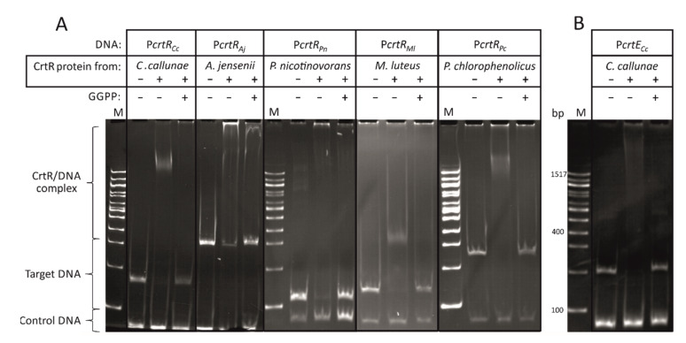
The CrtR orthologs from C. callunae (CrtRCc), A. jensenii (CrtRAj), P. nicotinovorans (CrtRPn), P. chlorophenolicus (CrtRPc) and M. luteus (CrtRMl) were fused with an N-terminal His-tag, and the proteins were purified by Ni-NTA affinity chromatography. In order to test if crtR autoregulation—as observed for CrtR from C. glutamicum—is conserved, each protein was tested for binding to the intergenic DNA sequences between its own gene and the divergently transcribed gene (Figure 2). Indeed, all CrtR orthologs analyzed bound to the DNA sequences upstream of their own crtR gene (Figure 2). This indicated crtR autoregulation and/or regulation of the respective divergently transcribed genes (mmpl for C. callunae, M. luteus and P. chlorophenolicus, crtE for A. jensenii and idi for P. nicotinovorans) (Figure 1). The finding that A. jensenii CrtR, P. nicotinovorans CrtR and M. luteus CrtR did not shift all target DNA may either indicate that the CrtR protein–target DNA interaction is less tight in these bacteria, or it may be due to technical reasons, e.g., due to the purification of the tagged proteins that may differ between the five CrtR proteins analyzed.
Figure 2.
In vitro characterization of CrtR orthologs. (A) Bandshift assays of CrtR orthologs from C. callunae, A. jensenii, P. nicotinovorans, M. luteus and P. chlorophenolicus with their respective own putative promoter region and the inhibition of the binding by GGPP. (B) Bandshift assays of CrtR from C. callunae with a putative crtE promoter region and the inhibition of the binding by GGPP. The presense or absence of CrtR protein and GGPP are indicated by “+” and “−“, respectively.
Since GGPP inhibits the binding of CrtR from C. glutamicum to its target promoter [23], it was tested if GGPP could also inhibit the binding of the other CrtR orthologs to their respective target DNA. Indeed, GGPP inhibited the interaction between the tested CrtR proteins and their target DNA (Figure 2A).
Thus, these in vitro results revealed that the binding of CrtR orthologs, from several actinobacteria, to their own upstream DNA sequences, and the inhibitory effect of GGPP, are conserved.
For C. callunae, a second putative target DNA sequence was tested, namely the intergenic region between its carotenogenic gene cluster crtEBIYe/fEb, which is located a few genes upstream of crtR in this bacterium, and the divergently transcribed gene epi (Figure 1). This intergenic DNA sequence from C. callunae was bound by a CrtR protein from C. callunae (CrtRCc) unless GGPP was added (Figure 2B). Thus, the GGPP-dependent regulation of carotenoid biosynthesis genes by CrtR is conserved at least in the closely related C. callunae, and possibly in other actinobacteria.
2.2. CrtR from C. glutamicum Binds to Heterologous crtR Promoter DNA Sequences
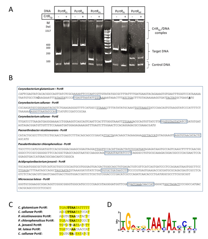
The binding of the C. glutamicum CrtR protein to heterologous crtR promoter DNA sequences was studied in order to (i) test if this specific DNA binding is conserved across the actinobacteria analyzed, and, if it is, to (ii) identify the putative DNA sequence motif. The intergenic DNA sequences between the five orthologous crtR genes and the respective divergently transcribed genes were used in a bandshift assay with His-tagged CrtR proteins from C. glutamicum (Figure 3A). The strong binding of CrtRCg to the crtR promoter sequences from C. callunae and P. nicotinovorans was detected, whereas the interactions between CrtRCg and the crtR promoter sequences from A. jensenii, M. luteus and P. chlorophenolicus were weak (Figure 3A).
Figure 3.
Characterization of CrtR from C. glutamicum in vitro. (A) Bandshift assays of His-tagged CrtR protein from C. glutamicum (CrtRCg) and the intergenic DNA sequences between the crtR orthologs from C. callunae, A. jensenii, P. nicotinovorans, M. luteus and P. chlorophenolicus, and the respective divergently transcribed genes. (B) Putative -10 and -35 promoter DNA sequences (underlined), translation start codons (italics) and the putative conserved CrtR binding sequences (boxed). The mapped transcriptional start sites of C. glutamicum crtR and crtE are given in bold. (C) Putative conserved CrtR binding sequences (conserved nucleotides are given in yellow; the TTAA sequence that was shown previously to be required for C. glutamicum CrtR binding by mutational analysis is depicted in bold face). (D) The graphical representation of the derived consensus DNA binding motif of the CrtR proteins from C. glutamicum, C. callunae, A. jensenii, P. nicotinovorans, M. luteus and P. chlorophenolicus (designed using WebLogo).
Previously, we narrowed down the target DNA sequence, to which CrtR from C. glutamicum binds, to 19 bp (5′-CCCATGAGAATTTATTTTT-3′), and mutational analysis revealed that exchanging the central four nucleotides (TTAA) simultaneously interfered with binding [23]. Inspection of the DNA sequences upstream of the crtR genes from C. callunae, A. jensenii, P. nicotinovorans, M. luteus and P. chlorophenolicus revealed that this motif was present in the intergenic DNA regions in these species (Figure 3B), and conserved to some extent (Figure 3C). It is evident that conservation of the central TTAA sequence is not sufficient to explain the observed binding preferences of CrtRCg and the variuos crtR promoter sequences studied. It remains to be elucidated how specific nucleotides of the 15 bp motif (other than the central TTAA) affect the binding of CrtRCg protein to DNA.
The derived consensus DNA binding motif of CrtR proteins from the studied species is depicted in Figure 3D, with sequence conservation and relative frequency for each nucleotide position.
2.3. CrtR Orthologs from Actinobacteria Affected Carotenogenesis and Expression of a CrtE Transcriptional Fusion in C. glutamicum In Vivo
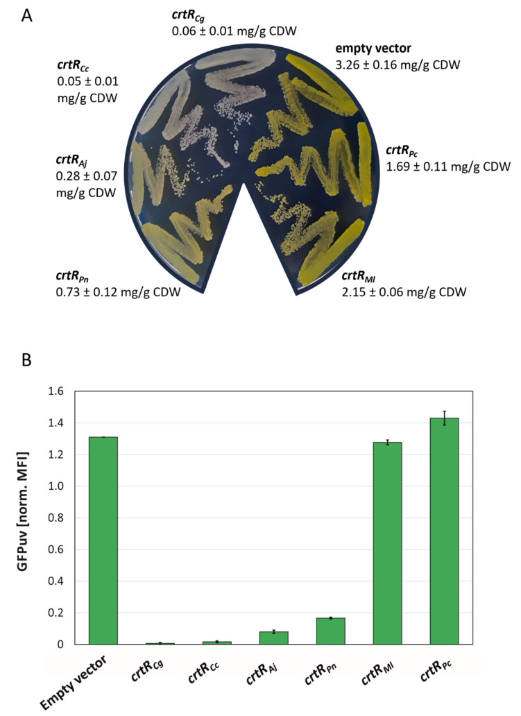
To monitor the promoter activity of the carotenogenic gene cluster of C. glutamicum in vivo, the promoter probe vector pEPR1 was used [37]. This reporter system was employed to determine the promoter activity of the carotenogenic promoter (PcrtE) from C. glutamicum in the absence of endogenous chromosomally encoded CrtR, but in the presence of CrtR orthologs from other actinobacteria. The CrtR orthologs were at different phylogenetic distances compared to CrtR from C. glutamicum, and therefore different protein identities: C. callunae (62% identity), A. jensenii (57% identity), P. nicotinovorans (53% identity), M. luteus (35% identity) and P. chlorophenolicus (35% identity). To this end, the crtR orthologs from C. callunae, A. jensenii, P. nicotinovorans, M. luteus and P. chlorophenolicus, as well as the crtR from C. glutamicum as reference, were expressed from the strong, constitutive promoter Pgap in divergent orientation to the PcrtE_gfpuv transcriptional fusion. The respective vectors were named pTEST_CrtRCg, pTEST_CrtRCc, pTEST_CrtRAj, pTEST_CrtRPn, pTEST_CrtRMl and pTEST_CrtRPc (Figure S1). First, maximal repression using the vector pTEST-crtRCg was determined and compared to a two-vector system, in which the expression of crtR and the reporter gene fusion PcrtE_gfpuv were decoupled; crtR was expressed by IPTG-inducible pEC-XT_crtRCg, while pEPR1_PcrtE contained the reporter gene fusion PcrtE_gfpuv (Figure S1). In the strains carrying the PcrtE_gfpuv fusion, but lacking crtR (WTΔcrtR(pTEST) and WTΔcrtR(pEPR1_PcrtE), pigmentation due to decaprenoxanthin accumulation (3.2–3.3 mg/g CDW) and GFPuv fluorescence (1.3 normalized MFI) were high (Figure S2). The IPTG-inducible expression of crtR in the two-vector system reduced decaprenoxanthin accumulation and GFPuv fluorescence 38- and 32-fold, respectively (Figure S2). Reduction of decaprenoxanthin accumulation and GFPuv fluorescence using pTEST_crtRCg (200- and 55-fold, respectively; Figure S2) was even higher (crtRCg is transcribed from the strong and constitutive Pgap, shown above). Next, the in vivo effects of the CrtR orthologs were tested. Constitutive overexpression of the crtR orthologs from the gap promoter revealed differential effects on carotenogenesis and expression of the PcrtE_gfpuv transcriptional fusion (Figure 4). WTΔcrtR carrying the empty vector pTEST showed the expected intense yellow pigmentation due to derepression of the chromosomal carotenoid biosynthesis genes (Figure 4A), as well as the derepressed expression of the PcrtE_gfpuv transcriptional fusion (Figure 4B). Upon plasmid-borne expression of CrtR repressor genes from C. glutamicum, C. callunae, A. jensenii and P. nicotinovorans, pigmentation was strongly reduced to less than 1 mg/g CDW (Figure 4A), which corresponded to the strongly reduced expression of the PcrtE_gfpuv transcriptional fusion (Figure 4B). Repression by CrtR from C. callunae was nearly as tight as that by endogenous CrtR, leading to a relative GFPuv signal of less than 0.1 (Figure 4B). CrtR orthologs from A. jensenii and P. nicotinorovans also repressed the crtE promoter fusion very efficiently, resulting in a GFPuv signal of less than 0.2 (Figure 4B). Upon expression of M. luteus crtR, carotenoid biosynthesis was reduced to a much lesser extent (Figure 4A), while the expression of the PcrtE_gfpuv transcriptional fusion was as high as in the empty vector control (Figure 4B). CrtR from P. chlorophenolicus reduced pigmentation, but expression of the PcrtE_gfpuv transcriptional fusion was unaffected (Figure 4A,B).
Figure 4.
In vivo characterization of CrtR orthologs in C. glutamicum WTΔcrtR. (A) Phenotypes on LB plates after incubation at 30 °C for 24 h and carotenoid concentration in mg/g CDW (β-carotene equivalents) of WTΔcrtR strains harboring pTEST derivatives expressing crtR genes from the indicated bacteria. (B) Flow cytometry analysis of the strains depicted in (A) during exponential growth in LB. Mean fluorescence intensities (MFI) of GFPuv signals were normalized to autofluorescence and shown for at least two biological replicates.
Taken together, the repression of carotenogenesis, and of the expression of crtE transcriptional fusion, in C. glutamicum in vivo by CrtR orthologs from other actinobacteria was possible. The efficacy was highest for closely related species, and it decreased with phylogenetic distance.
2.4. Construction and Analysis of a GGPP Biosensor
The expression control of PcrtE_gfpuv transcriptional fusion by CrtR from C. glutamicum has been described previously [19], and the results from above prompted us to consider its use in combination with chromosomally encoded crtR as a GGPP biosensor, i.e., using C. glutamicum WT(pEPR1_PcrtE) as a biosensor strain. Targeted metabolic engineering typically addresses four modules: carotenoid biosynthesis, precursor supply, central carbon metabolism and redox cofactor regeneration. A CrtR-based biosensor may allow us to simultaneously optimize the latter three modules via fluorescence reporter output. While GGPP inhibits the DNA binding of CrtR [23], no feeding regimen is known to predictably alter the intracellular GGPP concentration. Therefore, a genetic approach was chosen, and two C. glutamicum strains, expected to accumulate GGPP to intracellular concentrations different from the wild type, were constructed (Figure 5).
Figure 5.
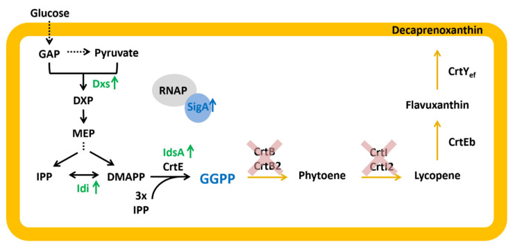
GGPP and decaprenoxanthin biosynthesis pathway in C. glutamicum. GAP: glyceraldehyde 3-phosphate, DXP: 1-deoxy-1-xylulose-5-phosphate; MEP: methylerythritol phosphate; IPP: isopentenyl pyrophosphate; DMAPP: dimethylallyl pyrophosphate; GGPP: geranylgeranyl pyrophosphate; RNAP: RNA-Polymerase core enzyme; SigA: housekeeping primary sigma factor A; Dxs: 1-deoxy-1-xylulose5-phosphate synthase; Idi: isopentenyl pyrophosphate isomerase; IdsA/CrtE: GGPP synthase; CrtB/CrtB2: phytoene synthase; CrtI/I2: phytoene desaturase; CrtEb: lycopene elongase; CrtYef: C50 ε-cyclase; genes overexpressed in strains GGPPA and GGPPB are shown in green, genes deleted on both strains are indicated by red crosses; blue shows genes overexpressed only in strain GGPPB.
First, the conversion of GGPP by the endogenous phytoene synthases CrtB [24] was prevented through the deletion of crtB and crtB2I’I2 from the chromosome of the C. glutamicum WT yielding strain WTΔcrtBΔcrtB2I’I2 (Table 1). Second, the supply of the precursor molecules DMAPP and IPP was increased by overexpression of the genes encoding the MEP pathway enzymes 1-deoxy-D-xylulose 5-phosphate synthase (Dxs) and Idi. It is known for C. glutamicum [28] and other organisms that the first enzymatic step in the MEP pathway strongly limits the flux [38,39,40]. Dxs is supposed to be feedback-regulated by isoprenoid pyrophosphates [38], and the overexpression of dxs was shown to increase the flux towards terpenoid biosynthesis [28,30,33]. In addition, the ratio of DMAPP to IPP was shown to be important for optimized isoprenoid production, and idi overexpression equilibrates intracellular concentrations of DMAPP and IPP [28,33,39]. Third, GGPP synthesis was improved by plasmid-driven overexpression of the major GGPP synthase gene idsA from C. glutamicum [26] (pEC-XT_idsA; Table 1). Combining the three strategies resulted in strain GGPPA (WTΔcrtBΔcrtB2I’I2 (pEKEx3-dxs_idi) (pECXT-idsA)) (Table 1) (Figure 5). Since engineering of the RNA polymerase sigma factor A improved isoprenoid carotenoid production [41], sigA from C. glutamicum was overexpressed in a synthetic operon with idsA (pEC-XT_idsA_sigA) (Table 1). The resulting strain GGPPB (WTΔcrtBΔcrtB2I’I2 (pEKEx3-dxs_idi) (pECXT-idsA_sigA)) differs from strain GGPPA only by the additional overexpression of sigA (Table 1) (Figure 5). The intracellular GGPP concentrations of the C. glutamicum strains WT, GGPPA and GGPPB were expected to differ.
Table 1.
Strains, genomic DNA and plasmids used in this study.
| Strain, gDNA or Plasmid | Relevant Characteristics or Sequence | Reference |
|---|---|---|
| E. coli strains | ||
| E.coli DH5α | F-thi-1 endA1 hsdr17(r-, m-) supE44 ΔlacU169 (Φ80lacZΔM15) recA1 gyrA96 | [42] |
| S17-1 | recA pro hsdR RP4-2-Tc::Mu-Km::Tn7 integrated into the chromosome | [43] |
| E.coli BL21 (DE3) | F– ompT gal dcm lon hsdSB(rB–mB–) λ(DE3 [lacI lacUV5-T7p07 ind1 sam7 nin5]) [malB+]K-12(λS) | [44] |
| E.coli BL21 (DE3) (pLysS) | F– ompT gal dcm lon hsdSB(rB–mB–) λ (DE3 [lacI lacUV5-T7p07 ind1 sam7 nin5]) [malB+]K-12( λ S) pLysS[T7p20 orip15A](CmR) | Promega |
| C. glutamicum strains | ||
| C. glutamicum WT | ATCC 13032, wild type | [45] |
| WTΔcrtR | ATCC 13,032 with deletion of crtR (cg0725) | [23] |
| WTΔcrtBΔcrtB2I’I2 | ATCC 13,032 with deletion of crtB (cg0721) and crtB2I’I2 (OP_cg2672) | this work |
| GGPPA | WTΔcrtBΔcrtB2I’I2 derivative with plasmid-driven IPTG-inducible expression of MEP pathway genes dxs (cg2083) and idi (cg2531) from pEKEx3 and the GGPP synthase gene idsA (cg2384) from pEC-XT. | this work |
| GGPPB | WTΔcrtBΔcrtB2I’I2 derivative with plasmid-driven IPTG-inducible expression of MEP pathway genes dxs (cg2083) and idi (cg2531) from pEKEx3 and the GGPP synthase gene idsA (2384) and primary sigma factor gene sigA (cg2092) from pEC-XT. | this work |
| Genomic DNA | ||
| Acidipropionibacterium jensenii | Wild type, DSM 20535, ATCC 4868 | [46], DSMZ |
| Corynebacterium callunae | Wild type, DSM 20147, ATCC 15991 | [47] |
| Micrococcus luteus | Wild type, DSM 20030, ATCC 4698 | [48], DSMZ |
| Paenarthrobacter nicotinovorans | Wild type, DSM 420, ATCC 49919 | [49], DSMZ |
| Pseudarthrobacter chlorophenolicus | Wild type, DSM 12829, ATCC 700700 | [50], DSMZ |
| Plasmids | ||
| pEPR1 | KmR, pCG1 oriVCG, gfpuv, promoterless, C. glutamicum/E.coli shuttle promoter-probe vector | [37] |
| pEPR1_PcrtE | pEPR1 derivate containing the promoter of crtE (PcrtE) | [23] |
| pTEST | pEPR1_PcrtE derivate containing an additional expression cassette for expression of crtR orthologs from the gap promoter | this work |
| pTEST_crtRCg | pTEST derivate for expression of the crtR from C. glutamicum | this work |
| pTEST_crtRCc | pTEST derivate for heterologous expression of the crtR orthologs from C. callunae | this work |
| pTEST_crtRAj | pTEST derivate for heterologous expression of the crtR ortholog from A. jensenii | this work |
| pTEST_crtRPn | pTEST derivate for heterologous expression of the crtR ortholog from P. nicotinovorans | this work |
| pTEST_crtRPc | pTEST derivate for heterologous expression of the crtR ortholog from P. chlorophenolicus | this work |
| pTEST_crtRMl | pTEST derivate for heterologous expression of the crtR ortholog from M. luteus | this work |
| pET16b | Expression plasmid for production of His-tagged proteins | Novagen |
| pET16b_crtRCg | pET16b derivate for production of His-tagged CrtR from C. glutamicum | this work |
| pET16b_crtRCc | pET16b derivate for production of His-tagged CrtR C. callunae | this work |
| pET16b_crtRAj | pET16b derivate for production of His-tagged CrtR A. jensenii | this work |
| pET16b_crtRPn | pET16b derivate for expression of the crtR from P. nicotinovorans | this work |
| pET16b_crtRPc | pET16b derivate for production of His-tagged CrtR P. chlorophenolicus | this work |
| pET16b_crtRMl | pET16b derivate for production of His-tagged CrtR M. luteus | this work |
| pEKEx3 | SpecR, Ptac lacIq, pBL1 oriVCg, C. glutamicum/E. coli expression shuttle vector | [51] |
| pEKEx3_dxs_idi | pEKEx3 derivate for IPTG-inducible expression of dxs and idi from C. glutamicum containing an artificial ribosome binding site | this work |
| pEC-XT99A | TetR, Ptrc lacIq, pGA1 oriVCg, C. glutamicum/E. coli expression shuttle vector | [52] |
| pEC-XT_idsA | pEC-XT99A derivate for IPTG-inducible expression of idsA from C. glutamicum containing an artificial ribosome binding site | this work |
| pEC-XT_idsA_sigA | pEC-XT99A derivate for IPTG-inducible expression of idsA and sigA from C. glutamicum containing an artificial ribosome binding site | this work |
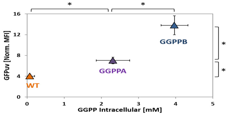
After transformation with the biosensor plasmid pEPR1_PcrtE, the strains were grown in CGXII minimal medium with glucose. Samples were taken during exponential growth 12 h after inoculation and analyzed by LC-MS (Figure 6). As expected, WT (pEPR1_PcrtE) accumulated the lowest GGPP concentration, with less than 0.1 mM GGPP (Figure 6). In comparison, GGPPA (pEPR1_PcrtE) accumulated about 23-fold more GGPP (2.3 ± 0.5 mM; Figure 6). With 4.0 ± 0.4 mM, strain GGPPB (pEPR1_PcrtE) exhibited the highest concentration of GGPP, i.e., about 1.7-fold higher than GGPPA (pEPR1_PcrtE). Thus, it was confirmed that the genetic approach altered the intracellular GGPP concentrations as anticipated.
Figure 6.
Biosensor-based differentiation between strains accumulating different GGPP concentrations. The intracellular GGPP concentrations are given in mM, and the GFPuv signals in mean fluorescence intensities were normalized to autofluorescence. Strains WT (pEPR1_PcrtE), GGPPA (pEPR1_PcrtE) and GGPPB (pEPR1_PcrtE) were cultivated in CGXII (100 mM Gluc + 100 µM IPTG) and data were taken after 12 h. Statistical significance was calculated with paired Student t-test (two-tailed); *: p-value < 0.05.
Since the strains harbored plasmid pEPR1_PcrtE, the sensing of intracellular GGPP by chromosomally encoded CrtR could be tested. The normalized mean fluorescence intensity (MFI) of the PcrtE_gfpuv fusion observed after 12 h growth in glucose minimal medium was lowest for WT (pEPR1_PcrtE) (MFI of 4.0 ± 0.3). The biosensor signal of GGPPB (pEPR1_PcrtE), of 14.0 ± 1.8, was about two-fold higher than that of GGPPA (pEPR1_PcrtE) (MFI of 7.0 ± 0.7; Figure 6). Thus, biosensor signal output correlated with intracellular GGPP concentration.
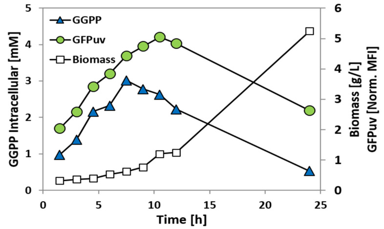
In order to determine if the PcrtE_gfpuv fusion can be used to monitor variations in the GGPP concentration during growth, strain GGPPB (pEPR1_PcrtE) was analyzed in a time-course experiment in CGXII minimal medium supplemented with 100 mM glucose and 100 µM IPTG (Figure 7). Samples were taken every 1.5 h for determination of the intracellular GGPP concentration and flow cytometry analysis (Figure 7). Both the intracellular GGPP concentration and the GFPuv signal strongly increased in the first 7.5–10.5 h after inoculation. An offset between the GGPP concentration and the GFPuv signal was observed, with the increase in the latter being delayed by about 4 h (Figure 7). This offset may be explained by the time required to synthesize GFPuv after CrtR has sensed an increased GGPP concentration and PcrtE_gfpuv has been derepressed. The intracellular GGPP concentration reached its maximum approximately after 7.5 h, with about 3 mM GGPP, and decreased afterwards, while the GFPuv reached its maximum after 10.5 h of cultivation (Figure 7).
Figure 7.
GGPP concentration and GFPuv fluorescence during growth of GGPP accumulating C. glutamicum strain GGPPB (pEPR1_PcrtE). Intracellular GGPP concentration (blue triangles; in mM) and GFPuv signal (green circles; mean fluorescence intensities normalized to autofluorescence) were monitored during growth in CGXII (100 mM Gluc + 100 µM IPTG). Biomass concentrations are given in gCDW/L (empty squares).
3. Discussion
This study revealed the conserved functions of CrtR orthologs in six actinobacteria with respect to GGPP-dependent regulation. The repression of carotenogenesis, and of the expression of a crtE transcriptional fusion, in C. glutamicum in vivo was highest for closely related species, and it decreased with phylogenetic distance. The PcrtE_gfpuv transcriptional fusion was suitable for monitoring intracellular GGPP concentrations in a strain with chromosomally encoded crtR as a genetically encoded biosensor system.
The conserved role of CrtR orthologs as GGPP-dependent transcriptional regulators suggested that they are relevant for the control of carotenogenesis and/or mmpL genes. Although not tested, it is tempting to speculate that these proteins may be used to monitor intracellular GGPP concentrations in their native hosts. C. glutamicum and C. callunae are close relatives, synthesizing the C50 carotenoid decaprenoxanthin and its glycosides as pigments [25,27]. P. nicotinovorans is pigmented most probably due to the accumulation of carotenoids [35]. M. luteus is a yellow-pigmented bacterium due to the accumulation of the C50 carotenoid sarcinaxanthin and its glycosides [53]. By contrast, A. jensenii does not synthesize a carotenoid, but the polyene pigment granadaene [54], while P. chlorophenolicus is a non-pigmented soil bacterium [55]. Interestingly, relatives of P. chlorophenolicus, such as Arthrobacter arilaitensis, are pigmented most probably due to the accumulation of C50 carotenoids, and are found on the surface of smear-ripened cheeses [55,56]. Thus, carotenogenesis is not conserved in actinobacteria possessing CrtR orthologs that control the expression of their own gene and/or the divergently oriented gene(s) in a GGPP-dependent manner. Besides carotenogenesis genes, mmpL genes are also transcribed divergently to crtR, and are presumably controlled by CrtR. MmpL transporters are considered candidate targets for the development of anti-tuberculosis drugs [57], as they couple lipid synthesis and the export of bulky, hydrophobic substrates [58]. Thus, MmpL proteins are essential for the cell envelope, and support the infectivity and persistence of M. tuberculosis in its host [59]. Moreover, MmpL proteins of M. tuberculosis are involved in the oxidative stress response [60].
MarR-type transcriptional regulators, including CrtR and its orthologs, are found in all bacteria, and are natural sensors that allow adaptation to environmental stresses such as ROS, toxic compounds or antibiotics (hence the name multiple antibiotic resistance regulators) [61]. As shown for the CrtR orthologs studied here (see Figure 1 and Figure 2), MarR-type regulators typically repress genes in the close vicinity [34]. MarR-type regulators typically bind and respond to low-molecular-weight compounds [34,62], such as GGPP for the CrtR orthologs. C. glutamicum possesses nine MarR-type regulators, eight of which have been characterized in some detail. RosR [63], CosR [64], OhsR [65] and OsmC [66] play roles in the response to ROS stress, whereas CarR [67], MalR [68] and PhdR [69] deal with other environmental stresses, such as toxic compounds or cell-membrane associated stress. The GGPP-dependent control of carotenogenesis by CrtR from C. glutamicum and its orthologs studied here may be considered a stress response, since the antioxidative properties of carotenoids counteract oxidative stress. This function would be in line with the CrtR-mediated control of mmpL genes that are involved in the oxidative stress response (see above).
MarR-type regulators typically bind to a palindromic 16–20 bp target site that overlaps with the -10 or -35 promoter regions for steric inhibition of RNA polymerase binding [34]. Previously, a 19 bp DNA sequence with a central TTAA motif was shown to be essential for the binding of CrtR from C. glutamium to its DNA target site [23]. Here, we showed that CrtR from C. glutamicum bound to DNA sequences upstream of crtR from other actinobacteria, and inspection of the DNA sequences revealed a conserved 15 bp binding motif, including the central TTAA base pairs (see Figure 3). The binding of CrtRCg decreased with increasing deviation from the consensus motif (see Figure 3). The CrtR binding motif is typical for the MarR-type family of transcriptional regulators [34]. The observed graded effect of CrtRCg binding to promoter sequences with increasing deviation from the consensus motif in vitro was congruent with the in vivo finding that different CrtR orthologs affected the pigmentation and expression of the PcrtE_gfpuv transcriptional fusion more weakly when their phylogenetic distance to C. glutamicum was greater (see Figure 4). This is in line with the phylogenetic analysis of CrtR orthologs [23].
The application of biosensors has become a prominent tool in strain development over the last few years [5,70]. In this study, CrtR from C. glutamicum was demonstrated to be the first genetically encoded biosensor for the detection of GGPP that allows one to distinguish between C. glutamicum strains that have accumulated GGPP to different intracellular concentrations (Figure 6), and to monitor GGPP accumulation over time during growth (Figure 7). Since the CrtR-based biosensor system was suitable for the detection of intracellular GGPP concentrations between 0.1 and at least 4 mM (Figure 6), it is plausible that the described system is applicable to the screening of mutants accumulating GGPP well above wild type levels, and their enrichment/isolation, by flow cytometry. As an alternative application, the on-demand expression control of GGPP converting enzymes in response to intracellular GGPP concentration can be envisioned for strain optimization with respect to the production of GGPP-derived diterpenoids and/or carotenoids. On-demand production may be established by transcriptional fusion of the PcrtE to the gene of interest, e.g., a diterpenoid synthase. This approach may improve production as the terminal biosynthesis pathway is initiated only in the presence of high concentrations of the precursor GGPP, which may prevent the accumulation of toxic GGPP concentrations. Biosensor approaches to on-demand expression control have been successfully applied, e.g., to improve lysine production by C. glutamicum [71,72].
The central role of GGPP as a terpenoid and carotenoid precursor suggests a wide application range for the GGPP-based biosensor developed here [10], since the tens of thousands of terpenoids derived from GGPP represent one of the biggest sources of valuable natural products for human use [73].
4. Materials and Methods
4.1. Bacterial Strains, Media and Growth Conditions
Strains and plasmids that were used in this study are listed in Table 1. C. glutamicum ATCC 13,032 [45] served as the wild type and was used as the basic strain for genetic engineering. Modifications aimed at higher production levels of GGPP and the establishment of the biosensor system. Precultures of C. glutamicum were performed in LB/BHI medium with 50 mM glucose as carbon and energy source [74] supplemented with the appropriate antibiotic at 30 °C and 120 rpm. The main cultures of C. glutamicum consisted of 50 mL CGXII medium with 100 mM glucose and 100 µM IPTG and were inoculated to an initial optical density (OD600) of 1. The OD600 of the cultures was measured with the Shimadzu UV-1202 spectrophotometer (Duisburg, Germany).
4.2. Recombinant DNA Work and Gene Expression
Cloning of plasmids was done in E. coli DH5α using PCR-generated fragments that were purified using the NucleoSpin kit (Macherey-Nagel, Düren, Germany). Oligonucleotides were ordered from Metabion GmbH (Planegg/Steinkirchen, Germany) (Table 2). For plasmid construction standard PCR, restriction and dephosphorylation reactions [75] were performed as well as Gibson Assembly [76]. Transformation of E. coli was performed via the RbCl method [42]. Cloned DNA insert fragments were verified by sequencing. Transformation of C. glutamicum was performed via electroporation using a Gene Pulser Xcell™ (Bio-Rad Laboratories GmbH, Munich, Germany) at 2.5 kV, 200 Ω and 25 µF [74]. For expression of CrtR in the pTEST vector (NA, Table 1 and Table 2) and the production of His-tagged CrtR from various organisms in the pET16b vector (HN, Table 1 and Table 2), the respective genes were amplified from chromosomal DNA using primer pairs NA25/26 and HN83/HN84 for C. glutamicum, NA27/28 and HN85/HN86 for C. callunae, NA31/32 and HN87/HN88 for A. jensenii, NA33/34 and HN89/HN90 for P. nicotinovorans, NA39/40 and HN93/HN94 for M. luteus and NA41/42 and HN95/HN96 for P. chlorophenolicus, respectively. The purified PCR products were cloned into pTEST restricted with BamHI and pET16b restricted with NdeI using Gibson assembly [76], respectively. Chromosomal DNA was extracted from DSMZ (see Table 1).
Table 2.
Oligonucleotides used in this study.
| Oligonucleotide (5′→3′) | |
|---|---|
| NH45 | CATGCCTGCAGGTCGACTCTAGAGGAAAGGAGGCCCTTCAGATGGGAATTCTGAACAGTATTTCAA |
| NH46 | GTTCGTGTGGCAGTTTTATTCCCCGAACAGGGAATC |
| NH47 | AACTGCCACACGAACGAAAGGAGGCCCTTCAGATGTCTAAGCTTAGGGGCATG |
| NH48 | ATTCGAGCTCGGTACCCGGGGATCTTACTCTGCGTCAAACGCTTC |
| NH49 | ATGGAATTCGAGCTCGGTACCCGGGGAAAGGAGGCCCTTCAGATGGCTTACTCCGCTATGGCTA |
| NH50 | GCATGCCTGCAGGTCGACTCTAGAGGATCTTAGTTCTGGCGGAAAGCAA |
| NH51 | GTTCGTGTGGCAGTTTTAGTTCTGGCGGAAAGCAA |
| NH52 | ATGGAATTCGAGCTCGGTACCCGGGGAAAGGAGGCCCTTCAGATGGACTTTCCGCAGCAACTCG |
| NH53 | GCATGCCTGCAGGTCGACTCTAGAGGATCTTATTTATTACGCTGGATGATGTAGTCC |
| NH54 | GTTCGTGTGGCAGTTTTATTTATTACGCTGGATGATGTAGTCC |
| NH55 | ATGGAATTCGAGCTCGGTACCCGGGGAAAGGAGGCCCTTCAGATGAGCAGTTTCGATGCCCA |
| NH56 | GCATGCCTGCAGGTCGACTCTAGAGGATCTTACATCCGACGTTCGGTTGA |
| NH57 | GTTCGTGTGGCAGTTTTACATCCGACGTTCGGTTGA |
| NH58 | ATGGAATTCGAGCTCGGTACCCGGGGAAAGGAGGCCCTTCAGATGGTAGAAAACAACGTAGCAA |
| NH59 | GCATGCCTGCAGGTCGACTCTAGAGGATCTTAGTCCAGGTAGTCGCGAAG |
| NH60 | AACTGCCACACGAACGAAAGGAGGCCCTTCAGATGGTAGAAAACAACGTAGCAA |
| NH63 | GCAAAGTTGTTGTCGTAGTC |
| NH64 | ATGAAAACGTTGTTGCCAT |
| NH65 | ATGAAGACGCCACTGAC |
| NH66 | CGGTGAGCTCGGCATCT |
| NH67 | GTGCCTTGCGAGCTGTCT |
| TH17 | CTGTTGATGACGACGAGGAG |
| pE-CXT fw | AATACGCAAACCGCCTCTCC |
| pE-CXT rv | TACTGCCGCCAGGCAAATTC |
| crtE-E | GTGACCATGAGGGCGAAAGC |
| crtE-F | TCACATAGTCCGGCGTTTGC |
| idsA-E | GCAGCTTCGCCAGAGTGTAT |
| idsA-F | CAATGCGGACAATGCTCCAG |
| 581 | CATCATAACGGTTCTGGC |
| 582 | ATCTTCTCTCATCCGCCA |
| Pgap fw | TGGCCTTTTGCTGGCCTTTTGCTCACTGCGAAATCTTTGTTTCCCCG |
| Pgap rv | GGATCCGTTGTGTCTCCTCTAAAGATT |
| term fw | AATCTTTAGAGGAGACACAACGGATCCTTTTGGCGGATGAGAGAA |
| term rv | AATCAGGGGATAACGCAGGAAAGAACAAAAGAGTTTGTAGAA |
| NA25- Cg fw | TACAATCTTTAGAGGAGACACAACGGAAAGGAGGCCCTTCAGATGCTGAATATGCAGGAACCA |
| NA26- Cg rv | AAAATCTTCTCTCATCCGCCAAAAGTTACTCCGTGTTGAGCCATGG |
| NA27- Cc fw | TACAATCTTTAGAGGAGACACAACGGAAAGGAGGCCCTTCAGATGTCCGATCCGCAAGAACC |
| NA28- Cc rv | AAAATCTTCTCTCATCCGCCAAAAGTTAATGTGAGGAAGACTCGAAC |
| NA31- Aj fw | TACAATCTTTAGAGGAGACACAACGGAAAGGAGGCCCTTCAGATGAGTGAAGACCGCGATG |
| NA32- Aj rv | AAAATCTTCTCTCATCCGCCAAAAGTTACCGCGGGTGGCGC |
| NA33-An fw | TACAATCTTTAGAGGAGACACAACGGAAAGGAGGCCCTTCAGATGTCCAGTCTTGAAGAAATGC |
| NA34-An rv | AAAATCTTCTCTCATCCGCCAAAAGTTAGCGTGGAGCCGCAG |
| NA39- Ml fw | TACAATCTTTAGAGGAGACACAACGGAAAGGAGGCCCTTCAGATGACCACGCAGCCCC |
| NA40- Ml rv | AAAATCTTCTCTCATCCGCCAAAAGTTACGGGTCCTCCGGGG |
| NA41- Pc fw | TACAATCTTTAGAGGAGACACAACGGAAAGGAGGCCCTTCAGATGAACGGCAACAATCCG |
| NA42- Pc rv | AAAATCTTCTCTCATCCGCCAAAAGTTACCCGGCTGGACGC |
| HN83-Cg-fw | GCGGCCATATCGAAGGTCGTCATCTGAATATGCAGGAACCAG |
| HN84-Cg-rv | TAGCAGCCGGATCCTCGAGCATTACTCCGTGTTGAGCCATG |
| HN85-Cc-fw | GCGGCCATATCGAAGGTCGTCATTCCGATCCGCAAGAACCCC |
| HN86-Cc-rv | TAGCAGCCGGATCCTCGAGCATTAATGTGAGGAAGACTCGAAC |
| HN87-Aj-fw | GCGGCCATATCGAAGGTCGTCATAGTGAAGACCGCGATGC |
| HN88-Aj-rv | TAGCAGCCGGATCCTCGAGCATTACCGCGGGTGGCGC |
| HN89-Pn-fw | GCGGCCATATCGAAGGTCGTCATTCCAGTCTTGAAGAAATGCC |
| HN90-Pn-rv | TAGCAGCCGGATCCTCGAGCATTAGCGTGGAGCCGCAG |
| HN93-Ml-fw | GCGGCCATATCGAAGGTCGTCATACCACGCAGCCCCCC |
| HN94-Ml-rv | TAGCAGCCGGATCCTCGAGCATTACGGGTCCTCCGGGG |
| HN95-Pc-fw | GCGGCCATATCGAAGGTCGTCATAACGGCAACAATCCGGGC |
| HN96-Pc-rv | TAGCAGCCGGATCCTCGAGCATTACCCGGCTGGACGC |
| Pc-PcrtR-fw | TGCCTTCCATGCGGATGGTC |
| Pc-PcrtR-rv | TGCCCGGATTGTTGCCGTTC |
4.3. Extraction of Carotenoids from Bacterial Cells and HPLC Analysis
The carotenoid extraction from C. glutamicum was performed as described previously [30] using 1 mL of the cell cultures. Pigments were isolated from the cell pellets with a methanol:acetone mixture (7:3) at 60 °C for 15 min with shaking at 500 rpm. The clear supernatant was used for HPLC analysis after centrifugation of the extract for 10 min at 13,000× g. The carotenoid concentration of cell extracts was determined through absorbance at 471 nm by high performance liquid chromatography (HPLC) analysis, performed on an Agilent 1200 series HPLC system (Agilent Technologies Sales & Services GmbH & Co. KG, Waldbronn, Germany), including a diode array detector (DAD) for UV/visible (Vis) spectrum recording. Separation of the carotenoids was performed by application of a column system consisting of a precolumn (LiChrospher 100 RP18 EC-5, 40 × 4 mm, CS-Chromatographie, Langerwehe, Germany) and a main column (LiChrospher 100 RP18 EC-5, 125 × 4 mm, CS-Chromatographie, Langerwehe, Germany) with methanol/water (9:1) (A) and methanol (B) as the mobile phase. The following gradient was used at a flow rate of 1.5 mL/min: 0 min B—0%; 10 min B—100%; 32.5 min B—100%. The quantification of decaprenoxanthin was calculated based on a β-carotene standard (Merck, Darmstadt, Germany) and reported as β-carotene equivalents.
4.4. Analysis of Fluorescence via Flow Cytometry
Cell cultures were analyzed regarding their fluorescent intensity. Samples were diluted to a final OD600 of 0.1 with pure CGXII medium and immediately analyzed with the FACS GalliosTM (Beckman Coulter GmbH, Krefeld, Germany). Alternatively, samples for fluorescence analysis were harvested and stored at 4 °C. C. glutamicum (pEPR1) was used as the autofluorescence reference. The GFPuv signal was measured with a blue solid-state laser at 405 nm excitation and fluorescence was detected using a 525/50 nm band-pass filter.
4.5. Overproduction and Purification of the Transcriptional Regulator CrtR
After transformation of the pET16b derivatives in E. coli BL21(DE3) or E. coli BL21(DE3) (pLysS) transformants carrying the respective plasmids pET16b-crtRCg, pET16b-crtRCc, pET16b-crtRAj, pET16b-crtRPn, pET16b-crtRMl and pET16b-crtRPc were grown at 37 °C in 500 mL LB medium with 10 µg/mL ampicillin to an OD600 of 0.5 before adding IPTG (0.5 mM) for induction of the gene expression. After induction, cells were cultivated at 21 °C for an additional 4 h and were harvested by centrifugation. Pellets were stored at −20 °C. Crude extract preparation and protein purification via Ni-NTA chromatography was performed as described elsewhere [23]. The purified regulator proteins were used for EMSA experiments without removing the N-terminal His-tag.
4.6. Electrophoretic Mobility Shift Assay (EMSA)
To analyze the physical protein–DNA interaction between the different CrtR proteins and their putative native target DNA, bandshift assays were performed [77]. The His-tagged CrtR proteins were mixed in varying molar excess with 30–90 ng of PCR amplified and purified promoter fragments of the target genes in bandshift (BS) buffer (50 mM Tris–HCl, 10% (v/v) glycerol, 50 mM KCl, 10 mM MgCl2, 0.5 mM EDTA, pH 7.5) in a total volume of 20 µL. The 5′ UTR of crtR genes were PCR-amplified and purified with NucleoSpin kit (MACHEREY-NAGEL GmbH & Co. KG, Düren, Germany). Promoter fragments were amplified using the respective oligonucleotide pairs (Table 2). A 78 bp-fragment of the upstream region of cg2228 was added in every sample as a negative control using oligonucleotides cg2228_fw and cg2228_rv. After 30 min of incubation at room temperature, gel shift samples were separated on a native 6% (w/v) polyacrylamide. Additionally, the binding affinity in the presence of 100–650 µM GGPP as effector was analyzed by incubation of the protein with the effector under buffered conditions for 15 min at room temperature prior to the addition of the promoter. Subsequently, the gel shift samples were separated on a 6% DNA retardation gel (Life Technologies GmbH, Darmstadt, Germany) at 100 V buffered in 44.5 mM Tris, 44.5 mM boric acid and 1 mM EDTA at pH 8.3. Staining of the DNA was achieved with ethidium bromide.
4.7. Extraction of GGPP and LC-MS Analysis
For isolation of GGPP, 10 mL of culture were harvested at 4000 rpm and 15 min. The supernatant was removed and the cells stored till further use (−80 °C). The cell pellet was defrosted on ice and resuspended in 600 µL acidified methanol (pH 5). Pyrophosphates were extracted by 3 × 30 s shaking in silamat (Ivoclar Vivadent AG, Schaan, Liechtenstein) in the presence of 300 µL silica beads. The clear supernatant was used for LC-MS analysis after subsequent centrifugation for 10 min at 13,000× g. LC-MS measurement was performed on a LaChrom ULTRA system (San Jose, CA, USA) using a SeQuant Zic-pHILIC column (5 µm 150 × 2.1 mm) (Merck Millipore, Darmstadt, Germany). As a buffer system, 10 mM ammonium bicarbonat pH 9.3 (A) and acetonitrile (B) was used with a flow rate of 0.2 mL/min; 0–5 min 5% A (const.), 5–20 min 35% A (gradient), 20–25 min 5% A (gradient), 25–35 min 5% A (const.); pre-run 15 min. with 2 µL. Isoprenoid pyrophosphates were identified using a micrOTOFQ (Bruker Daltonics, Billerica, MA, USA) according to their masses (GPP 313.0601; FPP 381.1227; GGPP 449.1853 [M-H]-) and elution time in accordance to a standard (Sigma-Aldrich, Merck, Darmstadt, Germany).
Acknowledgments
We thank Tim Treis for optimization of the GGPP extraction protocol.
Supplementary Materials
The following figures are available online at https://www.mdpi.com/1422-0067/21/15/5482/s1. Figure S1: Biosensor plasmids, Figure S2: Validation of the pTEST vector system for promoter activity assay and expression of crtR orthologs.
Author Contributions
N.A.H., P.P.-W. and V.F.W. planned and designed the experiments. N.A.H., S.A., I.L.G., S.G. and M.P. performed the experiments. N.A.H., M.P. and P.P.-W. analyzed the data. N.A.H. and P.P.-W. drafted the manuscript. V.F.W. coordinated the study and finalized the manuscript. All authors read and approved the final manuscript.
Funding
This research was funded by the European Regional Development Fund (ERDF) and the Ministry of Economic Affairs, Innovation, Digitalization and Energy of the State of North Rhine-Westphalia, grant number EFRE-0400184 (Bicomer). Support for the Article Processing Charge by the Deutsche Forschungsgemeinschaft and the Open Access Publication Fund of Bielefeld University is acknowledged.
Conflicts of Interest
The authors declare no conflict of interest. The funders had no role in the design of the study; in the collection, analyses, or interpretation of data; in the writing of the manuscript, or in the decision to publish the results.
References
- 1.Mahr R., Frunzke J. Transcription factor-based biosensors in biotechnology: Current state and future prospects. Appl. Microbiol. Biotechnol. 2016;100:79–90. doi: 10.1007/s00253-015-7090-3. [DOI] [PMC free article] [PubMed] [Google Scholar]
- 2.Rogers J.K., Church G.M. Genetically encoded sensors enable real-time observation of metabolite production. Proc. Natl. Acad. Sci. USA. 2016;113:2388–2393. doi: 10.1073/pnas.1600375113. [DOI] [PMC free article] [PubMed] [Google Scholar]
- 3.Liu Y., Liu Y., Wang M. Design, Optimization and Application of Small Molecule Biosensor in Metabolic Engineering. Front. Microbiol. 2017;8:2012. doi: 10.3389/fmicb.2017.02012. [DOI] [PMC free article] [PubMed] [Google Scholar]
- 4.Zhang J., Jensen M.K., Keasling J.D. Development of biosensors and their application in metabolic engineering. Curr. Opin. Chem. Biol. 2015;28:1–8. doi: 10.1016/j.cbpa.2015.05.013. [DOI] [PubMed] [Google Scholar]
- 5.De Paepe B., Peters G., Coussement P., Maertens J., De Mey M. Tailor-made transcriptional biosensors for optimizing microbial cell factories. J. Ind. Microbiol. Biotechnol. 2017;44:623–645. doi: 10.1007/s10295-016-1862-3. [DOI] [PubMed] [Google Scholar]
- 6.Rohmer M., Seemann M., Horbach H., Bringer-Meyer S., Sahm H. Glyceraldehyde 3-phosphate and pyruvate as precursors of isoprenic units in an alternative non-mevalonate pathway for terpenoid biosynthesis. J. Am. Chem. Soc. 1996;118:2564–2566. doi: 10.1021/ja9538344. [DOI] [Google Scholar]
- 7.Britton L.-J. Carotenoids Handbook. Birkhauser Verlag; Basel, Switzerland: 2004. Pfander. [DOI] [Google Scholar]
- 8.Henke N.A., Wichmann J., Baier T., Frohwitter J., Lauersen K.J., Risse J.M., Peters-Wendisch P., Kruse O., Wendisch V.F. Patchoulol Production with Metabolically Engineered Corynebacterium glutamicum. Genes (Basel) 2018;9 doi: 10.3390/genes9040219. [DOI] [PMC free article] [PubMed] [Google Scholar]
- 9.Schempp F.M., Drummond L., Buchhaupt M., Schrader J. Microbial cell factories for the production of terpenoid flavor and fragrance compounds. J. Agric. Food Chem. 2017 doi: 10.1021/acs.jafc.7b00473. [DOI] [PubMed] [Google Scholar]
- 10.Schrader J., Bohlmann J. Biotechnology of Isoprenoids. Springer International Publishing; New York, NY, USA: 2015. [DOI] [Google Scholar]
- 11.Misawa N. Pathway engineering for functional isoprenoids. Curr. Opin. Biotechnol. 2011;22:627–633. doi: 10.1016/j.copbio.2011.01.002. [DOI] [PubMed] [Google Scholar]
- 12.George K.W., Alonso-Gutierrez J., Keasling J.D., Lee T.S. Isoprenoid drugs, biofuels, and chemicals--artemisinin, farnesene, and beyond. Adv. Biochem. Eng. Biotechnol. 2015;148:355–389. doi: 10.1007/10_2014_288. [DOI] [PubMed] [Google Scholar]
- 13.Novoveská L., Ross M.E., Stanley M.S., Pradelles R., Wasiolek V., Sassi J.-F. Microalgal Carotenoids: A Review of Production, Current Markets, Regulations, and Future Direction. Mar. Drugs. 2019;17:640. doi: 10.3390/md17110640. [DOI] [PMC free article] [PubMed] [Google Scholar]
- 14.Rodriguez-Saiz M., de la Fuente J.L., Barredo J.L. Xanthophyllomyces dendrorhous for the industrial production of astaxanthin. Appl. Microbiol. Biotechnol. 2010;88:645–658. doi: 10.1007/s00253-010-2814-x. [DOI] [PubMed] [Google Scholar]
- 15.Tsubokura A., Yoneda H., Mizuta H. Paracoccus carotinifaciens sp. nov., a new aerobic gram-negative astaxanthin-producing bacterium. Pt. 1Int. J. Syst. Bacteriol. 1999;49:277–282. doi: 10.1099/00207713-49-1-277. [DOI] [PubMed] [Google Scholar]
- 16.Li C., Swofford C.A., Sinskey A.J. Modular engineering for microbial production of carotenoids. Metab. Eng. Commun. 2020;10:e00118. doi: 10.1016/j.mec.2019.e00118. [DOI] [PMC free article] [PubMed] [Google Scholar]
- 17.Pfleger B.F., Pitera D.J., Newman J.D., Martin V.J., Keasling J.D. Microbial sensors for small molecules: Development of a mevalonate biosensor. Metab. Eng. 2007;9:30–38. doi: 10.1016/j.ymben.2006.08.002. [DOI] [PubMed] [Google Scholar]
- 18.Tang S.Y., Cirino P.C. Design and application of a mevalonate-responsive regulatory protein. Angew. Chem. Int. Ed. Engl. 2011;50:1084–1086. doi: 10.1002/anie.201006083. [DOI] [PubMed] [Google Scholar]
- 19.Chou H.H., Keasling J.D. Programming adaptive control to evolve increased metabolite production. Nat. Commun. 2013;4:2595. doi: 10.1038/ncomms3595. [DOI] [PubMed] [Google Scholar]
- 20.Lee J.H., Wendisch V.F. Production of amino acids—Genetic and metabolic engineering approaches. Bioresour. Technol. 2017;245:1575–1587. doi: 10.1016/j.biortech.2017.05.065. [DOI] [PubMed] [Google Scholar]
- 21.Kinoshita S., Udaka S., Shimono M. Studies on the amino acid fermentation. Production of L-glutamic acid by various microorganisms. J. Gen. Appl. Microbiol. 1957;3:193–205. doi: 10.2323/jgam.3.193. [DOI] [PubMed] [Google Scholar]
- 22.Wendisch V.F. Metabolic engineering advances and prospects for amino acid production. Metab. Eng. 2020;58:17–34. doi: 10.1016/j.ymben.2019.03.008. [DOI] [PubMed] [Google Scholar]
- 23.Henke N.A., Heider S.A.E., Hannibal S., Wendisch V.F., Peters-Wendisch P. Isoprenoid Pyrophosphate-Dependent Transcriptional Regulation of Carotenogenesis in Corynebacterium glutamicum. Front. Microbiol. 2017;8:633. doi: 10.3389/fmicb.2017.00633. [DOI] [PMC free article] [PubMed] [Google Scholar]
- 24.Heider S.A., Peters-Wendisch P., Wendisch V.F. Carotenoid biosynthesis and overproduction in Corynebacterium glutamicum. BMC Microbiol. 2012;12:198. doi: 10.1186/1471-2180-12-198. [DOI] [PMC free article] [PubMed] [Google Scholar]
- 25.Krubasik P., Takaichi S., Maoka T., Kobayashi M., Masamoto K., Sandmann G. Detailed biosynthetic pathway to decaprenoxanthin diglucoside in Corynebacterium glutamicum and identification of novel intermediates. Arch. Microbiol. 2001;176:217–223. doi: 10.1007/s002030100315. [DOI] [PubMed] [Google Scholar]
- 26.Heider S.A., Peters-Wendisch P., Beekwilder J., Wendisch V.F. IdsA is the major geranylgeranyl pyrophosphate synthase involved in carotenogenesis in Corynebacterium glutamicum. FEBS J. 2014;281:4906–4920. doi: 10.1111/febs.13033. [DOI] [PubMed] [Google Scholar]
- 27.Heider S.A., Peters-Wendisch P., Wendisch V.F., Beekwilder J., Brautaset T. Metabolic engineering for the microbial production of carotenoids and related products with a focus on the rare C50 carotenoids. Appl. Microbiol. Biotechnol. 2014;98:4355–4368. doi: 10.1007/s00253-014-5693-8. [DOI] [PubMed] [Google Scholar]
- 28.Heider S.A., Wolf N., Hofemeier A., Peters-Wendisch P., Wendisch V.F. Optimization of the IPP precursor supply for the production of lycopene, decaprenoxanthin and astaxanthin by Corynebacterium glutamicum. Front. Bioeng. Biotechnol. 2014;2:28. doi: 10.3389/fbioe.2014.00028. [DOI] [PMC free article] [PubMed] [Google Scholar]
- 29.Heider S.A., Peters-Wendisch P., Netzer R., Stafnes M., Brautaset T., Wendisch V.F. Production and glucosylation of C50 and C40 carotenoids by metabolically engineered Corynebacterium glutamicum. Appl. Microbiol. Biotechnol. 2014;98:1223–1235. doi: 10.1007/s00253-013-5359-y. [DOI] [PubMed] [Google Scholar]
- 30.Henke N.A., Heider S.A., Peters-Wendisch P., Wendisch V.F. Production of the Marine Carotenoid Astaxanthin by Metabolically Engineered Corynebacterium glutamicum. Mar. Drugs. 2016;14:124. doi: 10.3390/md14070124. [DOI] [PMC free article] [PubMed] [Google Scholar]
- 31.Henke N.A., Wendisch V.F. Improved Astaxanthin Production with Corynebacterium glutamicum by Application of a Membrane Fusion Protein. Mar. Drugs. 2019;17:621. doi: 10.3390/md17110621. [DOI] [PMC free article] [PubMed] [Google Scholar]
- 32.Sprenger G.A., Schorken U., Wiegert T., Grolle S., de Graaf A.A., Taylor S.V., Begley T.P., Bringer-Meyer S., Sahm H. Identification of a thiamin-dependent synthase in Escherichia coli required for the formation of the 1-deoxy-D-xylulose 5-phosphate precursor to isoprenoids, thiamin, and pyridoxol. Proc. Natl. Acad. Sci. USA. 1997;94:12857–12862. doi: 10.1073/pnas.94.24.12857. [DOI] [PMC free article] [PubMed] [Google Scholar]
- 33.Frohwitter J., Heider S.A., Peters-Wendisch P., Beekwilder J., Wendisch V.F. Production of the sesquiterpene (+)-valencene by metabolically engineered Corynebacterium glutamicum. J. Biotechnol. 2014;191:205–213. doi: 10.1016/j.jbiotec.2014.05.032. [DOI] [PubMed] [Google Scholar]
- 34.Wilkinson S.P., Grove A. Ligand-responsive transcriptional regulation by members of the MarR family of winged helix proteins. Curr. Issues Mol. Boil. 2006;8:51–62. [PubMed] [Google Scholar]
- 35.Sumi S., Suzuki Y., Matsuki T., Yamamoto T., Tsuruta Y., Mise K., Kawamura T., Ito Y., Shimada Y., Watanabe E., et al. Light-inducible carotenoid production controlled by a MarR-type regulator in Corynebacterium glutamicum. Sci. Rep. 2019;9:13136. doi: 10.1038/s41598-019-49384-7. [DOI] [PMC free article] [PubMed] [Google Scholar]
- 36.Viljoen A., Dubois V., Girard-Misguich F., Blaise M., Herrmann J.L., Kremer L. The diverse family of MmpL transporters in mycobacteria: From regulation to antimicrobial developments. Mol. Microbiol. 2017;104:889–904. doi: 10.1111/mmi.13675. [DOI] [PubMed] [Google Scholar]
- 37.Knoppova M., Phensaijai M., Vesely M., Zemanova M., Nesvera J., Patek M. Plasmid vectors for testing in vivo promoter activities in Corynebacterium glutamicum and Rhodococcus erythropolis. Curr. Microbiol. 2007;55:234–239. doi: 10.1007/s00284-007-0106-1. [DOI] [PubMed] [Google Scholar]
- 38.Banerjee A., Wu Y., Banerjee R., Li Y., Yan H., Sharkey T.D. Feedback inhibition of deoxy-D-xylulose-5-phosphate synthase regulates the methylerythritol 4-phosphate pathway. J. Biol. Chem. 2013;288:16926–16936. doi: 10.1074/jbc.M113.464636. [DOI] [PMC free article] [PubMed] [Google Scholar]
- 39.Lv X., Xu H., Yu H. Significantly enhanced production of isoprene by ordered coexpression of genes dxs, dxr, and idi in Escherichia coli. Appl. Microbiol. Biotechnol. 2013;97:2357–2365. doi: 10.1007/s00253-012-4485-2. [DOI] [PubMed] [Google Scholar]
- 40.Lv X., Gu J., Wang F., Xie W., Liu M., Ye L., Yu H. Combinatorial pathway optimization in Escherichia coli by directed co-evolution of rate-limiting enzymes and modular pathway engineering. Biotechnol. Bioeng. 2016;113:2661–2669. doi: 10.1002/bit.26034. [DOI] [PubMed] [Google Scholar]
- 41.Taniguchi H., Henke N.A., Heider S.A.E., Wendisch V.F. Overexpression of the primary sigma factor gene sigA improved carotenoid production by Corynebacterium glutamicum: Application to production of β-carotene and the non-native linear C50 carotenoid bisanhydrobacterioruberin. Metab. Eng. Commun. 2017;4:1–11. doi: 10.1016/j.meteno.2017.01.001. [DOI] [PMC free article] [PubMed] [Google Scholar]
- 42.Hanahan D. Studies on transformation of Escherichia coli with plasmids. J. Mol. Biol. 1983;166:557–580. doi: 10.1016/S0022-2836(83)80284-8. [DOI] [PubMed] [Google Scholar]
- 43.Simon R., Priefer U., Puhler A. A Broad Host Range Mobilization System for In Vivo Genetic Engineering: Transposon Mutagenesis in Gram Negative Bacteria. Nat. Biotech. 1983;1:784–791. doi: 10.1038/nbt1183-784. [DOI] [Google Scholar]
- 44.Studier F.W., Moffatt B.A. Use of bacteriophage T7 RNA polymerase to direct selective high-level expression of cloned genes. J. Mol. Biol. 1986;189:113–130. doi: 10.1016/0022-2836(86)90385-2. [DOI] [PubMed] [Google Scholar]
- 45.Abe S., Takayarna K., Kinoshita S. Taxonomical studies on glutamic acid producing bacteria. J. Gener. Appl. Microbial. 1967;13:279–301. doi: 10.2323/jgam.13.279. [DOI] [Google Scholar]
- 46.Johnson J.L., Cummins C.S. Cell wall composition and deoxyribonucleic acid similarities among the anaerobic coryneforms, classical propionibacteria, and strains of Arachnia propionica. J. Bacteriol. 1972;109:1047–1066. doi: 10.1128/JB.109.3.1047-1066.1972. [DOI] [PMC free article] [PubMed] [Google Scholar]
- 47.Yamada K., Komagata K. Taxonomic Studies on Coryneform Bacteria. J. Gen. Appl. Microbial. 1970;16:103–113. doi: 10.2323/jgam.16.1_103. [DOI] [Google Scholar]
- 48.Schleifer K.H., Kloos W.E., Moore A. Toxonomic Status of Micrococcus luteus (Schroeter 1872) Cohn 1872: Correlation Between Peptidoglycan Type and Genetic Compatibility. Int. J. Syst. Evolut. Microbiol. 1972;22:224–227. [Google Scholar]
- 49.Kodama Y., Yamamoto H., Amano N., Amachi T. Reclassification of two strains of Arthrobacter oxydans and proposal of Arthrobacter nicotinovorans sp. nov. Int. J. Syst. Bacteriol. 1992;42:234–239. doi: 10.1099/00207713-42-2-234. [DOI] [PubMed] [Google Scholar]
- 50.Westerberg K., Elvang A.M., Stackebrandt E., Jansson J.K. Arthrobacter chlorophenolicus sp. nov., a new species capable of degrading high concentrations of 4-chlorophenol. Pt. 6Int. J. Syst. Evol. Microbiol. 2000;50:2083–2092. doi: 10.1099/00207713-50-6-2083. [DOI] [PubMed] [Google Scholar]
- 51.Stansen C., Uy D., Delaunay S., Eggeling L., Goergen J.L., Wendisch V.F. Characterization of a Corynebacterium glutamicum lactate utilization operon induced during temperature-triggered glutamate production. Appl. Environ. Microbiol. 2005;71:5920–5928. doi: 10.1128/AEM.71.10.5920-5928.2005. [DOI] [PMC free article] [PubMed] [Google Scholar]
- 52.Kirchner O., Tauch A. Tools for genetic engineering in the amino acid-producing bacterium Corynebacterium glutamicum. J. Biotechnol. 2003;104:287–299. doi: 10.1016/S0168-1656(03)00148-2. [DOI] [PubMed] [Google Scholar]
- 53.Netzer R., Stafsnes M.H., Andreassen T., Goksoyr A., Bruheim P., Brautaset T. Biosynthetic pathway for gamma-cyclic sarcinaxanthin in Micrococcus luteus: Heterologous expression and evidence for diverse and multiple catalytic functions of C(50) carotenoid cyclases. J. Bacteriol. 2010;192:5688–5699. doi: 10.1128/JB.00724-10. [DOI] [PMC free article] [PubMed] [Google Scholar]
- 54.Vanberg C., Lutnaes B.F., Langsrud T., Nes I.F., Holo H. Propionibacterium jensenii produces the polyene pigment granadaene and has hemolytic properties similar to those of Streptococcus agalactiae. Appl. Environ. Microbial. 2007;73:5501–5506. doi: 10.1128/AEM.00545-07. [DOI] [PMC free article] [PubMed] [Google Scholar]
- 55.Monnet C., Loux V., Gibrat J.-F., Spinnler E., Barbe V., Vacherie B., Gavory F., Gourbeyre E., Siguier P., Chandler M., et al. The arthrobacter arilaitensis Re117 genome sequence reveals its genetic adaptation to the surface of cheese. PLoS ONE. 2010;5:e15489. doi: 10.1371/journal.pone.0015489. [DOI] [PMC free article] [PubMed] [Google Scholar]
- 56.Sutthiwong N., Dufossé L. Production of carotenoids by Arthrobacter arilaitensis strains isolated from smear-ripened cheeses. FEMS Microbiol. Lett. 2014;360:174–181. doi: 10.1111/1574-6968.12603. [DOI] [PubMed] [Google Scholar]
- 57.Ma S., Huang Y., Xie F., Gong Z., Zhang Y., Stojkoska A., Xie J. Transport mechanism of Mycobacterium tuberculosis MmpL/S family proteins and implications in pharmaceutical targeting. Biol. Chem. 2020;401:331–348. doi: 10.1515/hsz-2019-0326. [DOI] [PubMed] [Google Scholar]
- 58.Chalut C. MmpL transporter-mediated export of cell-wall associated lipids and siderophores in mycobacteria. Tuberc. (Edinb. Scotl.) 2016;100:32–45. doi: 10.1016/j.tube.2016.06.004. [DOI] [PubMed] [Google Scholar]
- 59.Melly G., Purdy G.E. MmpL Proteins in Physiology and Pathogenesis of M. tuberculosis. Microorganisms. 2019;7 doi: 10.3390/microorganisms7030070. [DOI] [PMC free article] [PubMed] [Google Scholar]
- 60.Arumugam P., Shankaran D., Bothra A., Gandotra S., Rao V. The MmpS6-MmpL6 Operon Is an Oxidative Stress Response System Providing Selective Advantage to Mycobacterium tuberculosis in Stress. J. Infect. Dis. 2019;219:459–469. doi: 10.1093/infdis/jiy526. [DOI] [PubMed] [Google Scholar]
- 61.Schumacher M.A., Brennan R.G. Structural mechanisms of multidrug recognition and regulation by bacterial multidrug transcription factors. Mol. Microbiol. 2002;45:885–893. doi: 10.1046/j.1365-2958.2002.03039.x. [DOI] [PubMed] [Google Scholar]
- 62.Grove A. Regulation of Metabolic Pathways by MarR Family Transcription Factors. Comput. Struct. Biotechnol. J. 2017;15:366–371. doi: 10.1016/j.csbj.2017.06.001. [DOI] [PMC free article] [PubMed] [Google Scholar]
- 63.Bussmann M., Baumgart M., Bott M. RosR (Cg1324), a hydrogen peroxide-sensitive MarR-type transcriptional regulator of Corynebacterium glutamicum. J. Biol. Chem. 2010;285:29305–29318. doi: 10.1074/jbc.M110.156372. [DOI] [PMC free article] [PubMed] [Google Scholar]
- 64.Si M., Chen C., Su T., Che C., Yao S., Liang G., Li G., Yang G. CosR is an oxidative stress sensing a MarR-type transcriptional repressor in Corynebacterium glutamicum. Biochem. J. 2018;475:3979–3995. doi: 10.1042/BCJ20180677. [DOI] [PubMed] [Google Scholar]
- 65.Si M., Su T., Chen C., Liu J., Gong Z., Che C., Li G., Yang G. OhsR acts as an organic peroxide-sensing transcriptional activator using an S-mycothiolation mechanism in Corynebacterium glutamicum. Microb. Cell Fact. 2018;17:200. doi: 10.1186/s12934-018-1048-y. [DOI] [PMC free article] [PubMed] [Google Scholar]
- 66.Si M., Su T., Chen C., Wei Z., Gong Z., Li G. OsmC in Corynebacterium glutamicum was a thiol-dependent organic hydroperoxide reductase. Int. J. Boil. Macromol. 2019;136:642–652. doi: 10.1016/j.ijbiomac.2019.06.047. [DOI] [PubMed] [Google Scholar]
- 67.Si M., Chen C., Wei Z., Gong Z., Li G., Yao S. CarR, a MarR-family regulator from Corynebacterium glutamicum, modulated antibiotic and aromatic compound resistance. Biochem. J. 2019;476:3141–3159. doi: 10.1042/BCJ20190320. [DOI] [PubMed] [Google Scholar]
- 68.Hünnefeld M., Persicke M., Kalinowski J., Frunzke J. The MarR-Type Regulator MalR Is Involved in Stress-Responsive Cell Envelope Remodeling in Corynebacterium glutamicum. Front. Microbiol. 2019;10:1039. doi: 10.3389/fmicb.2019.01039. [DOI] [PMC free article] [PubMed] [Google Scholar]
- 69.Kallscheuer N., Vogt M., Kappelmann J., Krumbach K., Noack S., Bott M., Marienhagen J. Identification of the phd gene cluster responsible for phenylpropanoid utilization in Corynebacterium glutamicum. Appl. Microbiol. Biotechnol. 2016;100:1871–1881. doi: 10.1007/s00253-015-7165-1. [DOI] [PubMed] [Google Scholar]
- 70.Ng C.Y., Khodayari A., Chowdhury A., Maranas C.D. Advances in de novo strain design using integrated systems and synthetic biology tools. Curr. Opin. Chem. Biol. 2015;28:105–114. doi: 10.1016/j.cbpa.2015.06.026. [DOI] [PubMed] [Google Scholar]
- 71.Zhou L.B., Zeng A.P. Engineering a Lysine-ON Riboswitch for Metabolic Control of Lysine Production in Corynebacterium glutamicum. ACS Synth. Biol. 2015;4:1335–1340. doi: 10.1021/acssynbio.5b00075. [DOI] [PubMed] [Google Scholar]
- 72.Zhou L.B., Zeng A.P. Exploring lysine riboswitch for metabolic flux control and improvement of L-lysine synthesis in Corynebacterium glutamicum. ACS Synth. Biol. 2015;4:729–734. doi: 10.1021/sb500332c. [DOI] [PubMed] [Google Scholar]
- 73.Tetali S.D. Terpenes and isoprenoids: A wealth of compounds for global use. Planta. 2019;249:1–8. doi: 10.1007/s00425-018-3056-x. [DOI] [PubMed] [Google Scholar]
- 74.Eggeling L., Bott M., editors. Handbook of Corynebacterium Glutamicum. CRC Press Taylor & Francis Group; Boca Raton, FL, USA: 2005. [Google Scholar]
- 75.Sambrook J., Russell D. Molecular Cloning. A Laboratory Manual. 3rd ed. Cold Spring Harbor Laboratoy Press; Cold Spring Harbor, NY, USA: 2001. [Google Scholar]
- 76.Gibson D.G., Young L., Chuang R.Y., Venter J.C., Hutchison C.A., 3rd, Smith H.O. Enzymatic assembly of DNA molecules up to several hundred kilobases. Nat. Methods. 2009;6:343–345. doi: 10.1038/nmeth.1318. [DOI] [PubMed] [Google Scholar]
- 77.Krause J.P., Polen T., Youn J.W., Emer D., Eikmanns B.J., Wendisch V.F. Regulation of the malic enzyme gene malE by the transcriptional regulator MalR in Corynebacterium glutamicum. J. Biotechnol. 2012;159:204–215. doi: 10.1016/j.jbiotec.2012.01.003. [DOI] [PubMed] [Google Scholar]
Associated Data
This section collects any data citations, data availability statements, or supplementary materials included in this article.


