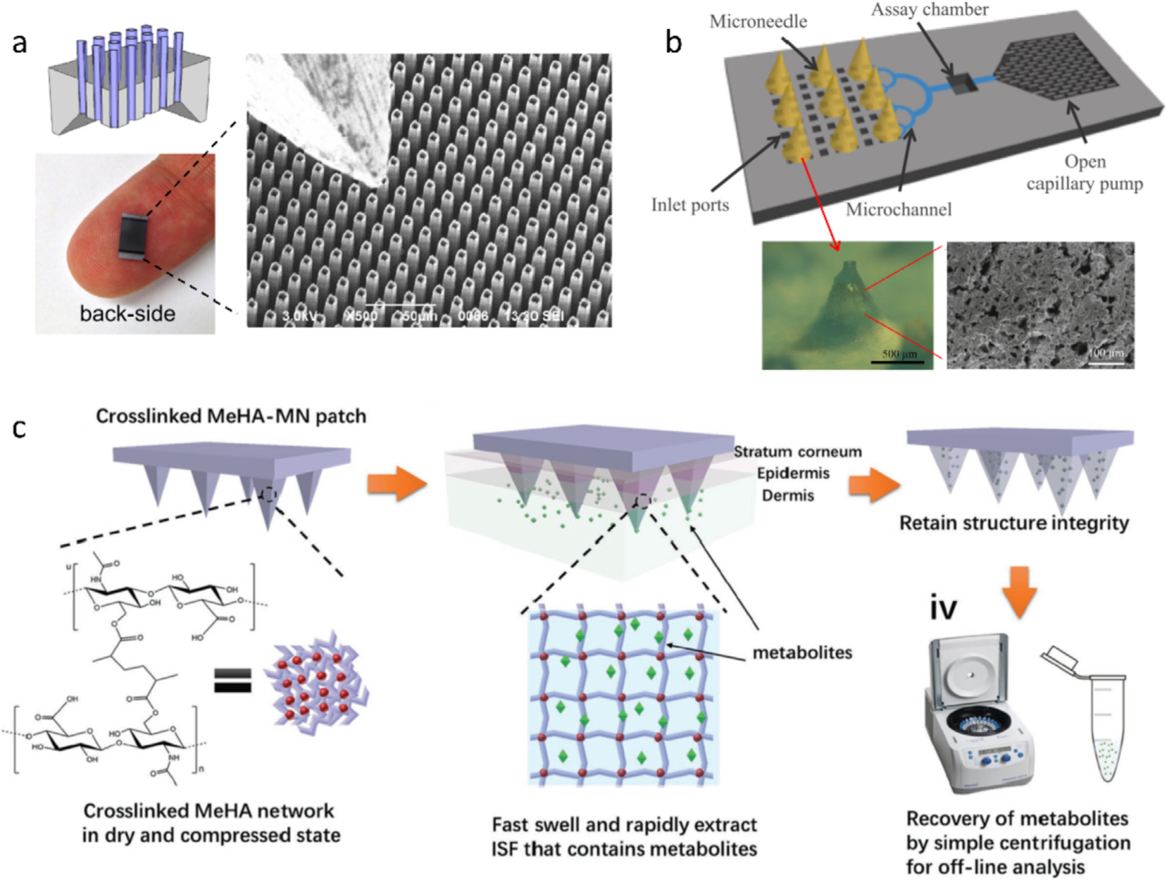
Fig. 4.

(a) Optical and SEM images of an HMN patch made of a silicon wafer. Protruding length: 100 μm; pitch: 16 μm; external diameter: 9 μm; internal diameter: 7 μm; HMN area: > 0.5 × 0.5 cm2 [32]. (b) Schematic of PMNs-based microfluidic chip for ISF extraction and direct analysis. The optical and SEM images show the PMN made of PDMS and hyaluronic acid [33]. (c) Schematic of hydrogel MNs made of a rapidly swelling hyaluronic acid crosslinked by methacrylic anhydride for ISF extraction [34]. Figures reprinted with permissions from Elsevier, Springer Nature, and John Wiley and Sons.
