Abstract
目的
研究肾癌伴下腔静脉瘤栓合并血栓患者的临床及影像资料,分析下腔静脉超声、泌尿系增强CT及增强磁共振3种影像学检查方法对下腔静脉瘤栓伴血栓的诊断效能。
方法
选择北京大学第三医院泌尿外科2014年1月至2018年7月的肾癌伴瘤栓病例56例进行回顾性分析,所有患者术前均同时行下腔静脉超声、泌尿系增强CT及增强磁共振检查并完成手术治疗,且术后病理诊断证实为肾癌伴下腔静脉瘤栓。
结果
根据术中观察及术后病理诊断证实下腔静脉瘤栓是否合并血栓为标准,将56例患者分为合并血栓组(n=18)及不合并血栓组(n=38)。比较发现,瘤栓合并血栓的患者,瘤栓长度更长[(10.50 ± 5.55) cm vs.(6.66 ± 3.73) cm,P = 0.014];瘤栓直径/下腔静脉(inferior vena cava, IVC)冠状最大径比值更接近1[1.0(0.7,1.0) vs. 0.9 (0.2,1.0), P= 0.004];出现下肢水肿的比例更高[66.7%(12/18) vs.5.3%(2/36),P = 0.005];行下腔静脉节段性切除或下腔静脉横断术的比例更高[66.7%(12/18) vs.15.8%(6/38), P<0.001]。对比下腔静脉超声、泌尿系增强CT及增强磁共振3种影像检查方法,鉴别瘤栓合并血栓,灵敏度最高的是增强磁共振(77.8%),特异性最高的是下腔静脉超声和增强CT(97.4%), 准确性最高的是增强CT及增强磁共振(83.9%),阳性预测值最高的是增强CT(90.9%),阴性预测值最高的是增强磁共振(89.2%)。
结论
肾癌伴下腔静脉瘤栓合并血栓的患者,下腔静脉瘤栓长度更长,瘤栓直径/IVC冠状最大径比值更接近1,更易出现下肢水肿。术前需综合多种影像方法,提高诊断的准确率。
Keywords: 肾癌, 下腔静脉瘤栓, 血栓, 影像诊断, 超声
Abstract
Objective
To analyse the clinical and imaging data of patients with renal cell carcinoma (RCC) with inferior vena cava tumor thrombus (IVCTT), and to assess the diagnostic efficacy of ultrasound, enhanced computed tomography (CT) and enhanced magnetic resonance imaging (MRI) in the diagnosis of RCC with IVCTT combined with bland thrombus was assessed.
Methods
We retrospectively analyzed the clinical and imaging data of 56 RCC patients with IVCTT who underwent radical nephrectomy and IVC thrombectomy between January 2014 and July 2018 in Department of Urology, Peking University Third Hospital. All the patients underwent US, enhanced CT and enhanced MRI preoperatively, and all the cases were confirmed with RCC with IVCTT by histological evaluation.
Results
The criteria of RCC with IVCTT combined with bland thrombus was confirmed by intraoperative observation and postoperative pathology. The 56 patients were divided into bland thrombus group (n=18) and non bland thrombus group (n=38). Compared the two groups, it was found that the length of IVCTT was longer [(10.50 ± 5.55) cm vs.(6.66 ± 3.73) cm, P=0.014]; the ratio of diameter of IVCTT to maximum coronal diameter of IVC was closer to 1 [1.0 (0.7, 1.0) vs. 0.9 (0.2, 1.0), P=0.004]; the proportion of lower limb edema was higher [66.7(12/8)% vs.5.3%(2/36), P=0.005];the proportion of segmental resection or interrupt of IVC was higher [66.7%(12/18) vs.15.8%(6/38), P<0.001], with statistical significance. Compared with the three imaging methods of US, enhanced CT and MRI, the highest sensitivity was MRI (77.8%), the highest specificity was enhanced MRI and enhanced CT (97.4%), the highest accuracy was enhanced CT and enhanced MRI (83.9%), the highest positive predictive value was enhanced CT (90.9%) and the highest negative predictive value was enhanced MRI (89.2%).
Conclusion
For the patients that RCC with IVCTT combined with bland thrombus, the length of IVCTT is longer, and the ratio of the diameter of IVCTT to the maximum corona diameter of IVC is closer to 1, and more likely to cause lower limb edema. Preoperative comprehensive evaluation of multiple images is needed to improve the accuracy of diagnosis.
Keywords: Renal cell carcinoma, Inferior vena cava tumor thrombus, Bland thrombus, Imaging diagnosis, Ultrasound
肾癌是泌尿系统常见的恶性肿瘤[1,2],具有进入静脉系统形成瘤栓的生物学特征,约4%~10%的肾癌患者伴下腔静脉瘤栓(inferior vena cava tumor thrombus, IVCTT)[3,4,5],1%瘤栓可进入右心房[6]。肾癌伴下腔静脉瘤栓是泌尿外科肿瘤领域最具挑战性的课题之一,根治性肾切除术和瘤栓切除术是当前相对彻底的治疗手段。
由于肾癌伴静脉瘤栓患者存在高凝状态、静脉瘀滞等,瘤栓常与血栓同时存在[7,8]。下腔静脉瘤栓合并血栓的出现,不仅会增加血栓术中脱落、肺栓塞等风险,也提示手术复杂程度更高[9],以及预后较差[10]。15%~20%的Mayo Ⅰ~Ⅳ级IVCTT需行下腔静脉节段性切除。下腔静脉节段切除患者术后易出现肾功能不全、下肢水肿等并发症,术中需全面评估确定是否行下腔静脉横断或节段切除,而下腔静脉血栓的存在是术中确定是否行下腔静脉节段切除的重要因素[9,11-12]。术前影像准确地鉴别血栓与瘤栓,对于手术方式的选择,评估患者预后具有重要意义。目前关于瘤栓合并血栓的研究尚少见报道,本文回顾性分析瘤栓合并血栓患者的临床特征,与下腔静脉血管切除的关系,并对其进行影像评估,通过分析超声、增强CT及增强磁共振3种影像方法对下腔静脉瘤栓伴血栓的诊断效能,研究影像在瘤栓伴血栓诊断中的价值。
1. 资料与方法
1.1. 病例资料
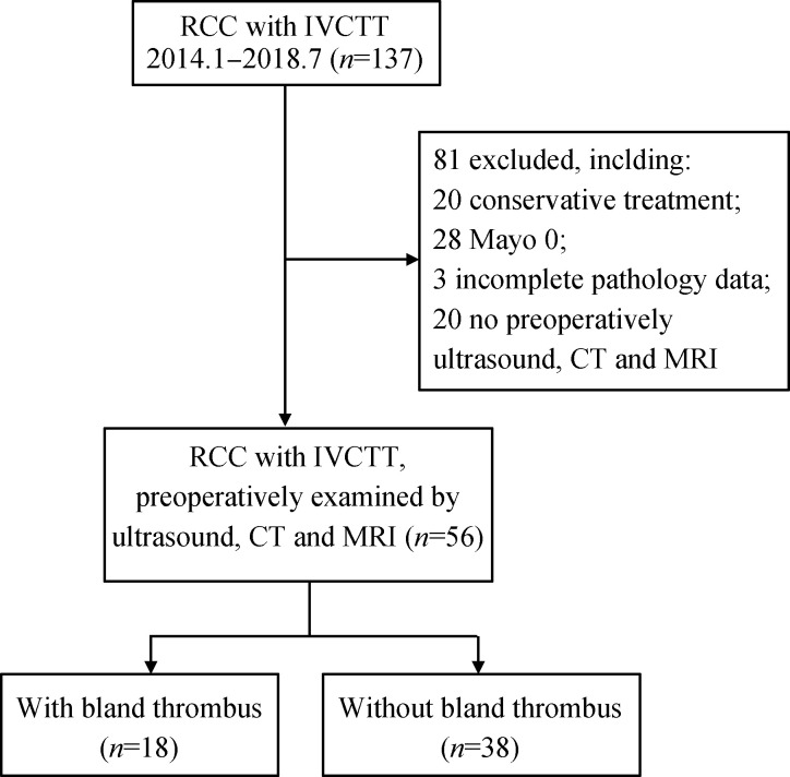
选择2014年1月至2018年7月北京大学第三医院泌尿外科因肾占位伴下腔静脉瘤栓住院患者共137例进行回顾性分析,排除因患者意愿或其他原因选择保守治疗者(n=20),Mayo 0级瘤栓者(n=28),无病理确诊结果者(n=3),术前未全部行超声、增强磁共振及增强CT患者(n=30), 最终56例Mayo Ⅰ~Ⅳ级肾癌伴下腔静脉瘤栓患者纳入本研究。入组患者均行术前腹部超声检查、泌尿系增强CT及增强磁共振检查,均行肾癌根治性切除及瘤栓切除术,术后病理诊断证实为肾癌伴下腔静脉瘤栓(图1)。
1.
研究流程及排除标准
Summary of the study cohort and exclusion criteria
分析患者临床、病理及影像学资料:(1)患者临床特征包括年龄、性别、肾肿瘤的侧别、是否伴下肢静脉水肿及术中下腔静脉处理方式;(2)病理诊断包括肾癌病理类型、核分级、是否伴横纹肌分化或肉瘤样变;(3)通过影像检查测量肾肿瘤的直径和瘤栓长度、瘤栓直径、下腔静脉冠状最大径及瘤栓直径/下腔静脉冠状最大径和下腔静脉瘤栓Mayo分级。
分析超声、增强磁共振及增强CT图像,分别记录3种影像检查诊断合并血栓的情况,以手术及病理为最终诊断标准,评估3种影像检查的敏感性、特异性、准确性、阳性预测值、阴性预测值。
1.2. 研究方法
1.2.1 超声仪器 常规行腹部超声及超声心动检查。下腔静脉超声检查使用凸阵探头C1-6MHz(LOGIQ E9, GE)、C5-1MHz(EPIQ 7,飞利浦)、C5-1MHz (Hivision Ascendus, 日立)、 CA1-7AMHz (RS80A, 三星)。对于超过膈肌的瘤栓,使用心脏探头X5-1、 S5-1 (EPIQ 7C, 飞利浦)和DM1-6A (RS80A, 三星), 观察瘤栓是否进入右心房。患者行仰卧位、侧卧位检查,以获得瘤栓清晰的图像,所有影像图像均储存,并记录瘤栓长度及是否伴血栓。
1.2.2 CT扫描序列及参数 患者行多排螺旋CT检查(GE Revolution, GE Healthcare, USA), 行能谱扫描GSI Abd模式,先行平扫。CT参数为80140 kV/190 mA, 层厚 5 mm,螺距 0.98,旋转时间 0.8 s, 窗宽 350 HU,窗位 40 HU,矩阵512×512,上界包括右心房。然后通过肘前静脉以4 mL/s 注射造影剂优维显,行增强扫描。监测升主动脉CT值,当CT值达到120 HU时,延迟10 s扫描动脉期,35 s为静脉期,50 s为延迟期。所有图像均上传至工作站。
1.2.3 磁共振扫描序列及参数 患者行全腹磁共振(MR)检查(平扫+增强),磁共振检查使用 3.0 T超导成像系统(Discovery MR750, GE Healthcare, USA), 使用腹部线圈。增强扫描经肘前静脉以23 mL/s注射造影剂钆特酸葡胺注射液(20 mL, 20 mL/9.39 g)或钆喷酸葡胺注射液(15 mL, 377 g/L),造影剂剂量(mL)依据患者体重计算[患者体重(kg)×0.4],之后生理盐水冲洗管,分别于20 s及40 s后行动脉期及门脉期扫描。
分析MRI及CT图像,记录是否合并血栓,测量相关影像参数。
1.3. 统计学方法
使用SPSS IBM 23.0软件进行分析,连续变量进行正态性检验,对于正态分布的数据用均数±标准差表示,采用两独立样本t检验分析合并血栓与不合并血栓两组患者的相应临床及影像特征;对于非正态分布的数据,采用中位数(最小值,最大值)表示,采用非参数检验比较两组分布是否存在差异;分类资料采用频数(百分比)表示,采用卡方检验比较组间比例的差异,不符合卡方检验者采用Fisher确切概率法;P<0.05认为差异有统计学意义。
以手术及病理为最终诊断标准,评估3种影像检查的敏感性、特异性、准确性、阳性预测值、阴性预测值。
2. 结果
2.1. 肾癌伴下腔静脉瘤栓合并血栓患者临床及影像特征
入组56例患者,以手术及病理诊断证实肾癌伴下腔静脉瘤栓合并血栓为标准,将患者分为伴血栓(n=18)及不伴血栓(n=38)两组,患者临床及病理资料见表1。对比两组间,临床及病理及影像特征,如性别(P=0.773)、年龄(P=0.369)、肿瘤侧别(P=0.419)、Mayo分级(P=0.203)、病理类型(P=0.447)、横纹肌分化或肉瘤样变(P=0.639)和核分级(P=0.829)、肾肿瘤直径(P=0.263)、瘤栓直径(P=0.105)、IVC冠状最大径(P=0.950),两组间差异均无统计学意义。
1.
两组患者临床及影像特征
Comparison of clinical and radiographic features of the two groups
| Features | n | With bland thrombus (n=18) |
Without bland thrombus (n=38) |
P |
| IVCTT, inferior vena cava tumor thrombus; IVC, inferior vena cava; ccRCC, clear cell renal cell carcinoma; pRCC, papillary renal cell carcinoma; *P < 0.05, statistically significance result. | ||||
| Age/years, x±s | 56 | 57.37±8.78 | 60.21±13.33 | 0.369 |
| Tumor diameter/cm, x±s | 56 | 8.82±3.75 | 9.04±3.34 | 0.263 |
| IVCTT length/cm, x±s | 56 | 10.50±5.55 | 6.66±3.73 | 0.014* |
| IVCTT diameter/cm, x±s | 56 | 3.54±0.68 | 2.76±1.04 | 0.105 |
| Maximum coronal IVC diameter/cm, x±s | 56 | 3.63±0.60 | 3.22±0.65 | 0.950 |
| IVCTT diameter/maximum coronal IVC diameter, median (min, max) | 56 | 1.0 (0.7,1.0) | 0.9 (0.2,1.0) | 0.004* |
| Gender, n (%) | 56 | 0.773 | ||
| Male | 13 (72.2) | 26 (68.4) | ||
| Female | 5 (27.8) | 12 (31.6) | ||
| Renal tumor side, n (%) | 56 | 0.419 | ||
| Right | 11 (61) | 28 (73.7) | ||
| Left | 7 (38.9) | 9 (23.7) | ||
| Blateral | 0 (0) | 1 (2.6) | ||
| Mayo classification, n (%) | 56 | 0.203 | ||
| Mayo Ⅰ | 1 (5.6) | 10 (26.3) | ||
| Mayo Ⅱ | 6 (33.3) | 14 (36.8) | ||
| Mayo Ⅲ | 6 (33.3) | 9 (23.7) | ||
| Mayo Ⅳ | 5 (27.8) | 5 (13.2) | ||
| Clinical symptoms, n (%) | 56 | 0.005* | ||
| Lower limb edema | 12 (66.7) | 2 (5.3) | ||
| No lower limb edema | 6 (33.3) | 36 (97.4) | ||
| Pathological type, n (%) | 56 | 0.447 | ||
| ccRCC | 16 (88.9) | 30 (78.9) | ||
| pRCC | 2 (11.1) | 5 (13.2) | ||
| Other | 0 (0) | 3 (7.9) | ||
| Rhabdomyosis or sarcomatoid degeneration, n (%) | 50 | 0.639 | ||
| Yes | 5 (27.8) | 7 (21.9) | ||
| No | 13 (72.2) | 25 (78.1) | ||
| Nuclear grading, n (%) | 53 | 0.829 | ||
| Ⅱ | 4 (23.6) | 8 (22.9) | ||
| Ⅲ | 9 (52.9) | 16 (45.7) | ||
| Ⅳ | 4 (23.5) | 11 (31.4) | ||
| Segmental resection or interuption IVC, n (%) | 58 | <0.001* | ||
| Yes | 12 (66.7) | 6 (15.8) | ||
| No | 6 (33.3) | 32 (84.2) | ||
伴血栓组对比不伴血栓组,瘤栓的长度更长[(10.50 ± 5.55) cm vs.(6.66 ± 3.73) cm,P = 0.014],临床出现下腔水肿的患者比例更高[66.7%(12/18)vs.5.3% (2/38),P =0.005],瘤栓直径/IVC冠状最大径比值更高[1.0(0.7,1.0)vs.0.9(0.2,1.0), P=0.004]。伴血栓对于临床手术的影响,合并血栓组中,66.8% (12/18)患者行下腔静脉节段性切除或横断下腔静脉,不伴血栓组下腔静脉切除或横断15.8% (6/38), 差异具有统计学意义(P<0.001),提示伴血栓可能是下腔静脉切除或横断的不良因素之一。
2.2. 不同影像学检查对鉴别是否伴血栓诊断效能评价
入组56例患者中,18例合并下腔静脉血栓,38例未合并血栓。合并血栓患者临床、手术处理及影像结果见表2,其中下腔静脉远心端血栓14例,对侧肾静脉血栓7例,下腔静脉近心端血栓1例。
2.
肾癌伴下腔静脉瘤栓合并血栓患者临床、病理及影像资料
Clinical, pathological and imaging features of RCC with IVCTT combined with bland thrombus
| No. | Gender | Age/ years |
Renal tumor side |
Renal tumor diameter/cm |
Pathology | Nuclear grading |
IVCTT length |
Mayo | IVC resection |
Bland thrombus location |
length in imaging/ cm |
Imaging diagnosis | |||
| US | MRI | CT | |||||||||||||
| RCC, renal cell carcinoma; IVCTT, inferior vena cava tumor thrombus; US, ultrasound; MRI, magnetic resonance imaging; CT, Computed tomography; M, male; F, famale; R, right; L, left; IVC, inferior vna cava; ccRCC, clear cell renal cell carcinoma. | |||||||||||||||
| 1 | M | 62 | L | 5.80 | ccRCC | 3 | 11.30 | Ⅳ | No | Proximal | - | 0 | 0 | 0 | |
| 2 | M | 67 | R | 3.60 | ccRCC | 2 | 16.40 | Ⅳ | Interruption | Distal | 12.94 | 0 | 1 | 1 | |
| 3 | M | 53 | R | 7.70 | ccRCC | 4 | 18.60 | Ⅱ | Segmental | Distal | 8.50 | 0 | 1 | 0 | |
| 4 | M | 53 | R | 6.40 | ccRCC | 4 | 20.00 | Ⅳ | Segmental | Distal | 2.00 | 0 | 1 | 1 | |
| 5 | F | 62 | R | 10.30 | ccRCC | 3 | 20.00 | Ⅲ | Interruption | Distal,contralateral | 12.25 | 0 | 1 | 1 | |
| 6 | M | 50 | R | 5.80 | ccRCC | 3 | 6.60 | Ⅲ | Segmental | Distal,contralateral | 2.70 | 1 | 1 | 1 | |
| 7 | M | 71 | R | 7.20 | ccRCC | 3 | 15.50 | Ⅲ | Interruption | Distal | 8.80 | 0 | 1 | 1 | |
| 8 | M | 45 | R | 10.30 | ccRCC | 2 | 10.00 | Ⅳ | Interruption | Distal | 2.13 | 0 | 1 | 1 | |
| 9 | M | 58 | L | 13.00 | ccRCC | 3 | 6.25 | Ⅱ | Interruption | Distal | 3.30 | 0 | 1 | 0 | |
| 10 | M | 47 | L | 15.50 | pRCC | 3 | 6.20 | Ⅲ | No | Contralateral, | - | 0 | 0 | 0 | |
| 11 | M | 58 | L | 14.50 | ccRCC | 4 | 5.00 | Ⅲ | Interruption | Distal | 1.30 | 0 | 1 | 1 | |
| 12 | F | 69 | L | 10.90 | ccRCC | 3 | 14.30 | Ⅳ | No | Distal | 1.30 | 0 | 1 | 0 | |
| 13 | F | 67 | R | 3.80 | pRCC | 3 | 9.40 | Ⅱ | Segmental | Distal,contralateral | 11.75 | 1 | 1 | 1 | |
| 14 | M | 62 | R | 4.50 | ccRCC | 2 | 5.48 | Ⅱ | Interruption | Distal, contralatera | 7.84 | 0 | 1 | 1 | |
| 15 | M | 48 | R | 12.00 | ccRCC | 4 | 6.90 | Ⅲ | Segmental | Distal | 2.00 | 0 | 1 | 0 | |
| 16 | F | 41 | R | 6.80 | ccRCC | 3 | 3.20 | Ⅱ | No | Contralateral | - | 0 | 0 | 0 | |
| 17 | M | 57 | L | 13.30 | ccRCC | 4 | 5.80 | Ⅱ | No | Contralateral | - | 0 | 0 | 0 | |
| 18 | F | 63 | R | 7.30 | ccRCC | 2 | 8.00 | Ⅰ | No | Distal | 1.13 | 1 | 1 | 1 | |
所有患者均行腹部超声、增强CT及增强磁共振。超声、增强磁共振及增强CT探及血栓的真阳性、假阳性及真阴性、假阴性结果见表3。分析3种影像检查评估合并血栓的敏感性、特异性、准确性、阳性预测值和阴性预测值见表4。
3.
超声、增强磁共振及增强CT诊断肾癌伴下腔静脉瘤栓合并血栓与临床病理诊断的对比
Comparison of US, enhanced MRI and enhanced CT finding with surgical and pathologic diagnosis of RCC with IVCTT combined with bland thrombus
| Imaging diagnosis result | Surgical pathologic diagnosis | |||
| With bland thrombus (n=18) | Without bland thrombus (n=38) | |||
| US, ultrasound; MRI, magnetic resonance imaging; CT, Computed tomography; RCC, renal cell carcinoma; IVCTT, inferior vena cava tumor thrombus. | ||||
| US | Positive | 3 | 1 | |
| Negative | 15 | 37 | ||
| Enhanced MRI | Positive | 14 | 5 | |
| Negative | 4 | 33 | ||
| Enhanced CT | Positive | 10 | 1 | |
| Negative | 8 | 37 | ||
4.
超声、增强磁共振及增强CT诊断肾癌伴下腔静脉瘤栓合并血栓的诊断效能
The diagnosis value of US, enhanced MRI and enhanced CT of RCC with IVCTT combined with bland thrombus /%
| Imaging diagnosis | Sensitivity | Specificity | Accuracy | Positive predictive value | Negative predictive value |
| *Among the evaluation indexes, the best diagnostic efficiency; US, ultrasound MRI, magnetic resonance imaging; CT, computed tomography; RCC, renal cell carcinoma; IVCTT, inferior vena cava tumor thrombus. | |||||
| US | 16.8 | 97.4* | 71.4 | 75.0 | 71.2 |
| Enhanced MRI | 77.8* | 86.8 | 83.9* | 73.7 | 89.2* |
| Enhanced CT | 55.7 | 97.4* | 83.9* | 90.9* | 82.2 |
在上述3种影像学检查中,灵敏度最佳的是增强磁共振检查(77.8%),特异性最佳的是超声检查和增强CT(97.4%),准确性最佳的是增强磁共振和CT(83.9%),阳性预测值最佳的是增强CT(90.9%),阴性预测值最佳的是增强磁共振(89.2%)。
3. 讨论
由于肿瘤伴静脉瘤栓患者血液常呈高凝状态,血液易在静脉内瘀滞,因此瘤栓常伴随血栓存在[13,14]。肿瘤患者易发生血栓原因有以下几点:(1)血管内皮损伤;(2)血流状态改变;(3)凝血因子激活等。瘤栓合并血栓会使手术难度增加:(1)瘤栓近心端合并血栓时,会增加栓子近心端的高度,增加手术难度,另外瘤栓近心端血栓可能不稳定,易出现血栓脱落,引起肺栓塞;(2)瘤栓的远心端存在血栓后,容易造成下腔静脉完全梗阻,形成静脉系统的侧支循环,肿瘤周围会出现代偿增生的粗大静脉,增加术中出血;(3)如下腔静脉内血栓范围较长,甚至延伸至双侧髂静脉,有时需要改变手术方式,如旷置下腔静脉。
如果下腔静脉瘤栓合并血栓,且血栓累及范围较长,术中无法全部取出,可能需要进行节段性切除下腔静脉[11,12]。下腔静脉瘤栓是否合并血栓,对于术式的选择具有重要意义。对于合并下腔静脉远心端长段血栓、侧枝循环建立充分的患者,为防止发生血栓脱落,可行下腔静脉横断术。本研究合并血栓组中,66.7%(12/18)患者行下腔静脉节段性切除或横断下腔静脉。有研究表明[11,15],合并血栓是下腔静脉横断的重要因素之一。对于肾癌合并下腔静脉瘤栓患者,如瘤栓梗阻程度较重,且尚未形成侧支循环时,可在肾静脉下方的下腔静脉内形成较长血栓,其血栓甚至可以延伸到双侧髂静脉,对于这些患者,术中切开下腔静脉后术者应尽量取净血栓,为了防止瘤栓取出后,瘤栓远心端剩余的血栓脱落,可选择行下腔静脉横断术。
Hutchinson等[10]研究血栓(主要由活化的血小板、巨噬细胞和纤维蛋白组成)对预后的影响,分析多中心446例行根治性肾切除伴下腔静脉瘤栓的患者,结果发现,伴血栓患者的肿瘤特异性死亡率增加。
术前影像学鉴别瘤栓和血栓的方法主要包括增强磁共振、CT及超声[10,16],可以通过栓子的形态、血管管腔及瘤栓血供进行判断:(1)栓子形态:瘤栓的近心端和远心端两端较为圆顿,而血栓的两端较为尖锐;(2)血管管腔:瘤栓多呈现膨胀性生长,使受累血管的管腔不均匀扩张,形成管腔的阻塞多为完全阻塞,而血栓所致的管腔扩张可表现为均匀扩张,管腔多为不完全阻塞,并且血栓在疾病的后期会缩小或机化;(3)瘤栓血供:瘤栓内部多可探及动脉血流,其原因在于瘤栓在下腔静脉血管管腔内生长、浸润需滋养动脉,增强CT有时可见瘤栓内的血管,血栓内部通常不会出现血流信号。需要注意的是,虽然有部分病例瘤栓内可探及动脉血流,但该检查手段特异性较高,敏感度相对较低,不足20%[17], 此外,有学者将PET-CT用于瘤栓与血栓的鉴别[18],含有瘤栓的静脉增宽,皮髓质期增强最为明显,瘤栓增强达高峰。Sharma等[19]研究24例IVCTT,使用最大标准化上升值(maximum standardized uptake value,SUVmax)鉴别瘤栓与血栓,血栓组平均SUVmax 值为 3.2,瘤栓组平均SUVmax值6.0(P= 0.013), 并得出鉴别血栓与瘤栓的SUVmax最佳界值为3.6,敏感度为72%,特异性为90%。
本研究伴血栓组,瘤栓直径与下腔静脉冠状最大径比值更接近1,提示下腔静脉完全被瘤栓阻塞时,更易继发血栓形成。
伴血栓组下肢水肿患者比例高于不伴血栓组(66.7% vs. 5.3%,P<0.05), 提示影像医生在诊断时,对于出现下肢静脉水肿的患者临床症状存在下肢水肿,同时影像学提示瘤栓较长时应注意瘤栓近心端、远心端及对侧肾静脉内是否合并血栓。
本研究对比超声、增强CT及增强磁共振3种影像学检查,对于肾癌伴下腔静脉瘤栓合并血栓的诊断。临床中,常规超声检查对于合并血栓尤其对侧肾静脉血栓的检出率相对较低。
本研究存在一定局限性,研究为回顾性研究,病例选择存在偏倚,同时术前均行超声、增强磁共振及CT的患者病例数量有限,今后需扩大样本进行进一步研究。
综上所述,肾癌伴下腔静脉瘤栓合并血栓的患者,瘤栓长度更长,瘤栓直径/下腔静脉冠状最大径更接近1,更易出现下腔静脉水肿,术前需多种影像综合评估,以提高诊断的准确率。
Funding Statement
国家重点研发计划专项(2016YFC0104700)
Supported by National Key Research and Development Program of China(2016YFC0104700)
Contributor Information
马 潞林 (Lu-lin MA), Email: malulin@medmail.com.cn.
王 淑敏 (Shu-min WANG), Email: shuminwang2014@163.com.
References
- 1.刘 茁, 马 潞林, 田 晓军, et al. 根治性肾切除术+下腔静脉癌栓取出术治疗Mayo 0~Ⅳ 级下腔静脉癌栓的临床经验. 中华泌尿外科杂志. 2017;38(11):842–847. [Google Scholar]
- 2.刘 茁, 马 潞林, 田 晓军, et al. 肾癌根治性切除加癌栓取出术治疗Mayo Ⅲ 级下腔静脉癌栓的手术技术及临床经验. 北京大学学报(医学版) 2017;49(4):597–602. [Google Scholar]
- 3.刘 茁, 田 晓军, 马 潞林. 根治性肾切除术联合Mayo 0-Ⅱ 级静脉癌栓取出术的临床麻醉管理. 现代肿瘤医学. 2017;25(16):2672–2677. [Google Scholar]
- 4.刘 茁, 马 潞林, 田 晓军, et al. 腹腔镜和开放肾癌根治性切除+Mayo Ⅱ 级下腔静脉癌栓取出术11例临床分析. 现代泌尿外科杂志. 2017;22(8):603–607. [Google Scholar]
- 5.马 潞林, 刘 茁. 肾癌并肝段和肝以上下腔静脉癌栓的诊治体会. 中华泌尿外科杂志. 2017;38(7):481–484. [Google Scholar]
- 6.Blute ML, Leibovich BC, Lohse CM, et al. The mayo clinic experience with surgical management, complications and outcome for patients with renal cell carcinoma and venous tumour thrombus. BJU Int. 2004;94(1):33–41. doi: 10.1111/j.1464-410X.2004.04897.x. [DOI] [PubMed] [Google Scholar]
- 7.Wang M, Ping H, Niu Y, et al. Pure conventional laparoscopic radical nephrectomy with level II vena cava tumor thrombectomy. Int Braz J Urol. 2014;40(2):266–273. doi: 10.1590/S1677-5538.IBJU.2014.02.18. [DOI] [PubMed] [Google Scholar]
- 8.程 艳, 蔡 欣, 刘 基巍. 恶性肿瘤与血栓形成. 临床肿瘤学杂志. 2010;15(4):376–379. [Google Scholar]
- 9.Ayyathurai R, Garciaroig M, Gorin MA, et al. Bland thrombus association with tumour thrombus in renal cell carcinoma: Analysis of surgical significance and role of inferior vena caval interruption. BJU Int. 2013;110(11b):E449–E455. doi: 10.1111/j.1464-410X.2012.11128.x. [DOI] [PubMed] [Google Scholar]
- 10.Hutchinson R, Rew C, Chen G, et al. The adverse survival implications of bland thrombus in renal cell carcinoma with venous tumor thrombus. Urology. 2018;115:119–124. doi: 10.1016/j.urology.2018.02.019. [DOI] [PubMed] [Google Scholar]
- 11.Blute ML, Boorjian SA, Leibovich BC, et al. Results of inferior vena caval interruption by greenfield filter, ligation or resection during radical nephrectomy and tumor thrombectomy. J Urol. 2007;178(2):440–445. doi: 10.1016/j.juro.2007.03.121. [DOI] [PubMed] [Google Scholar]
- 12.马 鑫. 机器人腹腔镜腔静脉瘤栓取出术:新的思考新的策略. 中华腔镜外科杂志: 电子版. 2017;10(5):272–273. [Google Scholar]
- 13.Quencer KB, Friedman T, Sheth R, et al. Tumor thrombus: Incidence, imaging, prognosis and treatment. Cardiovasc Diagn Ther. 2017;7(Suppl 3):S165–S177. doi: 10.21037/cdt.2017.09.16. [DOI] [PMC free article] [PubMed] [Google Scholar]
- 14.Mukai M, Oka T. Mechanism and management of cancer-associated thrombosis. J Cardiol. 2018;72(2):89–93. doi: 10.1016/j.jjcc.2018.02.011. [DOI] [PubMed] [Google Scholar]
- 15.肾癌伴静脉瘤栓北京专家共识 [J]. 微创泌尿外科杂志, 2017: 6(6): 321-327.
- 16.宋 奕宁, 赵 艺超, 李 建国. 下腔静脉肿瘤的超声影像诊断与鉴别. 中国超声医学杂志. 2018;34(1):37–39. [Google Scholar]
- 17.Tarantino L. Contrast-enhanced ultrasound in differentiating malignant from benign portal vein thrombosis in hepatocellular carcinoma. World J Gastroenterol. 2015;21(32):9457–9460. doi: 10.3748/wjg.v21.i32.9457. [DOI] [PMC free article] [PubMed] [Google Scholar]
- 18.Sonavane SN, Malhotra G, Asopa R, et al. Role of fluorine-18 fluorodeoxyglucose positron emission tomography in a case of renal cell carcinoma to differentiate tumor thrombus from bland thrombus. Indian J Nucl Med. 2015;30(4):355–357. doi: 10.4103/0972-3919.164016. [DOI] [PMC free article] [PubMed] [Google Scholar]
- 19.Sharma P, Kumar R, Jeph S, et al. 18F-FDG PET-CT in the diagnosis of tumor thrombus. Nucl Med Commun. 2011;32(9):782–788. doi: 10.1097/MNM.0b013e32834774c8. [DOI] [PubMed] [Google Scholar]


