

Abstract
目的
研究和比较初学者应用放大镜与应用显微镜进行瓷贴面牙体预备的效果,从操作效率、预备体质量、预备准确度以及喜好度等方面比较放大镜和显微镜的应用价值。
方法
从北京大学口腔医院修复科选择20名口腔修复医生进行前瞻性、单盲、自身对照试验,试验对象无使用放大镜或显微镜的经验。每人依次在常规视野下(空白对照组)、2.5倍头戴式放大镜下(放大镜组)和8倍医用显微镜下(显微镜组)在仿头模内完成右上中切牙开窗型瓷贴面牙体预备,试验过程中记录牙体预备所需的时间。操作完成后,由医生本人利用视觉模拟评分法(vi-sual analogue score,VAS)对操作效率、预备体质量和喜好度进行主观评分,由第三方专家在体视显微镜下对瓷贴面预备体的质量进行评分,并利用数字化方法对预备准确性进行评价。
结果
操作效率方面,对照组、放大镜组和显微镜组的主观VAS评分分别为7.15±1.73、8.10±0.91、5.40±2.04,放大镜组与显微镜组间的差异有统计学意义(P<0.05);客观操作时间三组分别为(430.10±163.04) s、(393.90±157.27) s、(441.95±164.18) s,放大镜组与显微镜组间的差异有统计学意义(P<0.05);放大镜组比显微镜组操作效率高。预备体质量方面,对照组、放大镜组和显微镜组的主观VAS评分分别为6.55±2.09、7.85±0.99、6.25±1.77,放大镜组与显微镜组间的差异有统计学意义(P<0.05);专家评分分别为12.20±1.67、12.50±1.70、11.35±2.60,放大镜组与显微镜组间的差异有统计学意义(P<0.05);放大镜组的预备体质量优于显微镜组。预备准确度方面,对照组、放大镜组和显微镜组的唇面切1/3分别为(0.107±0.097) mm、(0.142±0.118) mm、(0.123±0.087) mm,唇面中1/3分别为(0.128±0.073) mm、(0.113±0.105) mm、(0.125±0.077) mm,唇面颈1/3分别为(0.075±0.054) mm、(0.068±0.044) mm、(0.058±0.047) mm,三组间每个区域的差异均无统计学意义(P>0.05)。喜好度方面,对照组、放大镜组和显微镜组的主观VAS评分分别为6.55±2.31、8.60±1.10、5.80±2.07,放大镜组与显微镜组间的差异有统计学意义(P<0.05),放大镜组最受欢迎。
结论
针对初学者而言,放大镜比显微镜用于瓷贴面牙体预备的效果更好。
Keywords: 牙科设备, 牙科器械, 牙体预备, 放大镜, 显微镜
Abstract
Objective
To assess and compare the effects of loupes and microscope on laminate veneer preparation of the first practitioner from the aspects of efficiency, quality and accuracy of preparation, and preference.
Methods
Twenty young prosthodontists from the Department of Prosthodontics, Peking University School and Hospital of Stomatology were recruited into this study, which was prospective, single blind, self-control trials. The participants had no experience of using dental magnification devices. They prepared laminate veneers in the artificial dental model, under routine visual field (control group), 2.5× headwear loupes (loupes group), and 8× operating microscope (microscopic group) by turning. The time for tooth preparation was recorded. Thereafter, subjective assessments of efficiency, quality of preparation and preference were performed by themselves using visual analogue score (VAS). Expert assessments of quality and accuracy of preparation were performed by two professors using stereomicroscope and digital technique respectively.
Results
In terms of efficiency, the subjective scores for the control group, loupes group and microscopic group were 7.15±1.73, 8.10±0.91 and 5.40±2.04, respectively. There was significant difference between the loupes group and microscopic group (P<0.05). The time of tooth preparation for the control group, loupes group and microscopic group was (430.10±163.04) s, (393.90±157.27) s and (441.95±164.18) s, respectively. There was significant diffe-rence between the loupes group and microscopic group (P<0.05). The loupes group was more efficient than the microscopic group. In terms of the quality of preparations, the subjective scores for the control group, loupes group and microscopic group were 6.55±2.09, 7.85±0.99 and 6.25±1.77, respectively. There was significant difference between the loupes group and microscopic group (P<0.05). The expert evaluations for the control group, loupes group and microscopic group were 12.20±1.67, 12.50±1.70 and 11.35±2.60, respectively. There was significant difference between the loupes group and microscopic group (P<0.05). The loupes group had higher quality than the microscopic group. In terms of the accuracy of preparations, the control group, loupes group and microscopic group of incisal 1/3 were (0.107±0.097) mm, (0.142±0.118) mm and (0.123±0.087) mm, respectively, of middle 1/3 were (0.128±0.073) mm, (0.113±0.105) mm and (0.125±0.077) mm, respectively, and of cervical 1/3 were (0.075±0.054) mm, (0.068±0.044) mm and (0.058±0.047) mm, respectively. There was no significant difference among the three groups (P>0.05). In terms of the preference, the subjective scores for the control group, loupes group and microscopic group were 6.55±2.31, 8.60±1.10 and 5.80±2.07, respectively. There was significant difference between the loupes group and microscopic group (P<0.05). The participants had the highest preference for loupes.
Conclusion
For the first practitioners, loupes is better than microscope for laminate veneer preparation.
Keywords: Dental equipment, Dental instruments, Tooth preparation, Loupes, Microscopy
人眼在无辅助设备时能分辨的最小距离为200 μm,且距离目标物越近,越容易产生视觉疲劳,此外,随着年龄增长,人眼还会出现视力调节能力减退[1]。放大设备可以显著提高口腔临床操作中的近距离视野清晰度,补偿裸眼视力的不足[2,3]。
目前,口腔放大镜(loupes)已几乎成为修复医生日常工作的必备辅助设备。在许多国家,口腔放大镜已成为研究生必备的诊疗设备。除了放大镜以外,医用显微镜(microscope)在口腔医学当中的应用也越来越广泛,有些口腔医学院校已经将显微镜下口腔修复治疗纳入教学内容当中。但是,这些放大设备对口腔临床工作效果的影响主要见于一些专家意见[4,5,6,7]及病例报道等[8,9],仅有少量的相关对照研究,且多是牙体牙髓专业领域,而且其研究结果尚无定论[10,11,12,13,14]。对于初学者而言,放大镜与显微镜的临床应用效果如何,二者对操作效率、预备体质量、预备准确度方面有何影响,以及初学者对他们的喜好度如何,尚不明确。
本试验的主要目的是研究和比较初学者应用放大镜与显微镜进行瓷贴面牙体预备的效果,从操作效率、预备体质量、预备准确度以及喜好度等方面比较放大镜与显微镜的应用价值。研究的无效假设为:初学者应用放大镜与显微镜进行瓷贴面牙体预备的效果无明显差别。
1. 资料与方法
1.1. 研究对象
用随机数表法从北京大学口腔医院口腔修复科60名年轻医生(含住院医师)当中随机选择20人参加本项研究。
纳入标准:(1)裸眼视力或矫正视力达到1.0;(2)已有独立完成瓷贴面牙体预备10例以上的临床经验;(3)没有使用放大镜和显微镜的经验。
排除标准:(1)裸眼视力和矫正视力均未达到1.0;(2)瓷贴面牙体预备的临床经验少于10例;(3)已有使用放大镜或显微镜的经验。
1.2. 试验方法
本试验采用前瞻性、单盲、自身对照的试验方法。20名口腔修复医生依次在常规视野下(对照组)、2.5倍头戴式放大镜下(放大镜组)和8倍医用显微镜下(显微镜组)在仿头模内完成右上中切牙开窗型瓷贴面牙体预备,每次操作之间间隔5 min。牙体预备所用的钻针为具有深度指示功能的金刚砂钻针,用于指导临床牙体预备量。试验时不告知被试者本研究的真实目的,试验过程中记录完成牙体预备所需的时间。操作完成后,由医生本人利用视觉模拟评分法(visual analogue scale,VAS)对操作效率、预备体质量和喜好度进行主观评分,由第三方专家对瓷贴面预备体的质量进行评分,并利用数字化方法对预备准确性进行评价。
1.3. VAS主观评分
每名医生完成全部牙体预备后,利用VAS对操作效率、预备体质量和喜好度进行主观评分。将每个评价指标用0~10共11个数字表示,分值越低说明该项结果越不理想,分值越高说明该项结果越理想。
1.4. 预备体质量的专家评价
由两名北京大学口腔医院口腔修复教研室高级职称教师对瓷贴面预备体质量进行评价,评分标准见表1,共包括唇面预备量、表面光洁度、颈部终止线形态、颈部终止线位置、邻面终止线5项指标。每项分为3个等级:良好为3分、一般为2分、较差为1分,总分最高为15分、最低为5分。分值越高说明预备体质量越好,分值越低说明预备体质量越差。两名评价者首先回顾理想的预备体形态,浏览全部模型并了解可能的情况之后,再在16倍体视显微镜下进行独立评分,以两人的平均分为最终评分。
1.
瓷贴面牙体预备评分表
The criteria of preparation for porcelain veneer
| Parameter | Excellent (3 points) | Compromised (2 points) | Standard not met (1 point) |
| Facial reduction | Optimal reduction (incisal third: 0.7 mm, middle third: 0.5 mm, cervical third: 0.3 mm) | Moderately over-reduced or under-reduced | Severely over-reduced or under-reduced |
| Surface smoothness | Fine diamond texture | Catches with explorer tip | Horizontal or vertical steps |
| Cervical finish line configuration | Chamfer is continuous and well-defined | Chamfer is moderately nonconti-nuous or moderately lack of definition | Chamfer is noncontinuous or lack of definition or aggressively prepared |
| Cervical finish line position | Placed to specified target: 0.5-1.0 mm supragingivally | Even with gingival margin or <0.5 mm supragingivally | >1.0 mm supragingivally or subgingivally |
| Interproximal finish line | Mesial and distal finish lines are continuous and well-defined, extended to, but do not open the interproximal contact region | Mesial or distal finish line is mo-derately noncontinuous or mode-rately lack of definition, at interproximal surface but do not extend to contact region | Mesial and distal finish lines are noncontinuous or lack of definition or aggressively prepared, at labial surface or open the contact |
1.5. 牙体预备准确度评价
利用口内扫描仪扫描原始模型和预备体模型,将预备前后的数据进行配准,分别在唇面切1/3、中1/3和颈1/3三个区域各选择近中、中央和远中3个点测量牙体预备量(图1),以3个点的平均值为该区域的平均预备量。将切1/3、中1/3和颈1/3三个区域的预备量分别与各自区域的目标牙体预备量(切1/3: 0.7 mm、中1/3: 0.5 mm、颈1/3: 0.3 mm)相比,计算差值的绝对值,以此评价预备的准确度。
1.
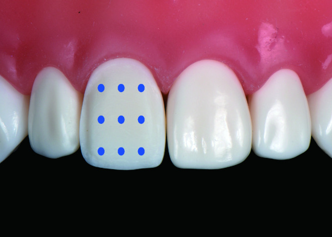
唇面牙体预备量测量点
Measurement points for measuring facial reduction
1.6. 材料及设备
研究所用设备有放大镜(速迈公司,中国)、医用显微镜(Leica公司,德国)、仿头模及人工牙颌模型(Nissin公司,日本)、牙体预备用金刚砂钻针(Coltene公司,瑞士)、扫描仪(3Shape公司,丹麦)、体视显微镜(Nikon公司,日本)等。
1.7. 统计学分析
计量数据以均数±标准差表示,使用SPSS Statistics 20.0软件(IBM公司,美国)对数据进行统计分析。对评分进行两因素方差分析,并通过Tukey HSD方法进行组间多重比较,以P<0.05为差异有统计学意义。
2. 结果
对照组、放大镜组和显微镜组的操作效率、预备体质量、预备准确度和喜好度评价结果见表2。
2.
三种操作条件下的牙体预备效果
Results of tooth preparation under three conditions
| Group | Efficiency of preparation | Quality of preparation | Accuracy of preparation/mm | Preference | |||||
| Subjective visual analogue score |
Time/s | Subjective visual analogue score |
Expert evaluation |
Incisal third | Middle third | Cervical third | Subjective visual analogue score |
||
| For each column, groups identified by different superscript letters (a, b) were significantly different (P<0.05). | |||||||||
| Control | 7.15±1.73a | 430.10±163.04ab | 6.55±2.09ab | 12.20±1.67ab | 0.107±0.097a | 0.128±0.073a | 0.075±0.054a | 6.55±2.31a | |
| Loupes | 8.10±0.91a | 393.90±157.27a | 7.85±0.99a | 12.50±1.70a | 0.142±0.118a | 0.113±0.105a | 0.068±0.044a | 8.60±1.10b | |
| Microscopic | 5.40±2.04b | 441.95±164.18b | 6.25±1.77b | 11.35±2.60b | 0.123±0.087a | 0.125±0.077a | 0.058±0.047a | 5.80±2.07a | |
| P | <0.001 | 0.017 | 0.018 | 0.015 | 0.299 | 0.637 | 0.467 | <0.001 | |
2.1. 操作效率
对照组、放大镜组和显微镜组的操作效率主观VAS评分分别为7.15±1.73、8.10±0.91、5.40±2.04,三组间差异有统计学意义(P<0.05);多重比较结果显示,放大镜组与显微镜组间的差异有统计学意义(P<0.05)。
对照组、放大镜组和显微镜组的操作时长分别为(430.10±163.04) s、(393.90±157.27) s、(441.95±164.18) s,三组间差异有统计学意义(P<0.05);多重比较结果显示,放大镜组与显微镜组间的差异有统计学意义(P<0.05)。
2.2. 预备体质量
对照组、放大镜组和显微镜组的预备体质量主观VAS评分分别为6.55±2.09、7.85±0.99、6.25±1.77,三组间差异有统计学意义(P<0.05);多重比较结果显示,放大镜组与显微镜组间的差异有统计学意义(P<0.05)。
对照组、放大镜组和显微镜组的预备体质量专家评分分别为12.20±1.67、12.50±1.70、11.35±2.60,三组间差异有统计学意义(P<0.05);多重比较结果显示,放大镜组与显微镜组间的差异有统计学意义(P<0.05)。
2.3. 预备准确度
对照组、放大镜组和显微镜组的预备准确度,切1/3分别为(0.107±0.097) mm、(0.142±0.118) mm、(0.123±0.087) mm,三组间差异无统计学意义(P>0.05);中1/3分别为(0.128±0.073) mm、(0.113±0.105) mm、(0.125±0.077) mm,三组间差异无统计学意义(P>0.05);颈1/3分别为(0.075±0.054) mm、(0.068±0.044) mm、(0.058±0.047) mm,三组间差异无统计学意义(P>0.05)。
2.4. 喜好度
对照组、放大镜组和显微镜组的喜好度主观VAS评分分别为6.55±2.31、8.60±1.10、5.80±2.07,三组间差异有统计学意义(P<0.05);多重比较结果显示,放大镜组与显微镜组间的差异有统计学意义(P<0.05)。
3. 讨论
本研究结果表明,对于初学者而言,放大镜比显微镜下进行瓷贴面牙体预备的效率更高、预备体质量更好、更受欢迎,因此拒绝本研究初始的无效假设。
关于口腔放大设备对牙体预备临床效果的影响尚无标准统一的研究模型和方案,本研究采用在仿头模上进行瓷贴面牙体预备来模拟临床情况。瓷贴面成功的关键之一是对牙釉质的保存[15],上中切牙颈部的牙釉质厚度仅为0.3~0.5 mm[16],瓷贴面预备需要对牙体预备量有非常准确的控制,因此,瓷贴面预备更能够体现口腔修复牙体预备对准确性的要求,更适于评价放大镜和显微镜对口腔修复专业初学者牙体预备准确性的影响。
本研究通过初学者主观VAS评分以及客观操作时间两个方面比较了放大镜和显微镜对牙体预备效率的影响。主、客观评分均表明,显微镜的操作效率最低,这可能是由于显微镜难以把握实际的空间距离,操作视野有限,操作幅度较大时容易超出视野范围,使用初期还有可能出现轻度视觉紊乱、一过性的模糊等,需要一定的适应过程,这些原因都有可能影响牙体预备效率,这也提示显微镜比放大镜的学习难度更大。
从预备体质量的评价结果来看,不论主观评价还是专家评价,放大镜组都优于显微镜组,这是由于本次研究的受试对象都是初学者,没有放大镜和显微镜的使用经验,显微镜更为复杂的操作技巧和适应能力要求,可能对最终的预备结果产生了负面影响。
从预备准确度来看,三组间差异并没有统计学意义,这可能与采用的预备模型为瓷贴面预备,预备量较小,初学者对放大镜和显微镜的使用经验不足,无法充分利用放大设备的优势有关系。
从主观喜好度来看,放大镜组优于显微镜组。可能由于放大镜对于初学者来说更容易从裸眼过渡,适应期短,容易学习。显微镜需要更多的时间学习操作方法,建立操作习惯,需要更长的学习曲线,适应期长。今后的研究将对放大镜和显微镜的学习曲线进行评价,评估在经过一定的学习之后二者对牙体预备效果及喜好度的影响。
综上,对初学者而言,放大镜比显微镜的应用效果更好,前者的瓷贴面牙体预备效率和预备质量更好,且更受欢迎,提示在初学者当中应首先使用放大镜而不是显微镜进行教学工作和临床操作。
Funding Statement
国家自然科学基金(81701003); 北京大学口腔医院教学改革项目基金(2017-PT-01)
Supported by the National Nature Science Foundation of China(81701003); and the Program for Educational Reform of Peking University School and Hospital of Stomatology(2017-PT-01)
Footnotes
The authors have declared that no competing interests exist.
作者已声明无竞争性利益关系。
References
- 1.Carr GB, Murgel CA. The use of the operating microscope in endodontics. Dent Clin North Am. 2010;54(2):191–214. doi: 10.1016/j.cden.2010.01.002. [DOI] [PubMed] [Google Scholar]
- 2.Eichenberger M, Perrin P, Ramseyer ST, et al. Visual acuity and experience with magnification devices in Swiss dental practices. Oper Dent. 2015;40(4):E142–E149. doi: 10.2341/14-103-C. [DOI] [PubMed] [Google Scholar]
- 3.Perrin P, Eichenberger M, Neuhaus KW, et al. A visual acuity and magnification devices in dentistry. Swiss Dent J. 2016;126(3):222–235. doi: 10.61872/sdj-2016-03-142. [DOI] [PubMed] [Google Scholar]
- 4.Sitbon Y, Attathom T. Minimal intervention dentistry II: Part 6. Microscope and microsurgical techniques in periodontics. British Dent J. 2014;216(9):503–509. doi: 10.1038/sj.bdj.2014.356. [DOI] [PubMed] [Google Scholar]
- 5.Sitbon Y, Attathom T, St-Georges AJ. Minimal intervention dentistry II: part 1. Contribution of the operating microscope to dentistry. British Dent J. 2014;216(3):125–130. doi: 10.1038/sj.bdj.2014.48. [DOI] [PubMed] [Google Scholar]
- 6.Mamoun J. Use of high-magnification loupes or surgical operating microscope when performing dental extractions. N Y State Dent J. 2013;79(3):28–33. [PubMed] [Google Scholar]
- 7.Malterud MI. Magnification: You can’t effectively practice minimally in biomimetic dentistry without it. Gen Dent. 2013;61(3):14–17. [PubMed] [Google Scholar]
- 8.Brito MJ, Moreira GJ, Normanha JA, et al. Midbuccal canals of maxillary molars evaluated by cone-beam computed tomography: Endodontic management of two cases. Braz Dent J. 2013;24(6):575–579. doi: 10.1590/0103-6440201302358. [DOI] [PubMed] [Google Scholar]
- 9.Albuquerque DV, Kottoor J, Dham S, et al. Endodontic management of maxillary permanent first molar with 6 root canals: 3 case reports. Oral Surg Oral Med Oral Pathol Oral Radiol Endod. 2010;110(4):e79–e83. doi: 10.1016/j.tripleo.2010.04.017. [DOI] [PubMed] [Google Scholar]
- 10.Lussi A, Kronenberg O, Megert B. The effect of magnification on the iatrogenic damage to adjacent tooth surfaces during class II preparation. J Dent. 2003;31(4):291–296. doi: 10.1016/s0300-5712(03)00029-0. [DOI] [PubMed] [Google Scholar]
- 11.Neuhaus KW, Jost F, Perrin P, et al. Impact of different magnification levels on visual caries detection with ICDAS. J Dent. 2015;43(12):1559–1564. doi: 10.1016/j.jdent.2015.09.002. [DOI] [PubMed] [Google Scholar]
- 12.Mitropoulos P, Rahiotis C, Kakaboura A, et al. The impact of magnification on occlusal caries diagnosis with implementation of the ICDAS II criteria. Caries Res. 2012;46(1):82–86. doi: 10.1159/000335988. [DOI] [PubMed] [Google Scholar]
- 13.Smadi L, Khraisat A. Detection of a second mesiobuccal canal in the mesiobuccal roots of maxillary first molar teeth. Oral Surg Oral Med Oral Pathol Oral Radiol Endod. 2007;103(3):e77–e81. doi: 10.1016/j.tripleo.2006.10.007. [DOI] [PubMed] [Google Scholar]
- 14.Donaldson ME, Knight GW, Guenzel PJ. The effect of magnification on student performance in pediatric operative dentistry. J Dent Educ. 1998;62(11):905–910. [PubMed] [Google Scholar]
- 15.Gurel G, Sesma N, Calamita MA, et al. Influence of enamel pre-servation on failure rates of porcelain laminate veneers. Int J Periodontics Restorative Dent. 2013;33(1):31–39. doi: 10.11607/prd.1488. [DOI] [PubMed] [Google Scholar]
- 16.Ferrari M, Patroni S, Balleri P. Measurement of enamel thickness in relation to reduction for etched laminate veneers. Int J Periodontics Restorative Dent. 1992;12(5):407–413. [PubMed] [Google Scholar]