

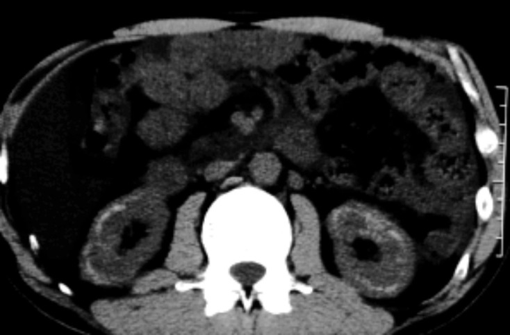
A 47-year-old woman presented at our clinic with a complaint of “longstanding bilateral lower leg edema.” For the past 10 years, she had been taking furosemide 10 mg/day to reduce the “edema.” Her medical history was otherwise normal. She was 158 cm tall and weighed 54.5 kg. A physical examination revealed subcutaneous fat but no edema in the legs. Abdominal ultrasound imaging showed linear hyperechoic corticomedullary junctions in the bilateral kidneys. Abdominal computed tomography showed linear high-density lesions along the renal corticomedullary junctions of both kidneys (Picture), which were compatible with nephrocalcinosis. Serum calcium, phosphate and creatinine levels were normal, while the potassium level was low at 3.5 mEq/L. Urinary pH was low at 4.5. Mineralization of the renal tubules at the corticomedullary junction is reported in cases of hypercalcemia or an excessive phosphate intake (1,2) but may also appear after the prolonged use of loop diuretics due to an elevated concentration of minerals in the renal tubules.
Picture.
The authors state that they have no Conflict of Interest (COI).
References
- 1.Rao GN. Diet and kidney diseases in rats. Toxicol Pathol 30: 651-656, 2002. [DOI] [PubMed] [Google Scholar]
- 2.Okubo K, Okubo H, Todani S, Shirotori K, Oguchi T, Higuchi K. Linear calcification in the cortico-medullary junction of the kidney. Intern Med 47: 565, 2008. [DOI] [PubMed] [Google Scholar]