Abstract
A 93‐year‐old woman was admitted with a 10‐day history of cough and prostration. Thoracic computed tomography revealed extensive ground‐glass opacities in both the lungs. The polymerase chain reaction test of sputum for severe acute respiratory syndrome‐coronavirus‐2 (SARS‐CoV‐2) was positive. She was treated with antiviral agents and steroid pulse therapy. However, her oxygen saturation gradually declined, and she died 10 days after hospitalization. The most important autopsy finding was fuzzily segmented diffuse alveolar damage (DAD) that expanded from the subpleural to the medial area. No remarkable changes were observed in organs other than the lungs. Therefore, pneumocytes were suggested as the primary target for SARS‐CoV‐2, which might explain why coronavirus infectious disease‐19 is a serious condition. Thus, early treatment is essential to prevent viral replication from reaching a level that triggers DAD.
Keywords: COVID‐19, diffuse alveolar damage, Japanese case
Abbreviations
- COVID‐19
coronavirus infectious disease‐19
- SARS‐CoV‐2
severe acute respiratory syndrome‐coronavirus‐2
INTRODUCTION
Coronavirus infectious disease‐19 (COVID‐19) has rapidly spread throughout the world since late 2019, with millions of infected patients and hundreds of thousands of deaths. 1 , 2 One crucial feature of COVID‐19 is a substantially higher mortality rate (~15%) than the seasonal influenza virus infectious disease (~0.1%). 1 , 2 Ethnic differences in mortality rates have been observed, with higher rates in Europe (~15%) than in East Asia (~5%). 1 , 2 , 3 , 4 To elucidate the underlying mechanism for these characteristics, autopsy cases from different countries need to be compared. Presently, less than 50 cases have been reported in the English language literature. 5 , 6 , 7 , 8 , 9 , 10 We present the first such case report of a Japanese COVID‐19 patient.
CLINICAL SUMMARY
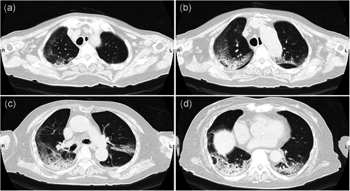
A 93‐year‐old Japanese woman was admitted with a 10‐day history of cough and prostration. Thoracic computed tomography (CT) revealed ground‐glass opacities in both the lungs with a fuzzy segmental distribution, predominantly seen in the subpleural area at the lateral‐dorsal side of the lower lobes (Fig. 1). The right lobe seemed predominantly affected (Fig. 1). She was treated with antibiotics (sulbactam, ampicillin and ceftriaxone), but the pneumonia did not improve. The polymerase chain reaction (PCR) test (LightMix, TIB MOLBIOL, Berlin, Germany) of the sputum for severe acute respiratory syndrome‐coronavirus‐2 (SARS‐CoV‐2) was performed on the third day after hospitalization, and positive results were obtained on the fifth day. A diagnosis of COVID‐19 was confirmed. Thereafter, she was treated with antiviral agents (favipiravir, hydroxychloroquine and ciclesonide) and steroid pulse therapy (methylprednisolone). Her systemic status worsened gradually, with declining oxygen saturation levels. She died of respiratory failure 10 days after hospitalization.
Figure 1.

Representative images of thoracic computed tomography (CT) reveal ground‐glass opacity, which shows segmental distribution, (a) predominant in the subpleural area of the lateral‐dorsal side (b and c) of the lower lobe (c and d). The right lobe seems predominantly affected (a–d).
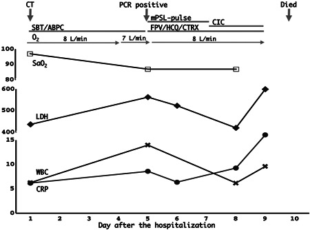
A summary of the clinical course with the essential laboratory findings is shown in Fig. 2. A notable finding was the rapid elevation of white blood cells, C‐reactive protein and lactate dehydrogenase levels 2 days before her death.
Figure 2.

A summary of fluctuations in arterial oxygen saturation (SaO2; %), white blood cell (WBC; 103/μL) count, C‐reactive protein (CRP; mg/dL) and lactate dehydrogenase (LDH; IU/L) levels. The WBC, CRP and LDH levels increased from 2 days before the death. ABPC, ampicillin; CIC, ciclesonide; CTRX, ceftriaxone; FPV, favipiravir; HCQ, hydroxychloroquine; mPSL, methylprednisolone; SBT, sulbactam.
PATHOLOGICAL FINDINGS
An autopsy was performed 13 h postmortem. The height and weight were average. No remarkable changes were observed on the body surface.
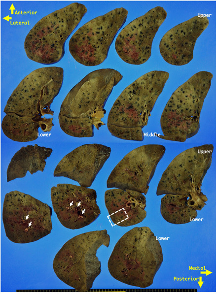
Both lungs were edematous and heavy (not measured to avoid contamination). The cut surfaces felt solid and firm, appeared partially glittering, brownish‐red in color and had viscous exudate (Fig. 3). The changes were unevenly distributed. The subpleural area on the lateral‐dorsal side felt solid and firm.
Figure 3.

Gross appearance of transverse sections from the right lung. The cut surfaces appear glittering and focally brownish‐red and are solid and firm. Adjacent to the brownish‐red areas, yellowish‐white thrombi are seen in interlobular vessels (arrows). The changes are unevenly distributed and appear fuzzy segmentally. The subpleural area on the lateral‐dorsal side appears more solid and firm. The area distant from the pleura is soft and has much more exudate. The area marked with a square is representative and has been used to present the histological appearances in Fig. 4.
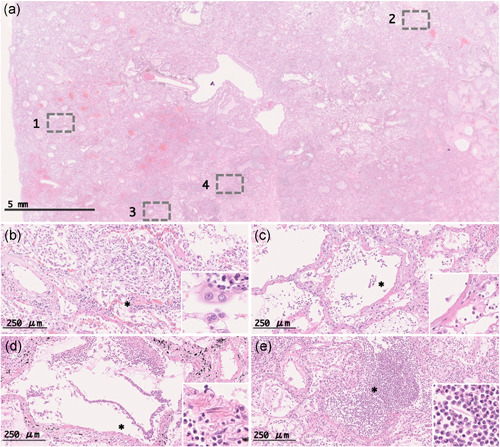
Microscopically, the subpleural area showed remarkable pathological changes (Fig. 4a), where most alveoli were covered or filled with a mixture of regenerating/desquamative pneumocytes, macrophages and fibroblasts (organizing stage) (Fig. 4b). Degenerated pneumocytes with markedly swollen eosinophilic cytoplasm were occasionally observed (Fig. 4b), which resembled SARS‐CoV‐2‐infected cells, as shown in a previous report. 6 In the area distant from the pleura, the alveolar lumina and alveolar ostia were covered with hyaline membrane (Fig. 4c), and focal alveolar hemorrhage was seen (acute stage). Thus, the findings confirmed diffuse alveolar damage (DAD), which expanded from the subpleural to the medial area. In addition, the severity and temporal phases of the DAD were segmentally different (Fig. S1), which could depend on the viral dose initially inhaled and could be due to secondary viral spreading via hematogenous or lymphatic routes during the current history.
Figure 4.

Representative microscopic appearances from the square in Fig. 3. The areas marked with squares 1, 2, 3 and 4 in the scanning view (a) have been magnified and demonstrated in the other panels (b–e). In area 1, alveoli covered or filled with a mixture of regenerating pneumocytes and fibroblasts, and degenerated pneumocytes with markedly swollen eosinophilic cytoplasm are seen (b). In area 2, alveoli covered with hyaline membrane are seen (c). In area 3, bronchiole with entirely desquamated epithelial cells and neutrophil infiltration is seen (d). In area 4, alveolar spaces filled with neutrophils containing a few fragments of keratinized materials are seen (e). In panels b–e, the areas marked with asterisks have been magnified and shown in insets.
On the other hand, the respiratory tract (trachea, bronchi and bronchioles) focally showed acute inflammation, with entirely desquamated epithelial cells and infiltration of numerous neutrophils (Fig. 4d). Additionally, acute bronchopneumonia was observed (Fig. 4e and Fig. S2), where a few fragments of keratinized material, giant cells and bacterial colonies were detected. Fibroblastic proliferation was not observed, suggesting that bronchitis, bronchiolitis and bronchopneumonia may have exacerbated shortly before her death, and likely resulted from aspiration and secondary bacterial infection, but not as a primary event caused directly by the viral infection. Acute thrombi were occasionally seen in different levels of pulmonary arteries (interlobular or more distal levels) and were restricted to the area adjacent to the bronchopneumonia and severe DAD. This suggests that these thrombi could be secondary events because of expansion of the purulent inflammation and localized circulation failure resulting from tissue destruction (Fig. S3). An obvious microthrombus indicating endothelial cell damage was not detected.
No remarkable change was observed in other organs, confirming that the cause of her death was respiratory failure mainly caused by the DAD.
DISCUSSION
The most essential pathological finding demonstrated by the autopsy was the fuzzy segmental DAD that expanded from the subpleural to the medial area, suggesting that pneumocytes are the primary target. SARS‐CoV‐2 could pass through the respiratory tract, reach alveoli, and directly and primarily infect pneumocytes. This behavior is quite different from most viruses that cause the common cold, which infect epithelial cells in the respiratory tract, and may be the reason why COVID‐19 is a serious condition.
SARS‐CoV‐2 is generally known to infect host cells through angiotensin converting enzyme 2 (ACE2) as the interface. 11 ACE2 is reportedly expressed in 15 organs, including the lungs, heart, and kidneys. 12 In fact, severe heart failure and renal failure have been reported in patients with COVID‐19. Once viremia occurs in severe cases, SARS‐CoV‐2 may affect organs other than the lungs.
Thus, we believe it is essential to treat COVID‐19 patients with potentially effective antiviral agents as early as possible in order to prevent viral replication from reaching pathogenic levels, triggering severe DAD, and subsequent viremia.
Regarding viral detection, the current PCR test that examines material swabbed from the nasopharyngeal mucosa may not be the best, as the sensitivity (0.6–0.7) seems unsatisfactory. 13 , 14 Sputum could be a more suitable choice to detect SARS‐CoV‐2.
We did not find any significant differences, which could be related to ethnicity, in the pathological findings between our case and previously reported cases. 5 , 6 , 7 , 8 , 9 , 10 However, only a small number of autopsy cases have been reported to address this issue. To understand the mechanism behind ethnic differences in the severity of COVID‐19, more autopsy cases and different research approaches are required.
In conclusion, we presented an autopsy case of COVID‐19. We hope that our findings will help in better understanding of COVID‐19 and improve therapeutic strategies.
ETHICS STATEMENT
None.
DISCLOSURE STATEMENT
None declared.
AUTHOR CONTRIBUTIONS
The contributions of each author were as follows: OK and HH wrote most of the manuscript; OK and OK designed this study and suggested the content of the manuscript; SH, HH, MN, YY and TN cared for the patient and suggested the content of the manuscript; HH, TY and MH contributed to the diagnostic process and the autopsy.
Supporting information
Additional Supporting Information may be found in the online version of this article at the publisher's website.
Supporting information.
Supporting information.
Supporting information.
Supporting information.
ACKNOWLEDGMENTS
The authors would like to thank the technicians at the Division of Pathology of Yokohama Municipal Citizen's Hospital for their assistance and Editage (www.editage.com) for English language editing.
REFERENCES
- 1. COVID‐19 , 2020. Available from: https://www.cebm.net/covid-19/global-covid-19-case-fatality-rates/. Accessed 17 May 2020.
- 2. COVID‐19 coronavirus pandemic , 2020. Available from: https://reliefweb.int/topics/covid-19. Accessed 6 May 2020.
- 3. Global Covid‐19 case fatality rates , 2020. Available from: https://www.worldometers.info/coronavirus/. Accessed 7 May 2020.
- 4. Khafaie MA, Rahim F. Cross‐country comparison of case fatality rates of COVID‐19/SARS‐COV‐2. Osong Public Health Res Perspect 2020; 11: 74–80. [DOI] [PMC free article] [PubMed] [Google Scholar]
- 5. Barton LM, Duval EJ, Stroberg E, Ghosh S, Mukhopadhyay S. COVID‐19 autopsies, Oklahoma, USA. Am J Clin Pathol 2020; 153: 725–33. [DOI] [PMC free article] [PubMed] [Google Scholar]
- 6. Fox SE, Akmatbekov A, Harbert JL, Li G, Brown JQ, Vander Heide RS. Pulmonary and cardiac pathology in Covid‐19: The first autopsy series from New Orleans. Lancet Respir Med 2020;87:681–686. [DOI] [PMC free article] [PubMed] [Google Scholar]
- 7. Karami P, Naghavi M, Feyzi A et al. Mortality of a pregnant patient diagnosed with COVID‐19: A case report with clinical, radiological, and histopathological findings. Travel Med Infect Dis 2020; 101665. [DOI] [PMC free article] [PubMed] [Google Scholar]
- 8. Konopka KE, Wilson A, Myers JL. Postmortem lung findings in an asthmatic with coronavirus disease 2019 (COVID‐19). Chest 2020. [DOI] [PMC free article] [PubMed] [Google Scholar]
- 9. Menter T, Haslbauer JD, Nienhold R et al. Post‐mortem examination of COVID19 patients reveals diffuse alveolar damage with severe capillary congestion and variegated findings of lungs and other organs suggesting vascular dysfunction. Histopathology 2020. [DOI] [PMC free article] [PubMed] [Google Scholar]
- 10. Wichmann D, Sperhake JP, Lütgehetmann M et al. Autopsy findings and venous thromboembolism in patients with COVID‐19: A prospective cohort study. Ann Intern Med 2020. [DOI] [PMC free article] [PubMed] [Google Scholar]
- 11. Vaduganathan M, Vardeny O, Michel T, McMurray JJV, Pfeffer MA, Solomon SD. Renin‐angiotensinaldosterone system inhibitors in patients with Covid‐19. N Engl J Med 2020; 382: 1653–59. [DOI] [PMC free article] [PubMed] [Google Scholar]
- 12. Hamming I, Timens W, Bulthuis ML, Lely AT, Navis G, van Goor H. Tissue distribution of ACE2 protein, the functional receptor for SARS coronavirus. A first step in understanding SARS pathogenesis. J Pathol 2004; 203: 631–37. [DOI] [PMC free article] [PubMed] [Google Scholar]
- 13. Fang Y, Zhang H, Xie J et al. Sensitivity of chest CT for COVID‐19: Comparison to RT‐PCR. Radiology 2020; 296: 200432. [DOI] [PMC free article] [PubMed] [Google Scholar]
- 14. Ai T, Yang Z, Hou H et al. Correlation of chest CT and RT‐PCR testing in coronavirus disease 2019 (COVID‐19) in China: A report of 1014 cases. Radiology 2020; 296: 200642. [DOI] [PMC free article] [PubMed] [Google Scholar]
Associated Data
This section collects any data citations, data availability statements, or supplementary materials included in this article.
Supplementary Materials
Additional Supporting Information may be found in the online version of this article at the publisher's website.
Supporting information.
Supporting information.
Supporting information.
Supporting information.


