

1. INTRODUCTION
In late 2019, a new virus called severe acute respiratory syndrome coronavirus 2 (SARS‐CoV‐2) led to many cases of severe respiratory tract infection in China. This virus was belonged to the Coronaviridae family and caused a wide range of symptoms and also death in patients. Although surveys confirmed that children could be infected with SARS‐CoV‐2, 1 , 2 , 3 there is evidence showing people more than 50 years old are more susceptible to the coronavirus disease 2019 (COVID‐19). In some recently published papers, researchers have reported the coinfection of SARS‐CoV‐2 with other viruses such as influenza. 4 In this study, we found SARS‐CoV‐2 in coinfection with human metapneumovirus (hMPV) in three SARS‐CoV‐2‐infected dead children.
2. CASE PRESENTATION
Blood tests, including blood cell differential count, erythrocyte sedimentation rate (ESR), and C‐reactive protein (CRP), were performed for all patients admitted to the hospital with suspicion of infection with SARS‐CoV‐2. Besides, all patients underwent a chest computed tomography (CT) scan that is the most sensitive test for COVID‐19 identification, 5 , 6 and SARS‐COV‐2 detection was performed using real‐time polymerase chain reaction (PCR). Finally, the detection of six respiratory viruses was done using PCR and reverse transcriptase (RT)‐PCR method on SARS‐CoV‐2 positive samples. We found three cases of coinfection with hMPV in children (Table 1).
Table 1.
The prevalence of viruses in various age range and sex of SARS‐CoV‐2 positive dead patients
| Virus | Type | Subtype | Age range (number/percent) | Sex (number/percent) | Total | ||||
|---|---|---|---|---|---|---|---|---|---|
| 0‐14 | 14‐40 | 40‐60 | >60 | Male | Female | ||||
| Human metapneumovirus | 3 (100%) | 0 | 0 | 0 | 2 (66.6%) | 1 (33.3%) | 3 | ||
| Human bocavirus | 0 | 1 (16.7%) | 3 (50%) | 2 (33.3%) | 2 (33.3%) | 4 (66.7%) | 6 | ||
| Adenovirus | 0 | 0 | 1 (50%) | 1 (50%) | 0 | 2 (100%) | 2 | ||
| Parainfluenza virus | 0 | 0 | 0 | 2 (100%) | 1 (50%) | 1 (50%) | 2 | ||
| Respiratory syncytial virus | 0 | 1 (16.7%) | 1 (16.7%) | 4 (66.7%) | 3 (50%) | 3 (50%) | 6 | ||
| Influenza virus | A | H1N1 | 0 | 0 | 3 (18.7%) | 13 (81.3%) | 11 (68.7%) | 5 (31.3%) | 16 |
| Non‐H1N1 | 0 | 0 | 1 (25%) | 3 (75%) | 2 (50%) | 2 (50%) | 4 | ||
| B | 0 | 0 | 0 | 0 | 0 | 0 | 0 | ||
Abbreviation: SARS‐CoV‐2, severe acute respiratory syndrome coronavirus 2.
2.1. Case 1
The 13‐month‐old toddler with a history of mild asthma, admitted to the hospital with chief complaints of cough, fever (38.7°C), and malaise. Laboratory blood tests showed a total white blood cell (WBC) 6 × 10³/L, lymphopenia, CRP 4+, and ESR 19 mm/h. She was hospitalized under ventilation on 20 Mach 2020 and died on the same day. The primary SARS‐CoV‐2 RT‐PCR test on her nasopharyngeal swab was positive. The chest CT scan revealed diffuse bilateral ground‐glass opacities with acute respiratory distress syndrome patterns (Figure 1). Further detection of other respiratory viruses demonstrated that its sample was positive for hMPV and negative for other viruses.
Figure 1.
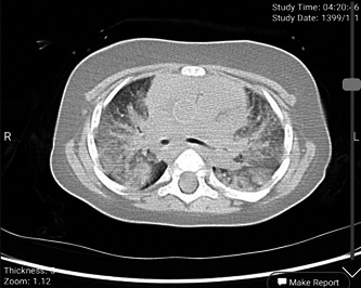
The chest computed tomography scan of case 1
2.2. Case 2
The second case was a 6‐year‐old child with symptoms of cough, 39°C fever, malaise, and diarrhea. Her blood count showed a total white cell count of 45.3 × 10³ (neutrophil 35.9%, lymphocyte 52.7%, and monocyte 11.4%). She had CRP test 3+ positive and ESR 9 mm/h with a high platelet count (493 × 103). The chest CT scan report emphasized bilateral ground‐glass opacity, but the image is not available. He died 2 days after hospitalization in the intensive care unit (ICU) under ventilation. Her nasopharyngeal swab test was positive for both SARS‐CoV‐2 and hMPV, while negative for other viruses.
2.3. Case 3
The third case was a 6‐year‐old child with cough, fever (38.1°C), and malaise as the chief complaints. He had a history of asthma. Laboratory blood tests showed total WBC 6.5 × 10³ (neutrophil 93%, lymphocyte 5%, and monocyte 2%) with the CRP 4+ positive and ESR 33 mm/h. The chest CT scan report confirmed COVID‐19, but the image is not available. He was hospitalized on 22 April 2020 and died 1 day after hospitalization in the ICU under mechanical ventilation. His sample was positive for both hMPV and SARS‐CoV‐2 and negative for other evaluated viruses.
3. DISCUSSION
In this study, we worked on samples of all 74 SARS‐CoV‐2 positive dead patients in North Khorasan Province, North‐Eastern Iran. We found the influenza virus, human bocavirus, respiratory syncytial virus, parainfluenza virus, and hMPV in some SARS‐CoV‐2 positive samples. Our exciting finding is that the hMPV merely found in children. It should be noted that between 74 dead patients, only three of them were children, and all of these children had SARS‐CoV‐2 and hMPV simultaneously. None of the adult patients' samples were positive for hMPV. In some papers, researchers hypothesized the role of chronic respiratory tract inflammation and asthma as a predisposing factor for other respiratory infections. 7 hMPV can affect the respiratory tract susceptibility to SARS‐CoV‐2 infection directly and indirectly. 8 hMPV infection can directly lead to inflammation and change of interferon secretion patterns in the respiratory tract of patients. 9 Interferons themselves increase the expression of angiotensin‐converting enzyme 2 receptors which is a SARS‐CoV‐2 receptor on respiratory epithelial cells. 10 , 11 , 12 The indirect role of hMPV may be through causing secondary diseases such as asthma that finally results in altering the secretion pattern of interferons. The relationship between hMPV infection and asthma has been proved in many articles. 13 , 14 , 15 , 16 As mentioned in the case presentation, two of our three cases had a history of asthma. Our findings are not enough, and further studies are needed to understand the correlation between the hMPV infection and increasing chance of infection and death due to the SARS‐CoV‐2 but detecting both SARS‐CoV‐2 and hMPV in dead children is an interesting finding and motivates us to evaluate the correlation between hMPV infection and susceptibility to SARS‐CoV‐2.
4. CONCLUSION
It would be beneficial to check SARS‐CoV‐2 positive children for other viral and bacterial pathogens since the identification of possible repetitious coinfections may lead to the change of diagnostic and therapeutic protocols.
CONFLICT OF INTERESTS
The authors declare that there are no conflict of interests.
AUTHOR CONTRIBUTIONS
SAH: Conceptualized and designed the study, coordinated and supervised data collection, and carried out the initial analyses. SS: Collected laboratory data, carried out the PCR and RT real‐time PCR tests, and edited and revised the final manuscript. HGZ‐M: Collected laboratory data, and carried out the PCR and RT real‐time PCR tests. MG: Participated in data collection and carried out the initial analyses. M‐sM‐H: Performed CT scan, data collection, and interpretation. HN‐A and HGZ‐M: Collected laboratory data and carried out the initial analyses. AA: Conceptualized and designed the study, carried out the PCR and RT real‐time PCR tests, drafted the initial manuscript, and revised the manuscript. All authors approved the final manuscript as submitted and agree to be accountable for all aspects of the study.
ACKNOWLEDGMENTS
This study is a part of the national COVID‐19 screening program of Iran. The primer and probes were supported by the Ministry of Health of Iran and other laboratory kits provided by the North Khorasan University of Medical Sciences (Grant Number 990002).
Hashemi S‐A, Safamanesh S, Ghasemzadeh‐Moghaddam H, et al. Report of death in children with SARS‐CoV‐2 and human metapneumovirus (hMPV) coinfection: Is hMPV the trigger? J Med Virol. 2021;93:579:579–581. 10.1002/jmv.26401
REFERENCES
- 1. Cao Q, Chen Y‐C, Chen C‐L, Chiu C‐H. SARS‐CoV‐2 infection in children: transmission dynamics and clinical characteristics. J Formos Med Assoc. 2020;119(3):670‐673. [DOI] [PMC free article] [PubMed] [Google Scholar]
- 2. Lee P‐I, Hu Y‐L, Chen P‐Y, Huang Y‐C, Hsueh P‐R. Are children less susceptible to COVID‐19? J Microbiol Immunol Infect. 2020;53:371‐372. [DOI] [PMC free article] [PubMed] [Google Scholar]
- 3. Liu W, Zhang Q, Chen J, et al. Detection of Covid‐19 in children in early January 2020 in Wuhan, China. N Engl J Med. 2020;382(14):1370‐1371. [DOI] [PMC free article] [PubMed] [Google Scholar]
- 4. Touzard‐romo F, Tapé C, Lonks JR. Co‐infection with SARS‐CoV‐2 and Human metapneumovirus. R I Med J. 2020;103:75‐76. [PubMed] [Google Scholar]
- 5. Ai T, Yang Z, Hou H, et al. Correlation of chest CT and RT‐PCR testing in coronavirus disease 2019 (COVID‐19) in China: a report of 1014 cases. Radiology. 2020;296:E32‐E40. [DOI] [PMC free article] [PubMed] [Google Scholar]
- 6. Fang Y, Zhang H, Xie J, et al. Sensitivity of chest CT for COVID‐19: comparison to RT‐PCR. Radiology. 2020;296:E115‐E117. [DOI] [PMC free article] [PubMed] [Google Scholar]
- 7. Céspedes PF, Palavecino CE, Kalergis AM, Bueno SM. Modulation of host immunity by the human metapneumovirus. Clin Microbiol Rev. 2016;29(4):795‐818. [DOI] [PMC free article] [PubMed] [Google Scholar]
- 8. Catanzaro M, Fagiani F, Racchi M, Corsini E, Govoni S, Lanni C. Immune response in COVID‐19: addressing a pharmacological challenge by targeting pathways triggered by SARS‐CoV‐2. Signal Transduct Target Ther. 2020;5:84. [DOI] [PMC free article] [PubMed] [Google Scholar]
- 9. Hossain FMA, Choi JY, Uyangaa E, Park SO, Eo SK. The Interplay between Host Immunity and Respiratory Viral Infection in asthma Exacerbation. Immune Netw. 2019;19:e31. [DOI] [PMC free article] [PubMed] [Google Scholar]
- 10. Ziegler CGK, Allon SJ, Nyquist SK, et al. SARS‐CoV‐2 receptor ACE2 is an interferon‐stimulated gene in human airway epithelial cells and is detected in specific cell subsets across tissues. Cell. 2020;181:1016‐1035.e19. [DOI] [PMC free article] [PubMed] [Google Scholar]
- 11. Bai TR, Knight DA. Structural changes in the airways in asthma: observations and consequences. Clin Sci. 2005;108(6):463‐477. [DOI] [PubMed] [Google Scholar]
- 12. Camiolo MJ, Gauthier M, Kaminski N, Ray A, Wenzel SE. Expression of SARS‐CoV‐2 receptor ACE2 and coincident host response signature varies by asthma inflammatory phenotype. J Allergy Clin Immunol. 2020;146:315‐324.e7. [DOI] [PMC free article] [PubMed] [Google Scholar]
- 13. Williams JV, Crowe JE Jr., Enriquez R, et al. Human metapneumovirus infection plays an etiologic role in acute asthma exacerbations requiring hospitalization in adults. J Infect Dis. 2005;192(7):1149‐1153. [DOI] [PMC free article] [PubMed] [Google Scholar]
- 14. García‐García ML, Calvo C, Casas I, et al. Human metapneumovirus bronchiolitis in infancy is an important risk factor for asthma at age 5. Pediatr Pulmonol. 2007;42(5):458‐464. [DOI] [PubMed] [Google Scholar]
- 15. Carroll KN, Wu P, Gebretsadik T, et al. The severity‐dependent relationship of infant bronchiolitis on the risk and morbidity of early childhood asthma. J Allergy Clin Immunol. 2009;123(5):1055‐1061.e1. [DOI] [PMC free article] [PubMed] [Google Scholar]
- 16. Carroll KN, Hartert TV. The impact of respiratory viral infection on wheezing illnesses and asthma exacerbations. Immunol Allergy Clin North Am. 2008;28(3):539‐561. [DOI] [PMC free article] [PubMed] [Google Scholar]