Abstract
Objective
The recent hydroxychloroquine (HCQ) shortage due to use in coronavirus disease 2019 (COVID‐19) has forced some rheumatic disease patients to choose between continuing their current dose of HCQ but exhaust their supply early or ration it in order to prolong its use. Blood HCQ concentrations are directly correlated with disease activity in rheumatic diseases such as systemic lupus erythematosus. We sought to model how changes in HCQ dosage will best maintain sufficient blood HCQ concentrations for the longest period of time in order to avoid potential future flares.
Methods
A one‐compartment pharmacokinetic model was used to predict mean blood HCQ concentrations. Monte Carlo simulations with 10‐fold inflated model parameter variance was utilized to assess the impact of variability.
Results
Maintenance of 400 mg/d resulted in mean therapeutic whole‐blood HCQ concentrations that exceeded 700 ng/ml for 10.5 days, whereas HCQ rationing by reducing the dose by half resulted in the mean concentration remaining above 700 ng/ml for 2.4 days (net gain = 8 days). Variability analysis demonstrates that results may differ at the individual level, dependent on baseline blood HCQ concentrations.
Conclusion
Although mean blood concentrations exceed 700 ng/ml for a longer time if patients maintain their full dose of HCQ, more information is needed to fully understand the elimination of HCQ at the patient level, particularly the contribution of tissue stores of HCQ transiting back into the blood.
INTRODUCTION
During the current coronavirus disease 2019 (COVID‐19) pandemic, studies have suggested a potential role of hydroxychloroquine (HCQ) in the treatment of COVID‐19 amid substantial controversy (1, 2). Intense interest of the drug has resulted in an immediate shortage of HCQ, putting patients on chronic HCQ treatment, such as those with systemic lupus erythematosus (SLE), rheumatoid arthritis (RA), and other rheumatic conditions, at increased risk of flares that may result in morbidity and mortality.
These patients, particularly those with SLE, depend on steady access to HCQ for disease stability, which is at least partly achieved by maintaining therapeutic HCQ drug concentrations. In a placebo‐controlled randomized clinical trial, discontinuation of HCQ in stable SLE patients increased the risk for flares, including severe ones, as early as 2 weeks after HCQ withdrawal (3). In an observational study, SLE patients with active disease (using a cutoff of SLE Disease Activity Index [SLEDAI] ≥ 6) had a mean whole‐blood HCQ concentration of 694 ng/ml versus SLE patients with inactive disease (SLEDAI < 6) who had a mean blood HCQ concentration of 1079 ng/ml (4). Thus, any threat to the supply of HCQ poses a risk of flare to patients with SLE and other rheumatic disease, which may result in morbidity and preventable hospitalizations at a time when some hospitals are already close to capacity due to COVID‐19.
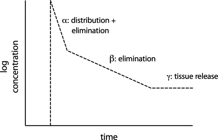
The terminal half‐life of HCQ, including tissue release, is approximately 40 days (5, 6). Thus, it is simple to assume that elimination of HCQ with uniform first‐order kinetics due to a one‐compartment model will yield a clear answer regarding the elimination of HCQ from the blood after cessation of drug. However, this assumes that HCQ is cleared at this rate from the blood; it does not take into account the dynamics of HCQ biodistribution. It is recognized that lysosome‐rich tissues retain a substantial amount of HCQ (7) and that the rapid elimination of HCQ in the blood yields a central compartment half‐life of ~40 hours in patients with RA (8). This creates a triphasic curve in the patient who is HCQ‐naïve, wherein both tissue and cellular absorption as well as renal excretion dominate the early kinetics of HCQ disappearance from the blood (ie, the first two phases) (Figure 1). Release of HCQ from various tissues back into the blood is the third phase. The prolonged half‐life that is frequently cited (5), ie, 40 days, is the summation of all of the elimination phases, including the very prolonged tissue release phase. Thus, there are separate kinetics for blood elimination and cellular/tissue elimination. At present, efficacy for rheumatologic diseases has been correlated to blood concentrations in patients that have presumably reached steady‐state concentrations of HCQ in both the blood and periphery (ie, tissues and cells). It is unknown which compartment HCQ concentrations directly link to efficacy, but blood is used as the present surrogate.
Figure 1.

Triphasic pharmacokinetics curve in a hydroxychloroquine‐naive patient.
Clinicians and patients are currently being confronted with strategies on how to manage HCQ dosing in the face of a massive HCQ shortage during the COVID‐19 pandemic.
With these caveats, we sought to model the blood concentrations of HCQ in the situation where patients continue a stable dose of HCQ (eg, 400 mg/d) but are forced to discontinue early or ration their supply by taking half the dose (200 mg daily or every other day alternating with 400 mg/d), thereby extending the number of days they are on HCQ but perhaps at risk of being subtherapeutic.
METHODS
We estimated the time it would take for SLE patients facing abrupt HCQ supply shortages to be at risk of flare due to subtherapeutic levels of HCQ. Mass transit of HCQ was modeled according to a scenario in which SLE patients taking HCQ 400 mg/d at baseline only had twenty 200‐mg tablets remaining due to HCQ shortages (day 0). The patient/provider had three options related to HCQ use: 1) continue taking the standard dose (400 mg/d) for 10 days, 2) ration their HCQ by alternating 200 mg and 400 mg every other day for 14 days, or 3) ration HCQ by taking 200 mg/d for 20 days, and then stop. We assumed a subtherapeutic threshold (below which the risk of flare increases) of a whole‐blood HCQ concentration at 700 ng/ml (and not return above 700 ng/ml within 24 hours) as a surrogate for potential increased disease activity.
To first demonstrate how traditional pharmacokinetic equations may be utilized to answer the question, we used the reported HCQ half‐life of 40 days and the elimination rate constant (K) = 0.693/(HCQ half‐life in hours), equaling 7.22*10−4. To calculate the number of hours needed to drop below 700 ng/ml, one can solve for time with the following equation:
For full pharmacokinetic modeling and simulation, we also employed the one‐compartment model published by Carmichael et al (8) based on patients with RA where the blood half‐life is ~40 hours. In brief, the mean parameter values for clearance, volume absorption constant, time lag, and bioavailability were employed (ie, identical structural model and inputs). Modeling was performed based on a 400 mg/d dose of HCQ sulfate (ie. 310 mg of HCQ base) given until steady‐state conditions were reached, then three scenarios for patient dosing were considered. Simulations were conducted with the Pmetrics for R as previously reported (9). Concentrations were simulated every 2 hours until concentrations less than 30 ng/ml were reached. Monte Carlo simulations (n = 1000) were performed for each scenario, and parameter variance and 10‐fold inflated model parameter variance were employed in order to assess the impact of variability. The number of individual simulations that resulted in concentrations exceeding 700 ng/ml were calculated for 24‐hour intervals (ie, concentration for the individual did or did not exceed 700 ng/ml in the 24‐hour period) and were reported as a fraction of total simulations performed.
RESULTS
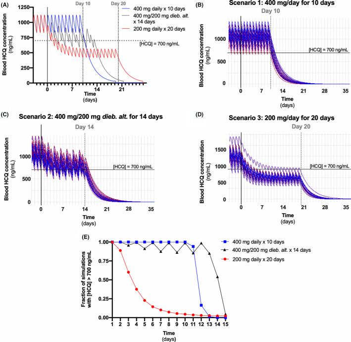
Using traditional first‐order pharmacokinetic equations and a HCQ half‐life of 40 days, concentrations were predicted to drop below 700 ng/ml in 17.8 and 36.0 days for baseline concentrations of 867 ng/ml (the low median end observed in Jallouli et al (10)) and 1079 ng/ml (the mean concentration observed in patients with inactive SLE (4)), respectively. This straightforward kinetics model is an oversimplification given the known rapid elimination of HCQ in blood. We then considered a model in which the rapid blood elimination of HCQ (half‐life of 43 hours (8)) exclusively drove the decrease in blood HCQ concentrations. This model makes the assumption that tissue release of HCQ is slow and only negligibly contributes to blood HCQ concentrations (thus the redistribution phase is not mathematically captured). Using the structural model and parameters determined by Carmichael et al (8), a baseline dose of 400 mg/d of HCQ sulfate (ie, 310 mg HCQ base) at steady state resulted in mean peak concentrations of ~1124 ng/ml and trough concentrations of ~829 ng/ml (Figure 2A, four days of steady state shown for visual appreciation of baseline). In the first model of the patient who continued to take 400 mg/d for 10 days (option 1), mean concentrations were below 700 ng/ml in 10.5 days versus 2.4 days if the dosing changed to 200 mg/d of HCQ sulfate for 20 days (option 3). In option 2 (alternating 200 mg and 400 mg doses every other day for 14 days), the mean concentrations consistently dropped below 700 ng/ml at 14 days. The peaks and troughs decrease with each cycle, to the point where mean concentrations are consistently below 700 ng/ml at day 13.3. Therefore, of the 20 days the patients were taking HCQ in option 2, they spent most of the time with concentrations of 700 ng/ml for 13 days, whereas in option 3, in only the first two days did they have a mean concentration greater than 700 mg/ml.
Figure 2.

Anticipated hydroxychloroquine (HCQ) concentrations for options 1, 2, and 3. Monte Carlo simulations using parameters identified in patients with rheumatoid arthritis (8) and a blood HCQ half‐life of 43 hours were performed for both scenarios. A, Mean blood HCQ concentrations for 400 mg/d for 10 days (blue), alternating 400 and 200 mg every other day for 14 days (grey), and 200 mg/d for 20 days (red). Ten‐fold inflated model parameter variance simulations for 400 mg/d for 10 days (B), alternating 400 and 200 mg every other day for 14 days (C), and 200 mg/d for 20 days (D). For visual clarity, 30 simulations are shown in panels B, C, and D, but the data in the text are from 1000 simulations. E, Fraction of the 1000 simulations wherein blood HCQ concentration greater than 700 ng/ml for each day for each scenario. dieb. alt. = deibus alternis (every other day).
To estimate what would happen with varying baseline blood HCQ concentrations, 10‐fold variance from the reported model was considered. The mean prediction is that the majority of patients would stay above 700 ng/ml longer in option 1 (400 mg/d for 10 days) (Figure 2B) compared with option 2 (alternating 400 and 200 mg doses every other day for 14 days) (Figure 2C) and option 3 (200 mg/d for 20 days) (Figure 2D). In this model that incorporates parameter variance (and not covariance), the main factor that drives subtherapeutic concentrations (ie, <700 ng/ml for over 24 hours) is the starting HCQ concentration in the blood. The model estimated the high (95th percentile) and low (5th percentile) baseline HCQ blood concentrations when taking 400 mg/d prior to day 0 (Supplementary Figure 1). When we compared the high and low baseline HCQ concentrations, there was a difference of 1.5 days in option 1 (low = 10 days, high = 11.5 days), 2.5 days in option 2 (low = 11.5 days, high = 14 days), and 7 days in option 3 (low = 0.5 days, high = 7.5 days) prior to consistently reach concentration lower than 700 ng/ml. If a patient has high HCQ blood concentrations at baseline, option 2 provides an extension of HCQ concentrations greater than 700 ng/ml (14 days) compared with option 1 (11.5 days) and option 3 (7.5 days). If a patient has low baseline concentrations, options 1 and 2 (11.5 and 14 days, respectively) clearly maintain a longer period of time with HCQ concentrations greater than 700 ng/ml than option 3 (0.5 days).
Similarly, when we examined what fraction of the simulations for each option were able to maintain a blood HCQ concentration greater than 700 ng/ml each day, options 1 and 2 maintained a substantially greater proportion of simulations over a longer period of time compared with option 3 (Figure 2E). Using a 0% target attainment, the number of days it took for option 1 to drop below 90% was 12 days versus 2 days for option 3. In option 2, it first dropped below 90% on day 7, then fluctuated above and below 90% until day 13.
HCQ concentrations dropped to near or below 700 ng/ml almost immediately following HCQ reduction or discontinuation in all models. Additionally, these models predicted that blood HCQ levels would be undetectable within 10 days after HCQ discontinuation.
Collectively, these data demonstrate how both HCQ dosing and baseline HCQ blood concentrations have an impact when subtherapeutic blood HCQ concentrations are reached.
DISCUSSION
We simulated trajectories of the elimination of whole‐blood HCQ levels related to three strategies of HCQ dose management applicable to a mass shortage of the drug: 1) continued treatment at full prescribed dose with early discontinuation, 2) alternating treatment at full prescribed doses with half doses, or 3) treatment at half dose with delayed discontinuation. We found that the baseline steady‐state blood HCQ concentration plays an important role in the time needed to fall below our surrogate concentration for increased flare, ie, 700 ng/ml. For those with higher baseline blood HCQ concentrations, the strategy of halving the dose (ie, 200 mg/d) to prolong duration of treatment was associated with an up to a seven‐day delay before falling below the presumed therapeutic threshold of HCQ. For those with lower baseline blood HCQ concentrations, the strategy of maintaining a full dose (i.e. 400 mg/d) was associated with up to a nine‐day benefit (compared with the 200 mg/d strategy). Understanding the strategy that maximizes the number of therapeutic days is important to provide longer population‐level disease stability for patients dependent on HCQ for rheumatic diseases and to allow for extra time for HCQ supply chains to be increased in the face of a mass drug shortage, such as with the current COVID‐19 pandemic. In addition, our simulations suggest that starting blood HCQ concentrations play an important role in the maintenance of an appropriate blood HCQ concentration. Importantly, no single strategy simulated herein was universally superior to its alternatives but needs to be considered in the context of baseline blood HCQ concentrations at steady state. This highlights the utility of therapeutic HCQ blood level monitoring in tailoring treatment strategies to the individual patient with rheumatic disease. The models also highlight differences between when certain blood HCQ concentration thresholds are met versus having any blood‐detectable HCQ.
Although we provide population‐level estimates of the possible trajectories of HCQ dosing strategies during a mass HCQ shortage due to the COVID‐19 pandemic, there are some limitations, particularly as they relate to individual‐level interpretation. First, these are mean estimations (and simulations based on 10‐fold variance) from a one‐compartment model derived from a target population (RA) (8). Our modeling does not take all interpatient variability into account, including differences in HCQ distribution and clearance. Second, all modeling was performed for HCQ in whole blood. Because little is known about the pharmacokinetics of HCQ in the periphery (tissue and cells), as well as the pharmacodynamic drivers of efficacy in the periphery, it was not possible to model the peripheral interface of drug concentrations and activity. For example, our model suggests undetectable blood HCQ concentrations within days of drug discontinuation, which seems unlikely. This suggests an important but unknown contribution of tissue HCQ stores to the blood compartment. Third, the assumptions of the models relied on HCQ thresholds related to typical and subtherapeutic blood levels in SLE derived from a relatively small sample at a single center (4). The particular HCQ level important to maintain disease stability likely varies substantially between individual patients due to heterogeneous SLE manifestations/severity, sex, age, weight, race, genetic polymorphisms, concomitant use of other immunosuppressants, among other possibilities. For example, patients with SLE with rash and inflammatory arthritis may be particularly reliant on HCQ for these manifestations due to known efficacy for these indications. Other SLE manifestations may be less strongly linked to acute changes in blood levels of HCQ. Many patients with SLE may already have subtherapeutic HCQ blood levels because of the known high levels of nonadherence in this patient population (11). Pharmacodynamic variability likely exists. Some patients may be exquisitely sensitive to even a few missed or lower doses of HCQ, whereas others may maintain clinical stability without the short‐term (or perhaps even long‐term) need for HCQ. However, it is currently not possible to determine the degree to which individual patients may be dependent on HCQ or to identify patients that could be amenable for a temporary drug holiday or even permanent cessation. Finally, there is likely variability in pharmacokinetics in the periphery (tissue, cells) that contributes to an unknown effect on pharmacodynamics.
Given these uncertainties, clinicians often rely on population‐level data to make individual treatment recommendations. Results from a previous clinical trial makes it clear that, on a population level, abrupt discontinuation of HCQ in patients with quiescent SLE increases the risk for flares, some of which can be severe, in as early as two weeks, increasing over time (3). Many patients with rheumatic disease take HCQ for other indications, such as RA, discoid lupus, dermatomyositis, and other forms of inflammatory arthritis and skin disease. Based on our modeling, there is a subset of patients that benefit from rationing HCQ to extend the duration of taking it, but this depends on the patient’s baseline blood HCQ concentration. Unfortunately, availability of blood HCQ concentration testing is not currently available or is financially infeasible for the vast majority of the world (including most of Latin and South America, Asia, Australia and New Zealand, Africa, Europe, Canada, and Mexico), limiting the usefulness of this approach. Nevertheless, it is certain that our understanding of the pharmacokinetics and pharmacodynamics of HCQ is insufficient and will require additional work especially at the tissue/cellular level. How peripheral tissue compartments influence blood HCQ concentrations or whether the peripheral compartments even drive clinical efficacy remain important unanswered questions.
Author Contributions
All authors contributed to the drafting and revision of the manuscript, and they provided final approval for publication.
Study conception and design
Scheetz, Konig, Robinson, Sparks, Kim.
Acquisition of data
Scheetz.
Analysis and interpretation of data
Scheetz, Konig, Robinson, Sparks, Kim.
Supporting information
Fig S1
The content is solely the responsibility of the authors and does not necessarily represent the official views of the National Institutes of Health.
The authors have no disclosures to report where potential benefit (including finanacial) or harm due to publication of this manuscript. MHS outside of this work is supported by NIH National Institute of Allergy and Infectious Disease (NIAID) (R21‐AI‐149026, K24‐AI‐104831, R01‐AI‐119446), Cystic Fibrosis Foundation, Nevakar, Allecra, and Merck (less than $10,000 each). PCR reports no competing interests related to this work, outside of this work he reports personal consulting fees from Abbvie, Eli Lilly, Janssen, Novartis, Pfizer and UCB (less than $10,000 each) and nonfinancial support from BMS and Roche. MFK outside of this work is supported by NIH National Institute of Arthritis and Musculoskeletal and Skin Diseases (NIAMS) T32‐AR‐048522 and reports personal fees unrelated to this work from Bristol‐Myers Squibb and Celltrion (less than $10,000 each). JAS reports outside of this work is supported by NIH NIAMS/NIAID/Autoimmune Centers of Excellence, the Rheumatology Research Foundation, the Brigham Research Institute, and the R. Bruce and Joan M. Mickey Research Scholar Fund as well as personal fees unrelated to this work from Bristol‐Myers Squibb, Gilead, Inova, Janssen, and Optum (less than $10,000 each). AHJK reports outside of this work is supported by NIH NIAMS P30‐AR‐073752, Rheumatology Research Foundation, and GlaxoSmithKline, along with personal fees from Exagen Diagnostics, Inc. and GlaxoSmithKline (less than $10,000 each).
Contributor Information
Marc H. Scheetz, Email: mschee@midwestern.edu.
Alfred H.J. Kim, Email: akim@wustl.edu.
References
- 1. Kim AH, Sparks JA, Liew JW, Putman MS, Berenbaum F, Duarte‐García A, et al. A rush to judgment? Rapid reporting and dissemination of results and its consequences regarding the use of hydroxychloroquine for COVID‐19 [corrected and republished in Ann Intern Med 2020;172:819–21]. Ann Intern Med 2020. [DOI] [PMC free article] [PubMed] [Google Scholar]
- 2. Yazdany J, Kim AH. Use of hydroxychloroquine and chloroquine during the COVID‐19 pandemic: what every clinician should know. Ann Intern Med 2020;172:754–5. [DOI] [PMC free article] [PubMed] [Google Scholar]
- 3. Canadian Hydroxychloroquine Study Group . A randomized study of the effect of withdrawing hydroxychloroquine sulfate in systemic lupus erythematosus. N Engl J Med 1991;324:150–4. [DOI] [PubMed] [Google Scholar]
- 4. Costedoat‐Chalumeau N, Amoura Z, Hulot JS, Hammoud HA, Aymard G, Cacoub P, et al. Low blood concentration of hydroxychloroquine is a marker for and predictor of disease exacerbations in patients with systemic lupus erythematosus. Arthritis Rheum 2006;54:3284–90. [DOI] [PubMed] [Google Scholar]
- 5. Schrezenmeier E, Dörner T. Mechanisms of action of hydroxychloroquine and chloroquine: implications for rheumatology. Nat Rev Rheumatol 2020;16:155–66. [DOI] [PubMed] [Google Scholar]
- 6. Furst DE. Pharmacokinetics of hydroxychloroquine and chloroquine during treatment of rheumatic diseases. Lupus 1996;Suppl 1:S11–5. [PubMed] [Google Scholar]
- 7. Tett SE, Cutler DJ, Day RO, Brown KF. Bioavailability of hydroxychloroquine tablets in healthy volunteers. Br J Clin Pharmacol 1989;27:771–9. [DOI] [PMC free article] [PubMed] [Google Scholar]
- 8. Carmichael SJ, Charles B, Tett SE. Population pharmacokinetics of hydroxychloroquine in patients with rheumatoid arthritis. Ther Drug Monit 2003;25:671–81. [DOI] [PubMed] [Google Scholar]
- 9. Miglis C, Rhodes NJ, Avedissian SN, Kubin CJ, Yin MT, Nelson BC, et al. Population pharmacokinetics of polymyxin B in acutely ill adult patients. Antimicrob Agents Chemother 2018;62:e01475–17. [DOI] [PMC free article] [PubMed] [Google Scholar]
- 10. Jallouli M, Galicier L, Zahr N, Aumaître O, Francès C, Le Guern V, et al. Determinants of hydroxychloroquine blood concentration variations in systemic lupus erythematosus. Arthritis Rheumatol 2015;67:2176–84. [DOI] [PubMed] [Google Scholar]
- 11. Feldman CH, Collins J, Zhang Z, Subramanian SV, Solomon DH, Kawachi I, et al. Dynamic patterns and predictors of hydroxychloroquine nonadherence among Medicaid beneficiaries with systemic lupus erythematosus. Semin Arthritis Rheum 2018;48:205–13. [DOI] [PMC free article] [PubMed] [Google Scholar]
Associated Data
This section collects any data citations, data availability statements, or supplementary materials included in this article.
Supplementary Materials
Fig S1


