Supplemental Digital Content is available in the text.
Keywords: coronavirus, mechanical ventilation, pulmonary embolism, venous thromboembolism
Objectives:
To assess the role of thromboprophylaxis regimens on the occurrence of pulmonary embolism in coronavirus disease 2019 patients.
Design:
Retrospective analysis of prospectively collected data on coronavirus disease 2019 patients, included between March 10, and April 30, 2020.
Setting:
ICU of an University Hospital in Belgium.
Patients and Interventions:
Critically ill adult mechanically ventilated coronavirus disease 2019 patients were eligible if they underwent a CT pulmonary angiography, as part of the routine management in case of persistent hypoxemia or respiratory deterioration. The primary endpoint of this study was the occurrence of pulmonary embolism according to the use of standard thromboprophylaxis (i.e. subcutaneous enoxaparin 4,000 international units once daily) or high regimen thromboprophylaxis (i.e. subcutaneous enoxaparin 4,000 international units bid or therapeutic unfractioned heparin).
Measurements and Main Results:
Of 49 mechanically ventilated coronavirus disease 2019, 40 underwent CT pulmonary angiography after a median of 7 days (4–8 d) since ICU admission and 12 days (9–16 d) days since the onset of symptoms. Thirteen patients (33%) were diagnosed of pulmonary embolism, which was bilateral in six patients and localized in the right lung in seven patients. D-dimers on the day of CT pulmonary angiography had a predictive accuracy of 0.90 (95% CIs: 0.78–1.00) for pulmonary embolism. The use of high-regimen thromboprophylaxis was associated with a lower occurrence of pulmonary embolism (2/18; 11%) than standard regimen (11/22, 50%—odds ratio 0.13 [0.02–0.69]; p = 0.02); this difference remained significant even after adjustment for confounders. Six patients with pulmonary embolism (46%) and 14 patients without pulmonary embolism (52%) died at ICU discharge (odds ratio 0.79 [0.24–3.26]; p = 0.99).
Conclusions:
In this study, one third of coronavirus disease 2019 mechanically ventilated patients have a pulmonary embolism visible on CT pulmonary angiography. High regimen thromboprophylaxis may decrease the occurrence of such complication.
As coronavirus disease 2019 (COVID-19) is spreading worldwide, a large number of patients develop acute respiratory failure, similar to the acute respiratory distress syndrome (ARDS), requiring the use of mechanical ventilation (1). The presence of relatively well-preserved lung mechanics and the increased dead space suggested other mechanisms involved in this disease, which could be related to altered lung perfusion and hypoxic vasoconstriction (2). Furthermore, the high mortality reported for COVID-19 patients undergoing mechanical ventilation, when compared with contemporary ARDS cohorts, raised serious concerns on potential additional complications occurring during this disease that may further compromise lung function and recovery.
A hallmark of severe COVID-19 is a severe hypercoagulable state. In one study, more than 70% of COVID-19 patients had criteria for disseminated intravascular coagulation; also, higher D-dimer levels and prolonged prothrombin (PT) and activated partial thromboplastin (aPTT) times were observed in nonsurvivors when compared with survivors on hospital admission (3). The risk of venous thromboembolism is poorly defined in hospitalized COVID-19 patients, but it is probably high. In one study from China, 25% of patients with severe COVID-19 pneumonia had lower limb venous thrombosis (4). More recently, acute pulmonary embolism (PE) was reported in 25 of 184 ICU patients and associated with spontaneous prolongation of the PT and aPTT (5). Nevertheless, CT pulmonary angiography (CTPA) was not routinely performed in all patients, and no data on the role of different thromboprophylaxis regimens on the occurrence of PE were reported.
We therefore studied the prevalence of PE in mechanically ventilated COVID-19 patients as well as the impact of high-dose thromboprophylaxis on its occurrence.
METHODS
Study Population
We performed a retrospective analysis of prospectively collected data after approval by the local ethics committee (P2020/252), which waived the need for an informed written consent, as data were anonymized, patients were unable to consent, and relatives were not allowed to visit. Patients were eligible for the study if they met all the following criteria: 1) an age of 18 years old or older; 2) diagnosed of COVID-19 using positive results on real-time polymerase chain reaction (RT-PCR) assay on the nasopharyngeal swab and/or bronchoalveaolar lavage (BAL) specimens; 3) being mechanically ventilated; 4) a CTPA was performed, as part of the routine management. Data collection and patient management are reported in the Supplemental Material (Supplemental Digital Content 1, http://links.lww.com/CCM/F716).
Follow-Up and Outcome Assessment
The primary endpoint of this study was the occurrence of PE according to the different thromboprophylaxis regimens. Clinical and biological differences between patients with and without PE were also analyzed. All patients were followed up to 30 April 2020 and were evaluated for mortality during the ICU stay, ICU discharge, and the occurrence of any bleeding events. Statistical analyses are reported in the Supplemental Material (Supplemental Digital Content 1, http://links.lww.com/CCM/F716).
RESULTS
Of the 82 patients admitted to the ICU over the study period, 49 (60%) were treated with mechanical ventilation; 40 of those underwent CTPA (excluded patients: n = 3 early deaths, n = 6 rapid improvement). The characteristics of the study population are shown in Supplemental Table 1 (Supplemental Digital Content 1, http://links.lww.com/CCM/F716); most of patients were male, suffered from chronic hypertension and obesity. Thirty-eight patients were diagnosed with positive RT-PCR assay on the nasopharyngeal swab, whereas two other patients tested positive only on BAL. Twenty of the 40 patients (50%) died during the ICU stay.
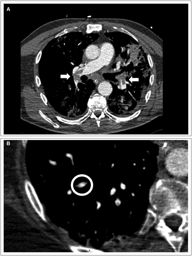
CTPA was performed 7 days (4–8 d) after ICU admission and 12 days (9–16 d) after the onset of symptoms; only one patient had acute hemodynamic instability before imaging. A total of 13 patients (33%) were found to have PE, which was bilateral in six patients and unilateral, in the right lung, in seven patients. Two patients had proximal bilateral PE (Fig. 1A), one patient had a subsegmental PE located in the right lower lobe (Fig. 1B), and all other patients had segmental PE. Cardiac echography was performed in 12 of the PE patients and revealed right ventricular dilation (n = 1) or acute right heart failure (n = 1). Lower limb echo-Doppler was performed in 11 of these patients on the same day than CTPA and revealed no deep venous thrombosis. Four of the patients without PE underwent a second CTPA during their ICU stay, which yielded no PE.
Figure 1.

Proximal bilateral partial occlusion of the pulmonary arteries (A—white arrows) and subsegmental embolism located in the right pulmonary artery (B—white circle).
Standard thromboprophylaxis was used in 22 patients; 11 of them (50%) developed PE; anti-Xa activity at the moment of CTPA was 0.34 (0.10–0.43) (n = 10). In the high-dose thromboprophylaxis group (i.e. six patients receiving continuous therapeutic infusion; dose ranges: 1,500–2,200 IU/hr—12 receiving subcutaneous enoxaparin 4,000 IU bid), only two of the 18 patients (11%) developed PE, 2 and 3 days after the implementation of such regimen (odds ratio [OR], 0.13 (0.02–0.69); p = 0.02). Anti-Xa activity at the moment of CTPA was 0.42 (0.39–0.52) (n = 18; p = 0.048 vs standard thromboprophylaxis). In the multivariate analysis, the probability of developing PE in patients receiving high-dose thromboprophylaxis remained significantly lower than others (OR 0.09 [0.02–0.57]; p = 0.01).
Patients with PE had lower WBCs count and higher Pao2/Fio2 at admission than the other patients (Supplemental Table 1, Supplemental Digital Content 1, http://links.lww.com/CCM/F716); also, on the day of CTPA, they had higher C-reactive protein and D-dimers levels and were ventilated with higher tidal volume and minute volume ventilation than those without PE. D-dimers on the day of CTPA had a high predictive accuracy (area under the receiver operating characteristics, 0.90; 95% CIs, 0.78–1.00) for PE; a D-dimers concentration greater than 3,647 ng/mL had 75% (95% CIs, 54–88%) sensitivity and 92% (95% CIs, 64–99%) specificity to predict PE.
All PE patients were eventually treated with continuous therapeutic infusion of unfractioned heparin; in one PE patient treated with ECMO, massive hemothorax developed, requiring interruption of anticoagulation and percutaneous thoracic drainage, whereas in another bleeding from gastric ulcer was detected on endoscopy. Among patients receiving enoxaparin, two patients on standard doses (one with hemoptysis and one with gastric bleeding) and three on high dose (two with gastric bleedings and one with bowel bleeding) had hemorrhagic complications. Six patients with PE (46%) and 14 patients without PE (52%) died at ICU discharge (OR 0.79 [0.24–3.26]; p = 0.99); four of the patients without PE underwent autopsy, which confirmed the absence of PE.
DISCUSSION
In this study, one third of patients (33%) who are mechanically ventilated for COVID-19 have PE diagnosed on CTPA. High-dose thromboprophylaxis was associated with a higher prevalence of PE when compared with standard regimen. No association of PE with mortality was observed.
COVID-19 can be associated with a massive release of inflammatory cytokines, which can promote the development of endothelial cell damage and the occurrence of thromboembolic events. We found that elevated D-dimers could be a marker of PE occurrence and may therefore help to identify the patients requiring full anticoagulation therapy; lack of PE diagnosis and specific therapies could explain the association of high D-dimers with poor outcome in COVID-19 patients (6). As such, daily monitoring of increasing D-dimers levels could be used to select patients at risk of PE, who could undergo chest CTA. These data are consistent with those from Cui et al (4) showing that the sensitivity and specificity of D-dimers greater than 3,000 ng/mL to predict lower limb venous thrombosis were 76.9% and 94.9%, respectively. In another study including hospitalized COVID-19 patients, D-dimers greater than 2,660 ng/mL had a sensitivity of 100% and a specificity of 67% to detect PE on CTPA (7).
Acute PE has been reported in 25 of 184 critically ill patients in one study, but with limited information and without systematic CTPA (5). In another series, 22 patients of 107 (21%) were diagnosed of PE after a median of 6 days after ICU admission (8); in this study, CTPA was performed only in case of suspected PE upon admission and/or acute degradation of hemodynamic/respiratory status. A large cohort including hospitalized COVID-19 patients showed that routine CTPA could identify PE in 22% of them; PE was more frequently observed in critically ill patients, in particular if treated with mechanical ventilation (9). As such, the occurrence of EP is significantly higher than in other critically ill patients population (i.e. around 3%) (10) and might be underestimated because hemodynamic instability, right ventricular dysfunction, or concomitant venous thrombosis are rarely observed.
The prevalence of PE appeared to depend on the thromboprophylaxis regimen. Higher thromboprophylaxis doses or continuous therapeutic infusion of unfractioned heparin was more common among patients without than with PE. Our findings suggest the need for increasing thromboprophylaxis doses in COVID-19 patients. In previous studies, the failure rate for standard thromboprophylaxis in an ICU setting is around 8% (11); in the study from Cui et al (4), 25% prevalence of deep venous thrombosis in the absence of any thromboprophylaxis. In another study, COVID-19 patients treated with UFH and with a high coagulopathy score had a lower 28-day mortality than untreated patients (12). No prospective studies have been published so far adequately evaluating the effects on anticoagulation on COVID-19 patients’ outcome.
This study has several limitations. First, this is a single-center, small cohort study; the results need to be confirmed in larger cohorts. Second, final outcome could not be fully assessed, since some patients are still in the hospital. Third, small subsegmental PE could have been undiagnosed in some patients, as this remains a diagnostic challenge in these patients in particular. Also, systematic research of associated deep venous thrombosis was not performed in all patients, as this would not have changed the indication for therapeutic anticoagulation. Finally, there is a risk of bias because the high frequency of PE observed in the standard thromboprophylaxis group might suffer from random fluctuation, which could explain a lower EP rate in the high dose thromboprophylaxis group, even without change in thromboprophylaxis regimen.
CONCLUSIONS
In this study, acute PE may occur in one third of mechanically ventilated COVID-19 patients. Higher thromboprophylaxis regimens should be considered in these patients.
ACKNOWLEDGMENTS
We thank all the ICU team for the wonderful job in the management of severe coronavirus disease 2019 patients.
Supplementary Material
Footnotes
Drs. Taccone, Peluso, Grimaldi, and Creteur prepared this article; Drs. Gevenois, Brasseur, Motte, Nobile, and Vincent collaborated in the design of the trial and organized the initial ethics applications; Drs. Pletchette, Garufi, and Talamonti are responsible for study governance and roll out of the trial. Dr. Taccone is responsible for logistical support of the study. Drs. Peluso, Pletchette, Garufi, and Talamonti collected the data; Drs. Taccone, Creteur, and Vincent supervised data collection. All authors contributed substantially to the design and methodology of this study and to the writing of this article. All authors have read and approved the final article.
Supplemental digital content is available for this article. Direct URL citations appear in the printed text and are provided in the HTML and PDF versions of this article on the journal’s website (http://journals.lww.com/ccmjournal).
The authors have disclosed that they do not have any potential conflicts of interest.
The datasets used and/or analyzed during the current study are available from the corresponding author on reasonable request.
REFERENCES
- 1.Grasselli G, Zangrillo A, Zanella A, et al. Baseline characteristics and outcomes of 1591 patients infected with SARS-CoV-2 admitted to ICUs of the Lombardy Region, Italy. JAMA 2020; 323:1574–1581 [DOI] [PMC free article] [PubMed] [Google Scholar]
- 2.Gattinoni L, Coppola S, Cressoni M, et al. Covid-19 does not lead to a “Typical” acute respiratory distress syndrome. Am J Respir Crit Care Med 2020; 201:1299–1300 [DOI] [PMC free article] [PubMed] [Google Scholar]
- 3.Tang N, Li D, Wang X, et al. Abnormal coagulation parameters are associated with poor prognosis in patients with novel coronavirus pneumonia. J Thromb Haemost 2020; 18:844–847 [DOI] [PMC free article] [PubMed] [Google Scholar]
- 4.Cui S, Chen S, Li X, et al. Prevalence of venous thromboembolism in patients with severe novel coronavirus pneumonia. J Thromb Haemost 2020; 18:1421–1424 [DOI] [PMC free article] [PubMed] [Google Scholar]
- 5.Klok FA, Kruip MJHA, van der Meer NJM, et al. Incidence of thrombotic complications in critically ill ICU patients with COVID-19. Thromb Res 2020; 191:145–147 [DOI] [PMC free article] [PubMed] [Google Scholar]
- 6.Wu C, Chen X, Cai Y, et al. Risk factors associated with acute respiratory distress syndrome and death in patients with coronavirus disease 2019 pneumonia in Wuhan, China. JAMA Intern Med 2020; 180:1–11 [DOI] [PMC free article] [PubMed] [Google Scholar]
- 7.Leonard-Lorant I, Delabranche X, Severac F, et al. Acute pulmonary embolism in COVID-19 Patients on CT angiography and relationship to D-Dimer levels. Radiology 2020:201561. [DOI] [PMC free article] [PubMed] [Google Scholar]
- 8.Poissy J, Goutay J, Caplan M, et al. ; Lille ICU Haemostasis COVID-19 Group: Pulmonary embolism in patients with COVID-19: Awareness of an increased prevalence. Circulation 2020; 142:184–186 [DOI] [PubMed] [Google Scholar]
- 9.Grillet F, Behr J, Calame P, et al. Acute pulmonary embolism associated with COVID-19 pneumonia detected by pulmonary CT angiography. Radiology 2020:201544. [DOI] [PMC free article] [PubMed] [Google Scholar]
- 10.Kaplan D, Casper TC, Elliott CG, et al. VTE incidence and risk factors in patients with severe sepsis and septic shock. Chest 2015; 148:1224–1230 [DOI] [PMC free article] [PubMed] [Google Scholar]
- 11.Lim W, Meade M, Lauzier F, et al. ; PROphylaxis for ThromboEmbolism in Critical Care Trial Investigators: Failure of anticoagulant thromboprophylaxis: risk factors in medical-surgical critically ill patients*. Crit Care Med 2015; 43:401–410 [DOI] [PubMed] [Google Scholar]
- 12.Tang N, Bai H, Chen X, et al. Anticoagulant treatment is associated with decreased mortality in severe coronavirus disease 2019 patients with coagulopathy. J Thromb Haemost 2020; 18:1094–1099 [DOI] [PMC free article] [PubMed] [Google Scholar]
Associated Data
This section collects any data citations, data availability statements, or supplementary materials included in this article.


