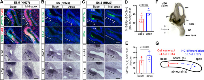
Fig. 1.
Characterization of terminal mitosis and HC differentiation in the developing chicken BP. (A-C) Adjacent cross-sections through the basal (proximal) and apical (distal) segment of wild-type BP stages (E5.5-E6.5; HH27-HH29). MYO7A and HCA immunostaining (green) marks HCs, Atoh1 in situ hybridization marks HC precursors and HCs, Lfng in situ hybridization and SOX2 immunostaining mark the pro-sensory/sensory domain within the developing BP. EdU was added at E4.5 and incorporation was analyzed at E5.5. Black and white lines mark the neural (n) to abneural (a) axis of the pro-sensory domain. (A) At E5.5 MYO7A+ HCs have not yet formed, but Atoh1+ HC precursors are present within the Lfng+ pro-sensory domain, with the highest number in the mid-apex. Pro-sensory cells (SOX2+, Lfng+) located at the base incorporated EdU at a lower rate than pro-sensory cells located more apically. (B) At E6, the number of Atoh1+ HC precursors/HCs increases and the sensory epithelium contains few scattered MYO7A + and HCA+ HCs. (C) At E6.5 the number of MYO7A+ HCA+ HCs increases, the majority of Atoh1(+) cells reside within the HC layer and Lfng expression is limited to the SC layer. (D) Quantification of EdU incorporation in SOX2+ basal and apical pro-sensory cells in A. Data are mean±s.d. (n=5 animals per group). (E) Quantification of MYO7A+ HCs in the base and apex of E6.0-E6.5 BPs. Data are mean±s.d. (n=5 animals per group). Individual data points in D,E represent value per animal. P-values were calculated using paired two-tailed Student's t-test (P≤0.05 is deemed significant). (F) Paint-filled chicken inner ear at ∼stage E6.0 indicating the position and orientation of tissue sections. (G) Summary model of terminal mitosis and differentiation in the developing BP. sac, sacculus; n, neural; a, abneural; psc, posterior semicircular canal; asc, anterior semicircular canal; lsc, lateral semicircular canal. Scale bars: 100 µm.

