Abstract
The neural mechanisms underlying emotional valence are at the interface between perception and action, integrating inputs from the external environment with past experiences to guide the behavior of an organism. Depending on the positive or negative valence assigned to an environmental stimulus, the organism will approach or avoid the source of the stimulus. Multiple convergent studies have demonstrated that the amygdala complex is a critical node of the circuits assigning valence. Here we examine the current progress in identifying valence coding properties of neural populations in different nuclei of the amygdala, based on their activity, connectivity, and gene expression profile.
Neural substrate of valence
The concept of valence
Across the animal kingdom, environmental stimuli can elicit a repertoire of behavioral responses ranging from approach to avoidance. Valence is the subjective value assigned to sensory stimuli which determines subsequent behavior. Positive valence leads to approach and consummatory behaviors while negative valence leads to defensive and avoidance behaviors [1,2]. For many sensory stimuli the assigned valence is innate, however, valence is weighted by the internal state of the organism and by its previous experiences [3,4] (Box 1). A simple example of state-dependence of valence is the value assigned to food, which strongly depends on the homeostatic needs of the animal [5]. Another internal state regulating valence assignment is basal anxiety. Indeed, high anxiety levels can induces a bias towards negative valence, even for stimuli that are normally rewarding [6].
Box 1 Innate versus learned valence in the amygdala.
Innate valence is attributed without requirement of learning and guides innate behavioral responses which have been selected during species evolution [5]. A clear example of innate valence is the unconditioned approach and avoidance exhibited by mice in presence of odorants of peanut oil and trimethylthiazoline (TMT), respectively. Neural ensembles in the BLA active during stimuli of positive or negative valence, also named ‘engrams’ [91,99], can drive behavioral responses such as approach for a ‘positive engram’ or avoidance for a ‘negative engram’ [99,100]. In the dentate gyrus of the hippocampus, the valence of a ‘positive engram’ (neurons in a male mouse activated during interaction with a female) can be reversed by reactivating this ‘engram’ during exposure to an experience of negative valence (electric footshock) [99]. Interestingly, the engram cells of the BLA do not exhibit such behavioral plasticity [99]. Nevertheless, pairing the activation of an innate positive or negative engram in the BLA with an olfactory stimulus can support learning and drive conditioned behaviors to the stimulus, even though it has not been paired with an aversive or rewarding experience [100]. This observation suggests that learned valence can be encoded by the same populations encoding innate valence. However, studies in projection-defined populations suggest that BLA-vHPC neurons specifically encode innate negative valence and not learned valence [12●●,56]. Similarly, in the CeA, direct electrophysiological recordings from neurons expressing the 5HT2A-R indicate that their activity is suppressed during innate fear but not during learned-fear, and that their inactivation upregulates innate-freezing response while downregulating learned-freezing response [101].
Despite the fundamental role of valence on animal survival and well-being, the underlying neurobiological substrate remains partially understood. One of the main working hypothesis postulates that specific neural circuits assign valence to stimuli in order to activate defined motor patterns and ensure an adaptive behavioral response [3,7]. In line with this hypothesis, human brain imaging has identified divergent networks activated in response to stimuli of positive or negative valence [8–10]. Multiple animal models including non-human primates [11], rodents [12●●], drosophila [13] and bees [14] have been used to decipher how neuronal populations composing these networks encode valence.
Defining valence circuits – role of the amygdala
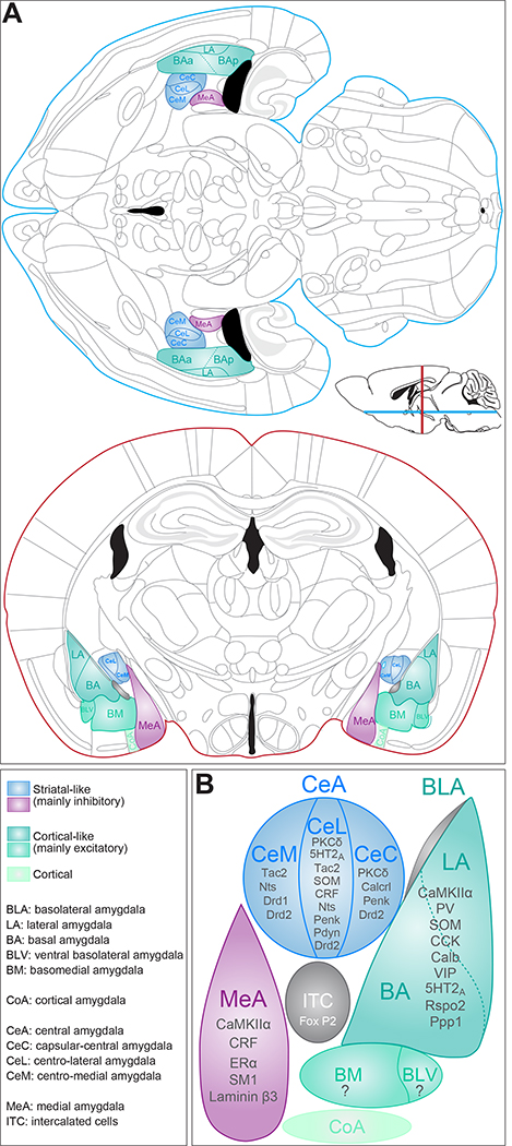
A vast body of literature reporting gain and loss of function experiments, as well as correlative measures of neuronal activity, has identified the amygdala complex as a central node to drive specific motor patterns in response to external stimuli [4,15–17]. The amygdala complex includes three main groups of nuclei: the basolateral amygdala (BLA), the central amygdala (CeA) and the medial amygdala (MeA) (Figure 1). Developmentally, the CeA and MeA arise from the same cell lineage, presenting a striatal-like organization compared to the BLA which originates from a different lineage and presents a cortical-like organization [18,19] (Figure 1a–b). Thus, the CeA is composed almost exclusively of inhibitory neurons, and the MeA is mainly composed of GABAergic neurons but contains one third of glutamatergic cells [20]. In contrast, the BLA is mainly composed of excitatory projection neurons (~85%) and of a small proportion of local inhibitory interneurons (~15%) [18,19]. Together with fast amino acid neurotransmitters, neurons of amygdala nuclei also produce numerous neuropeptides and express several neuromodulator receptors (Figure 1c). Despite increasing knowledge of anatomical and molecular properties of amygdala neurons, we are just starting to unravel their contribution to valence.
Figure 1.
The amygdala complex and genetically identified populations. (a) Atlas of horizontal and coronal sections of the adult mouse brain highlighting the different nuclei of the amygdala. (b) Schematic of the different amygdala nuclei and identified genetic markers.
Neural coding of valence
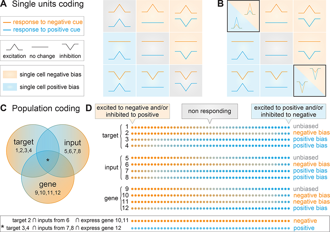
The role of neuronal populations in assigning valence has been studied using gain and loss of function experiments, and through analysis of neural activity recorded during tasks of opposite valences. Valence coding has been defined in terms of neural firing in response to at least two conditioned stimuli (CS), one of positive and one of negative valence [12●●,21–23]. Depending on their changes in firing rate in response to both CSs, neurons can be classified into nine coding populations [12●●,24] (Figure 2a). Among these nine classes, two include neurons responding similarly to cues of both positive and negative valence which could in principle support an arousal response. In addition, valence can also be defined as the differential response to positive and negative stimuli [21,25●,26,27●] (Figure 2b). In this case, neurons excited (or inhibited) by both positive and negative stimuli may still encode valence as they display a stronger response for one specific stimulus. In this framework, valence coding is rather defined by a coding preference or bias, than by an on/off coding pattern.
Figure 2.
Valence coding and population biases. (a) Definition of nine classes of neurons depending on their response to stimuli of positive and negative valence [12●●]. In this classification neurons responding to both stimuli in a similar way (excitation or inhibition to both stimuli) do not encode valence. (b) Alternative classification of units including the amplitude of the response. In this case, neurons responding to both stimuli in a similar way also encode valence as they exhibit a stronger response to one valence [12●●,21]. (c) Multidimensional definition of neurons encoding valence. (d) Each line represents a neuronal population and every dot corresponds to a single neuron. A single feature defines populations with valence coding biases, and combining multiple features could potentially reveal valence selective populations.
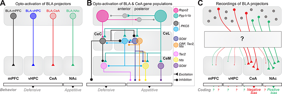
Recent advances in neurotechnologies allow us to analyze valence coding properties of single neurons in specific populations defined by other features including connectivity (inputs and outputs) and gene expression. This expands the experimental possibilities from local recordings within single brain regions, to circuit dissection at synaptic and molecular scales (Figure 3). The scope of this review is to synthesize the latest findings and future directions in identifying hallmarks of amygdala populations depending on their valence coding properties.
Figure 3.
Circuit diagram illustrating valence biases in BLA and CeA. (a) Optogenetic activation of three projection-defined BLA populations induces defensive behaviors [52,53,56] and activation of the last population induces appetitive behaviors [52,57,58] (b) Intra-amygdala circuit diagram of genetic populations in the anterior (a) and posterior (p) BLA, and CeA. Anterior BLA Rspo2+ and posterior BLA Ppp1r1b+ neurons drive opposite behavioral responses and reciprocally inhibit each other [62●●]. Rspo2+ neurons innervate CeC PKCδ+ neurons driving a defensive response. CeC PKCδ+ neurons inhibit CeL PKCδ+ neurons and Tac2+ CeM neurons, which mediate appetitive responses. Ppp1r1b+ neurons innervate all CeA neurons driving appetitive responses. CeC and CeL PKCδ+ neurons antagonize each other [29●●] (c) Recordings of BLA neurons defined by their projection have revealed coding biases for learned positive and negative valence. Although collateralization has been described at a population level, the relationship between collateralization pattern and valence coding at a single neuron level remains unknown.
Valence coding in populations of the central amygdala (CeA)
The CeA is the main output of the amygdala and has primarily been studied in the context of fear-related behaviors [28]. However, the CeA has also repeatedly been reported to promote appetitive behaviors [29●●,30,31]. Although contradictory, these results could be supported by divergent activity of distinct neural populations. Extensive research has been dedicated to identify the function of gene-defined and projection-defined populations within the capsular, lateral, and medial areas of the CeA (CeC, CeL and CeM, Figure 1). Pharmacological inhibition of CeL as well as optogenetic activation of CeM both induce unconditioned freezing suggesting that these different subregions differentially encode valence [32]. Subregions of CeA express selective genetic markers such as protein kinase-Cδ (PKCδ), somatostatin (SOM), corticotropin-releasing factor (CRF), tachykinin 2 (Tac2), neurotensin (Nts) or serotonin 2A receptor (5-HT2A, Figure 1c) [29●●,33●,34], and a complex microcircuit connectivity characterizes the interaction among these cells [35,36●].
PKCδ+ cells represent a prominent subpopulation in both CeC and CeL. Optogenetic inhibition of this population can suppress defensive behavior [29●●,37]. Nevertheless, while optogenetic activation of PKCδ+ cells in CeC can drive defensive behaviors, activation of CeL PKCδ+ cells does not drive valence-related behaviors and their suppression does not inhibit defensive behavior, but instead enhances drinking behavior [29●●] (Table 1). Consistently, it has been reported that neurons inhibited by a cue predicting a footshock (CeLOFF neurons) [32] largely overlap with PKCδ+ cells [35]. However, optogenetic activation of PKCδ+ cells in CeL can increase fear-cue generalization and can be anxiogenic [38]. Lastly, optogenetic activation of PKCδ+ cells in CeL suppresses food intake [39] whereas activation of 5HT2a+ cells, a marker for PKCδ− cells in CeL, promotes food intake [33●].
Table 1.
Studies manipulating and recording neural activity in gene-defined, input-defined and projection-defined populations.
| Experiment | Behavior or neural response | Bidirectional* | Valence | Reference | |||
|---|---|---|---|---|---|---|---|
| Cental amygdala (CeA) | |||||||
| Gene | PKCd+ | CeL | single-unit | inhibited by CS-shock (CeLOFF) | x | +? | Haubensak 201035 |
| CeL | ChR2 | no valence-related behavior | yes | +/− | Kim 201729 | ||
| CeC | ChR2 | freezing | yes | − | Kim 201729 | ||
| CeL | ChR2 | anxiogenic | yes | − | Botta 201538 | ||
| CeL | ChR2 | anxiolytic | x | + | Cai 201439 | ||
| CeL | ChR2 | food intake supression | yes | −? | Cai 201439 | ||
| CeL-SI | ChR2 | avoidance (CPA) | x | − | Cui 201785 | ||
| SOM | CeL | ChR2 | decrease CS avoidance | yes | + | Yu 201640 | |
| CeL | ChR2 | self-stimulation | yes | + | Kim 201729 | ||
| CeM | ChR2 | self-stimulation | yes | + | Kim 201729 | ||
| CeL | ChR2 | freezing | yes | − | Li 201342 | ||
| CeL | ChR2 | decrease CS-flight | no | + | Fadok 201741 | ||
| CeL | ChR2 | CS-freezing | no | − | Fadok 201741 | ||
| CeL | single-unit | excited during CS-shock and freezing | x | − | Fadok 201741 | ||
| CRF | CeL | ChR2 | CS flight | yes | − | Fadok 201741 | |
| CeL | ChR2 | decrease CS-freezing | no | − | Fadok 201741 | ||
| CeL | single-unit | excited during CS-flight | x | − | Fadok 201741 | ||
| 5HT2A | CeA | ChR2 | increase food intake | yes | + | Douglass 201733 | |
| CeA | ChR2 | decrease freezing to aversive US | yes | − | Isosaka 2015102 | ||
| Input | insula-CeA | ChR2 | avoidance (RTPA) | x | − | Wang 201848 | |
| PBNCGRP-CeC/L | ChR2 | appetite suppression | no | − | Carter 201345 | ||
| PBNCGRP-CeC/L | ChR2 | defensive behaviors | no | − | Han 201546 | ||
| PVT-CeLSOM | ChR2 | inhibition decrease FC | no | − | Penzo 201547 | ||
| BLARspo2-CeCPKCd | ChR2 | defensive response | yes | − | Kim 201729 | ||
| Output | CeA-vIPAG | ChR2 | hunting (prey approach) | no | + | Han 201749 | |
| CeL-SI | PKCd+ | ChR2 | avoidance (CPA) | x | − | Cui 201785 | |
| Basolateral amygdala (BLA) | |||||||
| Gene | Rspo2 (anterior) | ChR2 | freezing | yes | − | Kim 201662 | |
| Ppp1 (posterior) | ChR2 | self-stimulation | yes | + | Kim 201662 | ||
| PV | ChR2 | increase CS-shock freezing | yes | − | Wolff 201463 | ||
| single-unit | excited to the CS-shock | x | − | Wolff 201463 | |||
| SOM | ChR2 | decrease CS-shock freezing | yes | + | Wolff 201463 | ||
| single-unit | inhibited to the CS-shock | x | + | Wolff 201463 | |||
| Input | insula-BLA | ChR2 | approach (RTPP) | x | + | Wang 201848 | |
| ACC-BLA | NpHR | decrease observational fear retrieval | no | − | Allsop 201866 | ||
| Output | BLA-NAc | ChR2 | self-stimulation | x | + | Namburi 201552, Britt 201257, Stuber 201158 | |
| single-unit | preferentially excited to CS-sucrose | x | + | Beyeler 201612 | |||
| BLARspo2-NAc | ChR2 | freezing & stimulation avoidance (RTPA) | x | − | Kim 201729 | ||
| BLA-CeL | ChR2 | anxiolytic | yes | + | Tye 201196 | ||
| BLA-CeM | ChR2 | place aversion | x | − | Namburi 201552 | ||
| BLA-CeA | single-unit | preferentially excited to CS-quinine | x | − | Beyeler 201612 | ||
| BLA-vHPC | ChR2 | anxiogenic | yes | − | Felix-Ortiz 201354 | ||
| single-unit | unbiased | x | +/− | Beyeler 201612 | |||
| BLA-mPFC | ChR2 | anxiogenic | yes | − | Felix-Ortiz 201556, Lowery-Gionta 201897 | ||
| ChR2 | increase CS-shock freezing | yes | − | Burgos-Robles 201753 | |||
| single-unit | preferentially excited to CS-shock | x | − | Burgos-Robles 201753 | |||
| BLA-PL | single-unit | active during CS-shock (early extinction) | x | − | Senn 201459 | ||
| BLA-IL | single unit | active during CS-shock after extinction | x | + | Senn 201459 | ||
| BLA-adBNST | ChR2 | anxiogenic | x | − | Kim 201398 | ||
study reporting bidirectional manipulation of the tested behavior/s
As the PKCδ+ population, the SOM+ population represents about 40% of the neurons in CeL and the two populations interact through mutual inhibition [36●]. Optogenetic activation of SOM+ neurons in CeL and in CeM can drive appetitive behaviors [29●●], and their inhibition in CeL promotes defensive behaviors [40]. Consistent with this finding, activation of SOM+ neurons can decrease conditioned flight responses [41] but can also initiate passive freezing [41,42]. Finally, CRF+ cells are necessary for defensive behaviors [43], can increase conditioned flight responses, and the balance between conditioned flight and freezing behaviors is regulated by local inhibitory connections between CRF+ or activation of SOM+ neurons [41]. Altogether, activation of PKCδ+ and SOM+ neurons are able to drive both appetitive and defensive behaviors depending on the experimental conditions (Table 1).
This discrepancy might stem from divergent connectivity of genetically defined populations, including synaptic inputs and outputs. For example, PKCδ+ cells of CeC receive direct inputs from neurons of the parabrachial nucleus expressing calcitonin gene-related peptide (PBNcgRP) [44], and optogenetic activation of those inputs suppresses appetite and drives defensive responses [45,46]. On the other hand, inhibition of inputs from the paraventricular nucleus of the thalamus (PVT), which mainly targets SOM+ cells in CeL, strongly reduces fear conditioning [47]. Furthermore, inputs from the intermediate insular cortex (i.e. bitter gustatory cortex) in CeA promote avoidance behaviors [48]; however, the genetic identity of the CeA target population remains unknown. Further, projection neurons of CeL and CeM targeting the ventrolateral periaqueductal grey (vlPAG) strongly drive hunting behavior [49●]. Finally, a recent functional mapping study has shown that inhibitory projection form CeA suppresses activity of vlPAG local interneurons disinhibiting the excitatory cells, which in turn project to the cholinergic cells of the magnocellular nucleus of the medulla driving a defensive response [50●●]. Overall, despite in depth knowledge of genetic populations of the CeA, few studies have analyzed their single-unit activity in response to both positive and negative valence, leaving their valence coding properties elusive (Table 1).
Valence coding in populations of the basolateral amygdala (BLA)
Multiple studies have performed single-unit recordings in the BLA during stimuli of both positive and negative valence. Although direct optogenetic stimulation of the lateral amygdala (LA) can elicit a defensive response in a naïve mouse [51], recordings of BLA neurons in monkeys, rats and mice have shown that around 50% of the units respond to predictive cues of positive or negative valence [12●●,21,27●], with an overrepresentation of neurons responding to positive valence in monkeys [21] and mice [12●●], and an even distribution of neuron responding to both valences in rats [27●]. Additionally, pioneering work has shown that some BLA neurons track the value of a sensory stimulus during reversal of the CS-US association [21,22] emphasizing the critical role of the BLA in valence coding. Finally, a recent study has also identified that even if relatively few cells in the BLA cells encode valence, the valence assigned to a stimulus can be decoded at the population level of neural activity [27●].
As the BLA is mainly composed of glutamatergic projection neurons, the search for neuronal features defining the polarity of valence has been predominantly focused on post-synaptic targets (Table 1). Optogenetic activation has shown that projections to CeA (BLA-CeA) [52], medial prefrontal cortex (BLA-mPFC) [53–55] and ventral hippocampus (BLA-vHPC) [56] can drive defensive behaviors. On the contrary, optogenetic activation of projections to the nucleus accumbens (BLA-NAc) has repeatedly been shown to support reinforcement [52,57,58] (Figure 3a). This accumulation of results reporting regulation of valence-related behavior by BLA projections supports the hypothesis that anatomically divergent populations of the BLA differentially encode valence. Moreover, recordings combined with optogenetic photoidentification of specific neural subpopulations have shown that BLA-NAc units are preferentially excited by a positive CS and BLA-CeA units are preferentially excited by a negative one [12●●]. Further, synaptic inputs on BLA-NAc and BLA-CeA neurons are regulated in an opposite manner after learning associations of positive and negative valence [52]. Importantly, in vivo recordings have also revealed heterogeneity of single neuron activity within projection-defined populations [12●●,53,59]. This supports a model where valence coding of a projector population can be inferred from the projection target of its neurons, but the projection target of a single neuron is not sufficient to infer its valence coding properties (Figure 2).
BLA projection neurons are segregated in large neurons in the anterior part (magnocellular) and smaller neurons in the posterior part (parvocellular) [60]. Activity-dependent profiling combined with extensive gene screening [61,62●●] has shown that the magno-cellular and parvocellular populations are defined by the expression of the Rspo2 and Ppp1r1b genes, respectively. Interestingly, optogenetic stimulation of Rspo2+ cells elicits a defensive response in naive mice whereas stimulation of Ppp1r1b+ cells promotes an appetitive response [62●●]. Both populations send projections to the NAc and CeA, Rspo2+ cells monosynaptically contacting PKCδ+ cells of the CeC whereas Ppp1r1b+ cells innervate the other cellular subtypes of the CeM and CeL (Figure 3b) [29●●]. The anteroposterior topography of the Rspo2 and Ppp1r1b gene markers does not overlap with the distribution of BLA-NAc and BLA-CeA populations which are intermingled with mediolateral and dorsoventral gradients [25●]. The ability of genetically defined and anatomically defined populations to drive polarized behaviors combined with the coding heterogeneity recorded in BLA projectors raises the interesting possibility that defining populations using a combination of anatomical and genetic approaches may be instrumental in selecting populations sharply tuned to a specific valence.
Populations of local inhibitory interneurons of the BLA expressing SOM or parvalbumine (PV) have been shown to differentially drive behavioral responses to aversive cues [63]. However, their role in positive valence coding remains unexplored. Similarly, oscillatory activity, which is generated by local inhibitory interneurons [64], is known to causally regulate behaviors driven by negative valence [65●], but has not been investigated with positive valence (Box 2). In addition, the BLA receives inputs from a vast array of regions also involved in valence, including the mPFC, anterior cingulate cortex (ACC), auditory cortex and multiple nuclei of the thalamus — all of which have been almost exclusively analyzed during aversive states [55,66–69]. Albeit essential to understand the functional role of local interneurons and inputs to the BLA in fear and defensive behaviors, the studies leave their implication in reward processing uncharted.
Box 2 Oscillatory synchronization.
Cortical regions of the mammalian brain generate patterns of rhythmic oscillations of the local field potential (LFP) covering frequencies from 0.05 to 500 Hz [64]. Selective frequency bands are associated with different brain states and behaviors. Originally described in the neocortex and hippocampus, oscillations have also been observed in the rodent BLA [102], where changes in power of specific LFP frequencies have been correlated with learning of associations of negative valence [103,104]. Interestingly, it was shown that local oscillations can have different impact on neurons depending on their downstream target [103]. Consistent with the existence of distributed brain states, synchronous oscillations in the theta (7–12 Hz) and gamma (40–120 Hz) bands between the BLA and interconnected regions occur during consolidation and retrieval of emotional memories [104–106]. For example during retrieval of a fear memory, the hippocampus and amygdala are synchronized in the theta band [107], whereas prefrontal-amygdala circuits display synchronized 4-Hz oscillations [65●]. Interneurons are powerful regulators of this synchronized activity and are tightly controlled by several neuromodulators providing a gating mechanism for synaptic plasticity [108]. Whether oscillations of different frequencies, including gamma and theta display valence specific modulation remains unknown.
Valence in other amygdala nuclei
Most studies analyze the origin of valence in the CeA and BLA but surrounding amygdaloid nuclei also regulate valence. For example, direct optogenetic activation of the basomedial amygdala (BMA, Figure 1) is anxiogenic, as the optogenetic activation of the vmPFC inputs to this nucleus [70]. Interestingly, the BMA directly projects to the ventromedial hypothalamus (VMH) which regulates defensive and social behaviors [71].
Neurons in the medial amygdala (MeA, Figure 1) have repeatedly been shown to regulate social behaviors [20,72] and GABAergic neurons of the posterodorsal MeA promote social behaviors of both negative (e.g. aggression) and positive valence (e.g. mating and social grooming) [73]. Neurons in the MeA can be genetically identified by the unique marker laminin β3 [74] and express numerous receptors including oxytocin receptors [72], estrogen receptors and the CRF receptor 2 [75]. MeA cells expressing CRF-2 receptor mRNA are active during a social experience of negative valence (social defeat stress) [76]. In addition, a subpopulation of MeA neurons expressing kisspeptin protein modulates anxiety and sexual partner preference in male mice [77], whereas neurons expressing the alpha-estrogen receptor controls body weight [78]. When analyzed independently of projection or gene markers, neurons of the cortical amygdala (CoA, Figure 1) represent odor objects of both valences using distributive population codes [79]. Optogenetic inhibition of CoA reduces innate responses to odors of both positive and negative valence [80]. Interestingly, neurons activated by odors of positive or neutral valence are mainly recruited in the posterior section of the CoA, compared to neurons activated by an odor of negative valence which are equally distributed in the antero-posterior axis [80].
Importantly, the intercalated cells (ITC), which are clusters of GABAergic interneurons (Figure 1), were shown to relay negative valence to the BLA, including fear and pain information [81–84].
Moving forward to crack the valence code
Gain and loss of function experiments have demonstrated that neuronal subpopulations of the amygdala defined by their projection targets or gene expression can drive behaviors of opposite valence (Figure 3). Activity-dependent markers and electrophysiological recordings have revealed that average activity of a population is generally consistent with the driven behaviors (Figure 3c). Yet, recordings revealing single-unit heterogeneity in valence coding within populations [12●●,53] suggest the presence of functional subpopulations. Increasing the level of specificity by integrating multiple cell features, such as genetic identity and anatomical connectivity, could promote the identification of more uniform populations selectively encoding one valence [85●] (Figure 2c-d).
Anatomical complexity
Although the activity of BLA projection-defined populations can predict valence, these cells send collaterals to multiple brain regions [12●●]. The distribution of collaterals at a single cell level and its correlation with valence coding properties remains unexplored (Figure 3b). Projection collaterals, topography, and post-synaptic cell identity in the downstream region represent critical points that could reconcile conflicting results. For example, the NAc and the CeA both contain neurons expressing both dopamine D1 and D2 receptors, which have been shown to induce behaviors of opposite valence in the dorsal striatum [86]. Moreover it was shown that depending on the dorsal-to-ventral axis, optogenetic stimulation of NAc neurons induces preference or avoidance respectively [87]. To circumvent these limitations, systematic mapping of projection-defined, activity-defined or genetically defined populations in whole brain samples [88] will provide a greater level of understanding of the anatomo-functional organization of the amygdala. Micro-circuit connectivity including feedforward, feedback, and mutual inhibition also appears as a mechanism of population selection. For instance, optogenetic activation of the BLA-CeA population induces a stronger inhibition of neighboring neurons than activation of other projector populations [25●,89]. Moreover, mutual inhibition was described between functionally divergent populations such as PKCδ+ and SOM+ neurons in CeA [35], as well as Rspo2 and Ppp1rb1 neurons in BLA [62●●].
Genetic complexity
Unique transcriptional signatures of multiple immediate early genes have been identified in the amygdala after experiences of positive and negative valence [90]. Similarly, neurons with an increased expression of cyclic adenosine monophosphate response element-binding protein (CREB) in the LA are preferentially recruited to encode a memory of negative valence compared to neurons with a lower expression of CREB [91]. These studies highlight that beyond stable gene markers, dynamic gene expression is also a defining feature of neural populations encoding valence. Importantly, genetic identification of multiple genes is now possible in ‘intact’ fixed samples using new technologies such as MERFISH [92] or STARmap [93]. Interestingly, these techniques might also allow to identify the contribution of glial cells in valence coding which has so far only been described in CeM, for negative valence [94]. We expect the combination of these approaches with anatomical tracing and mapping of cellular activation to potentiate the progress of our understanding of valence coding in amygdala nuclei.
Conclusions
Over the last decade, the study of valence coding in the amygdala has made unprecedented progress by revealing elaborate genetic and anatomical circuits differentially involved in positive and negative valence (Figure 3). This exceptional leap forward is the fruit of technological advancements combined with the spread of systematic behavioral testing of both positive and negative valence in the same experiment. Beyond this experimental prerequisite, recent studies have even started to combine recordings in response to both positive and negative valence with recordings during anxiety-related behaviors [95], providing crucial data to understand the role of valence circuits in state anxiety. Future investigations into valence coding in animal models of neuropsychiatric disorders, might further advance our understanding of valence circuit dysregulations in the physiopathology of diseases including post-traumatic stress disorders, anxiety, depression, and addiction.
Acknowledgements
We thank Joanna Dabrowska, Mario Martin-Fernandez, Sebastien Delcasso, Xavier Leinekugel, Gwendolyn Calhoon, Caitlin Vander Weele and Praneeth Namburi for critical reading of the manuscript. We acknowledge support by the Région Nouvelle-Aquitaine and INSERM-Avenir to the Beyeler Lab and by the Brain and Behavior Research Foundation NARSAD young investigator grant to AB.
Footnotes
Conflict of interest statement
Nothing declared.
References and recommended reading
Papers of particular interest, published within the period of review, have been highlighted as:
● of special interest
●●of outstanding interest
- 1.Lewin K: The Conceptual Representation and the Measurement of Psychological Forces. Duke University Press; 1938. [Google Scholar]
- 2.Russell JA: A circumplex model of affect. J Pers Soc Psychol 1980, 39:1161–1178. [Google Scholar]
- 3.Anderson DJ, Adolphs R: A framework for studying emotions across species. Cell 2014, 157:187–200. [DOI] [PMC free article] [PubMed] [Google Scholar]
- 4.Morrison SE, Salzman CD: Re-valuing the amygdala. Curr Opin Neurobiol 2010, 20:221–230. [DOI] [PMC free article] [PubMed] [Google Scholar]
- 5.Tinbergen N: The Study of Instinct. Clarendon Press; 1951. [Google Scholar]
- 6.Samuels BA, Hen R: Novelty-suppressed feeding in the mouse In Mood and Anxiety Related Phenotypes in Mice, vol. 63 Edited by Gould TD. Humana Press; 2011:107–121. [Google Scholar]
- 7.Hebb DO: The Organization of Behavior: A Neuropsychological Theory. Psychology Press; 2002. [Google Scholar]
- 8.Borchardt V et al. : Echoes of affective stimulation in brain connectivity networks. Cereb Cortex 2017:1–14 10.1093/cercor/bhx290. [DOI] [PubMed] [Google Scholar]
- 9.Kragel PA, LaBar KS: Decoding the nature of emotion in the brain. Trends Cogn Sci 2016, 20:444–455. [DOI] [PMC free article] [PubMed] [Google Scholar]
- 10.Pessoa L: Understanding emotion with brain networks. Curr Opin Behav Sci 2018, 19:19–25. [DOI] [PMC free article] [PubMed] [Google Scholar]
- 11.Zhang W et al. : Functional circuits and anatomical distribution of response properties in the primate amygdala. J Neurosci 2013, 33:722–733. [DOI] [PMC free article] [PubMed] [Google Scholar]
- 12.Beyeler A et al. : Divergent routing of positive and negative information from the amygdala during memory retrieval. Neuron 2016, 90:348–361.●● This study analyzes the firing response of three subpopulations of BLA projector neurons to positive and negative associated auditory stimuli in head restrained behaving mice. Neurons are classified into nine groups according to the response to sounds associated to sucrose or quinine. The study suggests that a population code may determine the correct attribution of a specific valence.
- 13.Yamazaki D et al. : Two parallel pathways assign opposing odor valences during Drosophila memory formation. Cell Rep 2018, 22:2346–2358. [DOI] [PubMed] [Google Scholar]
- 14.Roussel E, Sandoz J-C, Giurfa M: Searching for learning-dependent changes in the antennal lobe: simultaneous recording of neural activity and aversive olfactory learning in honeybees. Front Behav Neurosci 2010, 4. [DOI] [PMC free article] [PubMed] [Google Scholar]
- 15.Bucy PC, Kluver H: An anatomical investigation of the temporal lobe in the monkey (Macaca mulatta). J Comp Neurol 1955, 103:151–251. [DOI] [PubMed] [Google Scholar]
- 16.Janak PH, Tye KM: From circuits to behaviour in the amygdala. Nature 2015, 517:284–292. [DOI] [PMC free article] [PubMed] [Google Scholar]
- 17.Weiskrantz L: Behavioral changes associated with ablation of the amygdaloid complex in monkeys. J Comp Physiol Psychol 1956, 49:381–391. [DOI] [PubMed] [Google Scholar]
- 18.Sah P, Faber ESL, Lopez De Armentia M, Power J: The amygdaloid complex: anatomy and physiology. Physiol Rev 2003, 83: 803–834. [DOI] [PubMed] [Google Scholar]
- 19.Swanson LW, Petrovich GD: What is the amygdala? Trends Neurosci 1998, 21:323–331. [DOI] [PubMed] [Google Scholar]
- 20.Li Y et al. : Neuronal representation of social information in the medial amygdala of awake behaving mice. Cell 2017, 171:1176–1190. e17. [DOI] [PMC free article] [PubMed] [Google Scholar]
- 21.Paton JJ, Belova MA, Morrison SE, Salzman CD: The primate amygdala represents the positive and negative value of visual stimuli during learning. Nature 2006, 439:865–870. [DOI] [PMC free article] [PubMed] [Google Scholar]
- 22.Schoenbaum G, Chiba AA, Gallagher M: Neural encoding in orbitofrontal cortex and basolateral amygdala during olfactory discrimination learning. J Neurosci 1999, 19:1876–1884. [DOI] [PMC free article] [PubMed] [Google Scholar]
- 23.Shabel SJ, Janak PH: Substantial similarity in amygdala neuronal activity during conditioned appetitive and aversive emotional arousal. Proc Natl Acad Sci U S A 2009, 106:15031–15036. [DOI] [PMC free article] [PubMed] [Google Scholar]
- 24.Namburi P, Al-Hasani R, Calhoon GG, Bruchas MR, Tye KM: Architectural representation of valence in the limbic system. Neuropsychopharmacology 2016, 41:1697–1715 10.1038/npp.2015.358. [DOI] [PMC free article] [PubMed] [Google Scholar]
- 25.Beyeler A et al. : Organization of valence-encoding and projection-defined neurons in the basolateral amygdala. Cell Rep 2018, 22:905–918.● This study maps the location of more than a 1000 BLA cells recorded during a Pavlovian discrimination task. Furthermore, by using triple retrograde tracing, the study reveals the three-dimensional location of BLA cells projecting to NAc, CeA and vHPC.
- 26.O’Neill P-K, Gore F, Salzman CD: Basolateral amygdala circuitry in positive and negative valence. Curr Opin Neurobiol 2018, 49:175–183. [DOI] [PMC free article] [PubMed] [Google Scholar]
- 27.Kyriazi P, Headley DB, Pare D: Multi-dimensional coding by basolateral amygdala neurons. Neuron 2018, 99:1315–1328 e5.● Using a task where rats can produce different behaviors in response to the same CS, the authors identified similarities at the population level between valence coding for CS and behavioral response.
- 28.Duvarci S, Pare D: Amygdala microcircuits controlling learned fear. Neuron 2014, 82:966–980. [DOI] [PMC free article] [PubMed] [Google Scholar]
- 29.Kim J, Zhang X, Muralidhar S, LeBlanc SA, Tonegawa S: Basolateral to central amygdala neural circuits for appetitive behaviors. Neuron 2017, 93:1464–1479 e5.●● By using a genetic screening this study demonstrates the presence of several segregated populations in the CeA, controlling negative or positive attribution of valence.
- 30.Robinson MJF, Warlow SM, Berridge KC: Optogenetic excitation of central amygdala amplifies and narrows incentive motivation to pursue one reward above another. J Neurosci 2014, 34:16567–16580. [DOI] [PMC free article] [PubMed] [Google Scholar]
- 31.Tom RL, Ahuja A, Maniates H, Freeland CM, Robinson MJF: Optogenetic activation of the central amygdala generates addiction-like preference for reward. Eur J Neurosci 2018. 10.1111/ejn.13967. [DOI] [PubMed] [Google Scholar]
- 32.Ciocchi S et al. : Encoding of conditioned fear in central amygdala inhibitory circuits. Nature 2010, 468:277–282. [DOI] [PubMed] [Google Scholar]
- 33.Douglass AM et al.: Central amygdala circuits modulate food consumption through a positive-valence mechanism. Nature Neurosci 2017, 20:1384–1394.● This study shows that GABAergic 5HT2A+ cells of the CeA modulate food consumption, promote positive reinforcement and are active in vivo during eating.
- 34.McCullough KM, Morrison FG, Hartmann J, Carlezon WA, Ressler KJ: Quantified coexpression analysis of central amygdala subpopulations. eNeuro 2018, 5. [DOI] [PMC free article] [PubMed] [Google Scholar]
- 35.Haubensak W et al. : Genetic dissection of an amygdala microcircuit that gates conditioned fear. Nature 2010, 468:270–276. [DOI] [PMC free article] [PubMed] [Google Scholar]
- 36.Hunt S, Sun Y, Kucukdereli H, Klein R, Sah P: Intrinsic circuits in the lateral central amygdala. eNeuro 2017, 4.● By combining patch clamp recordings in acute slices with immunostaining of specific cell markers, this study analyzes the monosynaptic connectivity among different neuronal types in the CeL.
- 37.Yu K et al. : The central amygdala controls learning in the lateral amygdala. Nat Neurosci 2017, 20:1680–1685. [DOI] [PMC free article] [PubMed] [Google Scholar]
- 38.Botta P et al. : Regulating anxiety with extrasynaptic inhibition. Nat Neurosci 2015, 18:1493–1500. [DOI] [PMC free article] [PubMed] [Google Scholar]
- 39.Cai H, Haubensak W, Anthony TE, Anderson DJ: Central amygdala PKC-δ neurons mediate the influence of multiple anorexigenic signals. Nat Neurosci 2014, 17:1240–1248. [DOI] [PMC free article] [PubMed] [Google Scholar]
- 40.Yu K, da Silva PG, Albeanu DF, Li B: Central amygdala somatostatin neurons gate passive and active defensive behaviors. J Neurosci 2016, 36:6488–6496. [DOI] [PMC free article] [PubMed] [Google Scholar]
- 41.Fadok JP et al. : A competitive inhibitory circuit for selection of active and passive fear responses. Nature 2017, 542:96–100. [DOI] [PubMed] [Google Scholar]
- 42.Li H et al. : Experience-dependent modification of a central amygdala fear circuit. Nat Neurosci 2013, 16:332–339. [DOI] [PMC free article] [PubMed] [Google Scholar]
- 43.Sanford CA et al. : A central amygdala CRF circuit facilitates learning about weak threats. Neuron 2017, 93:164–178. [DOI] [PMC free article] [PubMed] [Google Scholar]
- 44.Lu Y-C et al. : Neurochemical properties of the synapses between the parabrachial nucleus-derived CGRP-positive axonal terminals and the GABAergic neurons in the lateral capsular division of central nucleus of amygdala. Mol Neurobiol 2015, 51:105–118. [DOI] [PubMed] [Google Scholar]
- 45.Carter ME, Soden ME, Zweifel LS, Palmiter RD: Genetic identification of a neural circuit that suppresses appetite. Nature 2013, 503:111–114. [DOI] [PMC free article] [PubMed] [Google Scholar]
- 46.Han S, Soleiman MT, Soden ME, Zweifel LS, Palmiter RD: Elucidating an affective pain circuit that creates a threat memory. Cell 2015, 162:363–374. [DOI] [PMC free article] [PubMed] [Google Scholar]
- 47.Penzo MA et al. : The paraventricular thalamus controls a central amygdala fear circuit. Nature 2015, 519:455–459. [DOI] [PMC free article] [PubMed] [Google Scholar]
- 48.Wang L et al. : The coding of valence and identity in the mammalian taste system. Nature 2018, 558:127–131. [DOI] [PMC free article] [PubMed] [Google Scholar]
- 49.Han W et al. : Integrated control of predatory hunting by the central nucleus of the amygdala. Cell 2017, 168:311–324 e18.This unique study shows that optogenetic and chemogenetic stimulation of CeA of mice can elicit predatory attacks on insects.
- 50.Tovote P et al. : Midbrain circuits for defensive behaviour. Nature 2016, 534:206–212.●● This study identify an inhibitory pathway from the CeA to the ventrolateral periaqueductal grey that elicits freezing though disinhibition of ventrolateral periaqueductal grey excitatory outputs to pre-motor cells in the magnocellular nucleus of the medulla.
- 51.Johansen JP et al. : Optical activation of lateral amygdala pyramidal cells instructs associative fear learning. Proc Natl Acad Sci U S A 2010, 107:12692–12697. [DOI] [PMC free article] [PubMed] [Google Scholar]
- 52.Namburi P et al. : A circuit mechanism for differentiating positive and negative associations. Nature 2015, 520:675–678. [DOI] [PMC free article] [PubMed] [Google Scholar]
- 53.Burgos-Robles A et al. : Amygdala inputs to prefrontal cortex guide behavior amid conflicting cues of reward and punishment. Nat Neurosci 2017, 20:824–835. [DOI] [PMC free article] [PubMed] [Google Scholar]
- 54.Felix-Ortiz AC, Burgos-Robles A, Bhagat ND, Leppla CA, Tye KM: Bidirectional modulation of anxiety-related and social behaviors by amygdala projections to the medial prefrontal cortex. Neuroscience 2015, 79:658–664 10.1016/j.neuroscience.2015.07.041. [DOI] [PMC free article] [PubMed] [Google Scholar]
- 55.Yizhar O, Klavir O: Reciprocal amygdala-prefrontal interactions in learning. Curr Opin Neurobiol 2018, 52:149–155. [DOI] [PubMed] [Google Scholar]
- 56.Felix-Ortiz AC et al. : BLA to vHPC inputs modulate anxiety-related behaviors. Neuron 2013, 79:658–664. [DOI] [PMC free article] [PubMed] [Google Scholar]
- 57.Britt JP et al. : Synaptic and behavioral profile of multiple glutamatergic inputs to the nucleus accumbens. Neuron 2012, 76:790–803. [DOI] [PMC free article] [PubMed] [Google Scholar]
- 58.Stuber GD et al. : Excitatory transmission from the amygdala to nucleus accumbens facilitates reward seeking. Nature 2011, 475:377–380. [DOI] [PMC free article] [PubMed] [Google Scholar]
- 59.Senn V et al. : Long-range connectivity defines behavioral specificity of amygdala neurons. Neuron 2014, 81:428–437. [DOI] [PubMed] [Google Scholar]
- 60.Hall E: The amygdala of the cat: a Golgi study. Z Zellforsch Mikrosk Anat 1972, 134:439–458. [DOI] [PubMed] [Google Scholar]
- 61.Allen Brain Atlas: Allen Brain Atlas: Mouse Connectivity, Projections. . Available at: 2016. (Accessed 17 October 2016) http://connectivity.brain-map.org/. [Google Scholar]
- 62.Kim J, Pignatelli M, Xu S, Itohara S, Tonegawa S: Antagonistic negative and positive neurons of the basolateral amygdala. Nat Neurosci 2016, 19:1636–1646.●● By using a genetic screening this study demonstrates the presence of two segregated populations in the basal amygdala. The Rspo2+ and the Ppp1rb1+ cells are located in the BLA and BLP and control negative and positive attribution of valence respectively. A mutual inhibition characterizes their synaptic relationship.
- 63.Wolff SBE et al. : Amygdala interneuron subtypes control fear learning through disinhibition. Nature 2014, 509:453–458. [DOI] [PubMed] [Google Scholar]
- 64.Buzsaki G: Neuronal oscillations in cortical networks. Science 2004, 304:1926–1929. [DOI] [PubMed] [Google Scholar]
- 65.Karalis N et al. : 4-Hz oscillations synchronize prefrontal-amygdala circuits during fear behavior. Nat Neurosci 2016, 19: 605–612.● By using multiple extracellular recordings in behaving mice this study shows that freezing coincided with internally generated 4-Hz oscillations in prefrontal-amygdala circuits. 4-Hz oscillations predict freezing onset and offset and optogenetic induction of prefrontal 4-Hz oscillations can elicit fear behavior.
- 66.Allsop SA et al. : Corticoamygdala transfer of socially derived information gates observational learning. Cell 2018, 173:1–14 10.1016/j.cell.2018.04.004. [DOI] [PMC free article] [PubMed] [Google Scholar]
- 67.Mátyás F, Lee J, Shin H-S, Acsády L: The fear circuit of the mouse forebrain: connections between the mediodorsal thalamus, frontal cortices and basolateral amygdala. Eur J Neurosci 2014, 39:1810–1823. [DOI] [PubMed] [Google Scholar]
- 68.McGarry LM, Carter AG: Prefrontal cortex drives distinct projection neurons in the basolateral amygdala. Cell Rep 2017, 21:1426–1433. [DOI] [PMC free article] [PubMed] [Google Scholar]
- 69.Nabavi S et al. : Engineering a memory with LTD and LTP. Nature 2014, 511:348–352. [DOI] [PMC free article] [PubMed] [Google Scholar]
- 70.Adhikari A et al. : Basomedial amygdala mediates top-down control of anxiety and fear. Nature 2015, 527:179–185. [DOI] [PMC free article] [PubMed] [Google Scholar]
- 71.Yamamoto R, Ahmed N, Ito T, Gungor NZ, Pare D: Optogenetic study of anterior BNST and basomedial amygdala projections to the ventromedial hypothalamus. eNeuro 2018, 5. [DOI] [PMC free article] [PubMed] [Google Scholar]
- 72.Yao S, Bergan J, Lanjuin A, Dulac C: Oxytocin signaling in the medial amygdala is required for sex discrimination of social cues. eLife 2017, 6. [DOI] [PMC free article] [PubMed] [Google Scholar]
- 73.Hong W, Kim D-W, Anderson DJ: Antagonistic control of social behaviors by inhibitory and excitatory neurons in the medial amygdala. Cell 2014, 158:1348–1361. [DOI] [PMC free article] [PubMed] [Google Scholar]
- 74.Zirlinger M, Kreiman G, Anderson DJ: Amygdala-enriched genes identified by microarray technology are restricted to specific amygdaloid subnuclei. Proc Natl Acad Sci U S A 2001, 98:5270–5275. [DOI] [PMC free article] [PubMed] [Google Scholar]
- 75.Frankiensztajn LM, Gur-Pollack R, Wagner S: A combinatorial modulation of synaptic plasticity in the rat medial amygdala by oxytocin, urocortin3 and estrogen. Psychoneuroendocrinology 2018, 92:95–102. [DOI] [PubMed] [Google Scholar]
- 76.Fekete ÉM et al. : Social defeat stress activates medial amygdala cells that express type 2 CRF receptor mRNA. Neuroscience 2009, 162:5–13. [DOI] [PMC free article] [PubMed] [Google Scholar]
- 77.Adekunbi DA et al. : Kisspeptin neurones in the posterodorsal medial amygdala modulate sexual partner preference and anxiety in male mice. J Neuroendocrinol 2018, 30 e12572. [DOI] [PMC free article] [PubMed] [Google Scholar]
- 78.Xu P et al. : Estrogen receptor-a in medial amygdala neurons regulates body weight. J Clin Invest 2015, 125:2861–2876. [DOI] [PMC free article] [PubMed] [Google Scholar]
- 79.Iurilli G, Datta SR: Population coding in an innately relevant olfactory area. Neuron 2017, 93:1180–1197 e7. [DOI] [PMC free article] [PubMed] [Google Scholar]
- 80.Root CM, Denny CA, Hen R, Axel R: The participation of cortical amygdala in innate, odour-driven behaviour. Nature 2014, 515:269–273. [DOI] [PMC free article] [PubMed] [Google Scholar]
- 81.Asede D, Bosch D, Lüthi A, Ferraguti F, Ehrlich I: Sensory inputs to intercalated cells provide fear-learning modulated inhibition to the basolateral amygdala. Neuron 2015, 86:541–554. [DOI] [PubMed] [Google Scholar]
- 82.Bienvenu TCM et al. : Large intercalated neurons of amygdala relay noxious sensory information. J Neurosci 2015, 35:2044–2057. [DOI] [PMC free article] [PubMed] [Google Scholar]
- 83.Kuerbitz J et al. : Loss of intercalated cells (ITCs) in the mouse amygdala of Tshz1 mutants correlates with fear, depression, and social interaction phenotypes. J Neurosci 2018, 38:1160–1177. [DOI] [PMC free article] [PubMed] [Google Scholar]
- 84.Strobel C, Marek R, Gooch HM, Sullivan RKP, Sah P: Prefrontal and auditory input to intercalated neurons of the amygdala. Cell Rep 2015, 10:1435–1442 10.1016/j.celrep.2015.02.008. [DOI] [PubMed] [Google Scholar]
- 85.Cui Y et al. : A central amygdala-substantia innominata neural circuitry encodes aversive reinforcement signals. Cell Rep 2017, 21:1770–1782.● This study demonstrates the role of the projections from PKCδ neurons of CeA projecting to the substantia innominata in the modulation of negative reinforcement learning.
- 86.Kravitz AV, Tye LD, Kreitzer AC: Distinct roles for direct and indirect pathway striatal neurons in reinforcement. Nat Neurosci 2012, 15:816–818. [DOI] [PMC free article] [PubMed] [Google Scholar]
- 87.Al-Hasani R et al. : Distinct subpopulations of nucleus accumbens dynorphin neurons drive aversion and reward. Neuron 2015, 87:1063–1077. [DOI] [PMC free article] [PubMed] [Google Scholar]
- 88.Ye L et al. : Wiring and molecular features of prefrontal ensembles representing distinct experiences. Cell 2016, 165:1776–1788. [DOI] [PMC free article] [PubMed] [Google Scholar]
- 89.Calhoon GG et al. : Acute food deprivation rapidly modifies valence-coding microcircuits in the amygdala. bioRxiv 2018, 285189 10.1101/285189. [DOI] [Google Scholar]
- 90.Mukherjee D et al. : Salient experiences are represented by unique transcriptional signatures in the mouse brain. eLife 2018, 7. [DOI] [PMC free article] [PubMed] [Google Scholar]
- 91.Han J-H et al. : Selective erasure of a fear memory. Science 2009, 323:1492–1496. [DOI] [PubMed] [Google Scholar]
- 92.Moffitt JR et al. : High-performance multiplexed fluorescence in situ hybridization in culture and tissue with matrix imprinting and clearing. PNAS 2016, 113:14456–14461. [DOI] [PMC free article] [PubMed] [Google Scholar]
- 93.Wang X et al. : Three-dimensional intact-tissue sequencing of single-cell transcriptional states. Science 2018, 361. [DOI] [PMC free article] [PubMed] [Google Scholar]
- 94.Martin-Fernandez M et al. : Synapse-specific astrocyte gating of amygdala-related behavior. Nat Neurosci 2017, 20:1540–1548. [DOI] [PMC free article] [PubMed] [Google Scholar]
- 95.Lee S-C, Amir A, Haufler D, Pare D: Differential recruitment of competing valence-related amygdala networks during anxiety. Neuron 2017, 96:81–88 e5. [DOI] [PMC free article] [PubMed] [Google Scholar]
- 96.Tye KM et al. : Amygdala circuitry mediating reversible and bidirectional control of anxiety. Nature 2011, 471:358–362. [DOI] [PMC free article] [PubMed] [Google Scholar]
- 97.Lowery-Gionta EG et al. : Chronic stress dysregulates amygdalar output to the prefrontal cortex. Neuropharmacology 2018, 139:68–75. [DOI] [PMC free article] [PubMed] [Google Scholar]
- 98.Kim S-Y et al. : Diverging neural pathways assemble a behavioural state from separable features in anxiety. Nature 2013, 496:219–223. [DOI] [PMC free article] [PubMed] [Google Scholar]
- 99.Redondo RL et al. : Bidirectional switch of the valence associated with a hippocampal contextual memory engram. Nature 2014, 513:426–430. [DOI] [PMC free article] [PubMed] [Google Scholar]
- 100.Gore F et al. : Neural representations of unconditioned stimuli in basolateral amygdala mediate innate and learned responses. Cell 2015, 162:134–145. [DOI] [PMC free article] [PubMed] [Google Scholar]
- 101.Isosaka T et al. : Htr2a-expressing cells in the central amygdala control the hierarchy between innate and learned fear. Cell 2015, 163:1153–1164. [DOI] [PubMed] [Google Scholar]
- 102.Paré D, Collins DR, Pelletier JG: Amygdala oscillations and the consolidation of emotional memories. Trends Cogn Sci 2002, 6:306–314. [DOI] [PubMed] [Google Scholar]
- 103.Amir A, Headley DB, Lee S-C, Haufler D, Paré D: Vigilance-associated gamma oscillations coordinate the ensemble activity of basolateral amygdala neurons. Neuron 2018, 97:656–669 e7. [DOI] [PMC free article] [PubMed] [Google Scholar]
- 104.Girardeau G, Inema I, Buzsáki G: Reactivations of emotional memory in the hippocampus-amygdala system during sleep. Nat Neurosci 2017, 20:1634–1642. [DOI] [PubMed] [Google Scholar]
- 105.Stujenske JM, Likhtik E, Topiwala MA, Gordon JA: Fear and safety engage competing patterns of theta-gamma coupling in the basolateral amygdala. Neuron 2014, 83:919–933. [DOI] [PMC free article] [PubMed] [Google Scholar]
- 106.Taub AH, Perets R, Kahana E, Paz R: Oscillations synchronize amygdala-to-prefrontal primate circuits during aversive learning. Neuron 2018, 97:291–298 e3. [DOI] [PubMed] [Google Scholar]
- 107.Seidenbecher T, Laxmi TR, Stork O, Pape H-C: Amygdalar and hippocampal theta rhythm synchronization during fear memory retrieval. Science 2003, 301:846–850. [DOI] [PubMed] [Google Scholar]
- 108.Bocchio M, Nabavi S, Capogna M: Synaptic plasticity, engrams, and network oscillations in amygdala circuits for storage and retrieval of emotional memories. Neuron 2017, 94:731–743. [DOI] [PubMed] [Google Scholar]


