. 2020 Aug 4;9:e59328. doi: 10.7554/eLife.59328

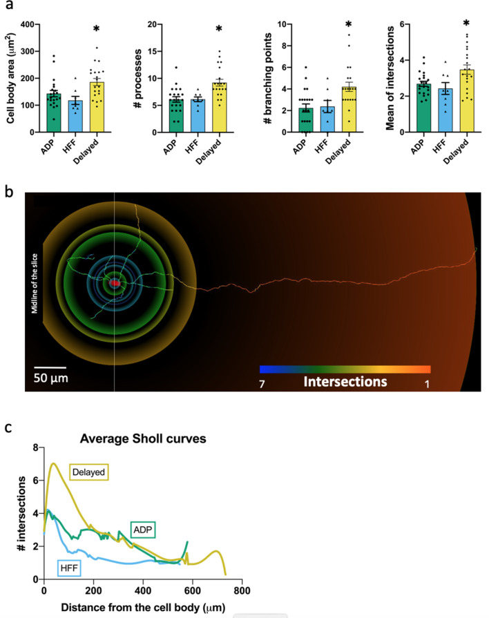
Figure 5. Morphological properties of the Sst-expressing neuron subtypes.

(a) Significantly different morphological characteristics between the three electrophysiological subtypes. Mean intersections, a parameter from Sholl analysis, shows how many times a circle of one radius intersects neuronal processes. All graphs show means ± SEM for ADP neurons (n = 21), HFF neurons (n = 8) and Delayed neurons (n = 20). Asterisks indicate values statistically different from two others (cell body area: F(2, 46)=5.565, p=0.0068; # processes: F(2, 46)=9.745, p=0.0003; # branching points: F(2, 46)=6.974, p=0.0023; mean intersections: F(2, 46)=5.280, p=0.0086). (b) Example of a Delayed neuron morphology with color-coded circles, produced by Sholl analysis: from red to blue for minimum to maximum number of intersections, respectively. White line crossing the cell body corresponds to the direction of the sagittal plane and ‘midline’ indicates center of the slice close to the interpeduncular nuclei (IPN, see also Figure 4a). (c) Average Sholl curves for the electrophysiological subtypes, showing that Delayed neurons had the largest number of intersections within 100 µm from the cell body. Morphology of the traced neurons and individual Sholl curves can be found in Additional files: Figure 5—source datas 12. Figure 5—figure supplements 12.

Figure 5—source data 1. raw data for Figure 5a.
Figure 5—source data 2. File Morphology_Source.zip: source files for morphology and location.
This zip archive contains morphological images of all traced neurons grouped according their electrophysiological profiles. Each subtype’s folder contains a PDF file (with the list of neurons, their original location within the VTA, images of the traced morphology and individual Sholl curves) and two subfolders: ‘3D_gif’ – with *.gif files of the listed neurons; and ‘WaveFront_3D_obj’ – with corresponding *.obj files. *.gif files can be opened by any image viewer. *.obj files save information about the 3D model of the neurons and can be opened/reused with any 3D viewer or graphic software.

Figure 5.

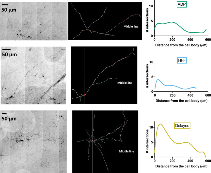
Figure 5—figure supplement 1. Examples of VTA Sst-neuron morphology.

Figure 5—figure supplement 1.

From the left, line by line: black and white-inverted copies of original micrographs of neurobiotin-filled Sst cells of the ADP, HFF and Delayed subtypes defined by clustering according to their electrophysiological features. Middle: reconstructed morphology of the same neurons. Right: the Sholl curves for the same cells.
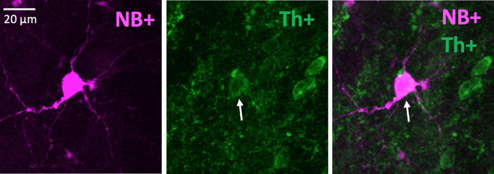
Figure 5—figure supplement 2. An example of a neurobiotin-filled (NB+) Delayed neuron with positive Th staining.

Figure 5—figure supplement 2.