Abstract
Objective The present study aimed to assess in vitro the antibacterial activity, cytotoxicity, and the expression of prostaglandin E 2 (PGE 2 ) of Bexident post topical gel (BP).
Materials and Methods The broth dilution test was performed to analyze the antimicrobial activity of BP against Staphylococcus aureus , Escherichia coli , and Streptococcus mutans . Minimal bactericidal concentrations (MBCs) and minimal inhibitory concentrations (MICs) were assessed. Cytotoxic activity was performed by the MTT (tetrazolium dye) method on human gingival fibroblast (HGF), human bone cells (HBC), and human pulp cells (HPC) (from primary cell culture) and HGF-1 from American Type Culture Collection. The expression of PGE 2 produced by RAW 264.7 cells was determined by ELISA utilizing an Enzyme Immuno-Assay Kit.
Statistical Analysis Shapiro–Wilks normality test and Mann–Whitney U test were performed for all data.
Results The MBCs of BP for S. aureus , E. coli , and S. mutans were found at 25, 50, and 12.5%, respectively. The MICs for the same strains were found at 12.5, 25, and 3.125%. The CC 50 of BP gel for HBC, HPC, and HGF, and HGF-1 were 12.5 ± 1.09, 0.37 ± 0.02, 0.35 ± 0.02, and 20.4 ± 0.02%, respectively. The levels of expression PGE 2 produced by RAW 264.7 cells treated with IL-1β exhibit an inverse dose-dependent effect on the concentrations of BP gel used.
Conclusion Our results indicate that the BP gel has a great antibacterial effect, adequate biocompatibility, showing a decrease in the expression of PGE 2 on cells with previously induced inflammation. Due to the above, its use as a healing agent after oral surgery seems to be adequate.
Keywords: chitosan, chlorhexidine, cytotoxicity, antibacterial properties, Bexident post
Introduction
The phases of the wound-healing process are hemostasis, inflammation, proliferation, and tissue remodeling or resolution. Such as phases are divided in that way only for study purposes; however, indeed are overlapping among them. 1 When the wound-healing process fails, nonhealing or delayed healing wounds, which can generate an increase in health care costs and a high risk of undesirables collateral effects as a secondary infection, excessive intake of nonsteroids analgesics or even opioid agents. Local delivery of drugs has several advantages to systemically administered agents. The dose or formulation concentration provided to the site can be much higher because the therapeutic agent need not be distributed throughout the body before reaching the site of action. 2
Recently, a new product in gel and mouthwash formulations for postexodontia has been introduced to Spain and the Latin American market. 3 The components of the Bexident Post topical gel (BP, ISDIN, Spain) are chitosan, chlorhexidine (CHX), panthenol, and allantoin. Chitosan at 0.5% (5 mg/mg) is constituent of BP gel formulation. Chitosan is a polysaccharide biomaterial composed of (1 → 4)―linked 2-acetamido-2-deoxy-β-D-glucan, (N-acetyl D glucosamine and 2-amino-2-deoxy-β-D-glucan), a natural carbohydrate, cheap, accessible, and obtained through deacetylation of chitin, the main constituent of the hard protective outer case of a mollusk or crustacean being a nontoxic, biodegradable, and biocompatible polymer. 4 5 Chitosan is unique due to the biological origin of polysaccharide that exhibits a cationic nature due to the protonation of its -NH 2 groups at low pH, and can interact with negatively charged compounds like those present in the bacterial membrane and other biological structures. 6 Chitosan also has been studied for its gel/film-forming properties and bioadhesion capacity due to potential applications in pharmaceuticals. 7 The pKa value of chitosan (6.3) is adequate to buffer the oral pH as much as necessary to avoid the harmful action of acids in the oral environment. 8 The antibacterial activity of chitosan is attributable to their polycationic structure. The electrostatic interaction between the chitosan structure and the anionic components from the surface of the microorganisms (proteins and lipopolysaccharides from bacteria membrane). The interactions are facilitated due to the surrounding pH is lower than the pKa of chitosan. 9
The formulation of BP also contains 0.2% (2 mg/mg) of CHX. CHX is a disinfectant widely used in dentistry which has not only a limited action over Gram-negative bacteria but also is active against most Gram-positive bacteria and also fungi. 10 Due to their dicationic nature (pH 5.5–6.0), CHX can donate protons. 11 That pH value could make it compatible with chitosan. CHX has a certain level of cytotoxicity; however, its topical application is tolerated. 12 Regarding panthenol, this compound is also known as Vitamin B 5 . It is a barrier to enhancing humectant. Allantoin is a compound extracted from a plant which is thought to stimulate cell growth and wound-healing process while also depressing inflammation. 13
To our best knowledge, there is no independent in vitro study to assess the biological effects of BP topical gel published so far. Due to that, the present study aimed to assess the antibacterial activity, cytotoxicity, and the expression of prostaglandin E 2 (PGE 2 ) of Bexident Post topical gel (BP) in vitro .
Materials and Methods
Antibacterial Activity
For this study, the bacterial strains used from the stock culture collection of the educative institution. The strains used were Staphylococcus aureus (American Type Culture Collection [ATCC] 29123), Escherichia coli (ATCC 35218), and Streptococcus mutans. S. mutans wild strain was previously isolated and stablished from one patient to the region from southwest Mexico and characterized properly by several tests. The experiments to determine the antimicrobial activity were performed as previously described for the broth dilution test. 14 Each strain was cultured in selective media. After overnight incubation, each strain was transferred to Mueller–Hinton agar and incubated for 24 hours. The starting suspension of each organism was matched with 0.5 McFarland standard. Then, each bacterial suspension was diluted at 1:20. In each 96-well microplate was placed Mueller–Hinton broth (100 µL) and twofold dilutions of the BP were carried out. Then, 5 µL were inoculated per well of the respective strain. For the validity of this test, sterility and bacterial growth controls were used. Tests were performed at two independent experiments for each strain. The inoculated microplates were incubated at 37°C for 24 hours. The presence or absence of turbidity in each well was recorded. Optical absorbance was determined at 595 nm. The results were expressed as a percentage of the viable bacteria compared with the controls. Subcultures on Mueller-Hinton agar plates of the wells without turbidity were performed. The minimal inhibitory concentration (MIC) and the minimal bactericidal concentration (MBC) were identified as it was previously reported. 15
Cytotoxic Assay
Cell Culture
The procedure was performed as previously described for the research group; further information of the technique could be consulted at the reference. 16 Prior approval from the bioethics committee, the tissues for primary culture of human bone cells (HBC), human pulp cells (HPC), and human gingival fibroblast (HGF) were obtained from a 26-year-old patient previously written informed consent. HGF-1 was obtained from ATCC (ATCC CRL-2014).
Assay for Cytotoxic Activity
The total amount of 1 × 10 5 cells/mL (HBC, HPC, HGF, and HGF-1) were placed into each well of the microplate for this test. The BP was inoculated at different dilutions and incubated for a further 24 hours. The MTT assay (Sigma–Aldrich, St Louis, Missouri, United States) was used to assess the cytotoxicity of BP. The details over the procedure are previously reported for the research group. 17 Total 50% of cytotoxic concentration (CC 50 ) was calculated. The experiments were conducted according to ISO 10990–5 Biological evaluation of medical devices—Part 5: Tests for in vitro cytotoxicity. 18
Prostaglandin E 2 Expression
RAW 264.7 cells (ATCC TIB-71 macrophages (1×10 5 cells/mL) were treated with different dilutions (%) of BP gel for 30 minutes in the fresh culture medium. The experiment was performed accordingly to the previous report for the research group by ELISA utilizing an Enzyme Immuno-Assay Kit (R&D Systems; Minneapolis, Minnesota, United States). 19 All experiments were performed in triplicate.
Statistical Analysis
Assessments are expressed as the mean ± standard deviation (SD). Shapiro–Wilks normality test and Mann–Whitney U test were performed for all data. SPSS (SPSS, Inc.; Chicago, Illinois, United States) software was used for the calculations. The p -values minor or equal to 0.05 are considered statically significant for the testing differences in medians from the data.
Results and Discussion
Antibacterial Activity
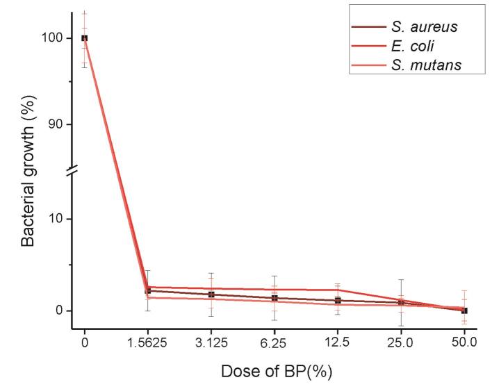
In the broth dilution test, the variability in the sensitivity results for the antimicrobial agents lies to different inoculum size, pH, and incubation time. In the current study, the highest dose tested was 50% of the BP gel (equivalent to chitosan at 25 mg/mg and CHX at 1 mg/mg) due to the procedure of the test is performed, it requires that the same amount of the gel (100 µL) and broth (100 µL) be placed at the initial dose wells, and then serial dilutions are made. However, the bacterial inhibition of all strains under study was complete, since no colony was observed in subcultures. The percentages of bacterial growth are enlisted in Table 1 and Fig. 1 , respectively. At the maximum dose tested, the strain that had the highest growth inhibition was S. aureus , showing statistical significance. However, there was no significant difference in the bacterial growth for S. aureus compared with S. mutans at 25, 6.25, and 3.125% of BP gel.
Table 1. Results of the microdilution broth test show the bacterial growth for the strains under study exposed to Bexident post topical gel.
| Dose of BP (%) | Staphylococcus aureus | Escherichia coli | Streptococcus mutans |
|---|---|---|---|
| Mean (%) ± SD | Mean (%) ± SD | Mean (%) ± SD | |
| Abbreviations: BP, Bexident post topical gel; SD, standard deviation. Note: Same letters are not statistical significant different. Uppercase letters represent differences between the doses and lowercase letters represent differences between the strains using in both Mann–Whitney U test. p ≤ 0.05. | |||
| 0.0 | 100.0 A,a ± 3.39 | 100.0 A,a ± 1.15 | 100.0 A,a ± 2.82 |
| 1.5625 | 2.1750 B,a ± 2.20 | 2.5609 B,a ± 0.10 | 1.4380 B,b ± 0.24 |
| 3.125 | 1.7528 B,a ± 2.35 | 2.4176 B,b ± 1.11 | 1.2500 B,a ± 0.98 |
| 6.25 | 1.3849 C,a ± 2.41 | 2.3062 B,b ± 0.38 | 0.9958 C,a ± 1.01 |
| 12.5 | 1.1254 C,a ± 1.54 | 2.2654 B,b ± 0.68 | 0.6507 C,c ± 0.56 |
| 25.0 | 0.8712 C,a ± 2.52 | 1.1380 C,b ± 0.51 | 0.5828 C,a ± 0.74 |
| 50.0 | 0.0035 D,a ± 0.17 | 0.0544 D,b ± 1.16 | 0.3580 D,c ± 1.81 |
Fig. 1.
Graph of broth dilution test against different strains.
Overall, E. coli showed less growth inhibition than S. aureus in all doses tested. For S. aureus , E. coli , and S. mutans , the MBCs were found at 25, 50, and 12.5% of the BP gel, respectively. The MICs for the same strains were found at 12.5, 25, and 3.125%. The chitosan and CHX mixture have been used in diverse research groups. In other work, it was found the MICs of the chitosan solution at 5 g/L and the CHX at 0.125 g/L against S. mutans . 20 The chitosan/CHX gel (at 2%, 1:1 ratio) was studied for the reduction of Enterococcus faecalis in vitro , showing good results for endodontic retreatment. 21 Besides, the combination of chitosan/CHX was used as a mouthwash in patients with periodontitis, and a significant reduction in the number of bacteria of those patients was found. 22 It has even been proposed that chitosan has a synergistic effect with CHX 23 since in different studies a better effect has been observed when both compounds are combined compared with CHX alone. 24 25 26
Cytotoxic Activity
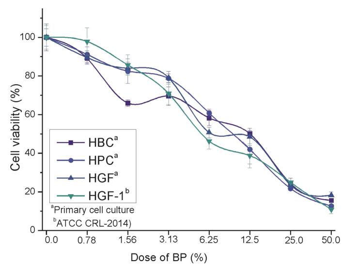
Table 2 Fig. 2 showed the results of the test. The CC 50 was 12.5 ± 1.09, 9.8 ± 0.01, 8.2 ± 0.08, and 5.7 ± 0.4% for HBC, HPC, HGF, and HGF-1, respectively. For HBC, there was a significant difference between all doses, except for 3.125% in comparison to 1.5625% of BP gel, while for HPC and HFG, both showed significant differences at each dose. For HFG, there were statically significant differences between all doses except the control (0%) compared with the lowest dose tested. The most susceptible cell line was HFG-1 at the highest dose tested. In general, there is no significant difference between the cytotoxicity exhibit from HPC and HGF. According to that, slight cytotoxicity and reduced side effects of chitosan have been reported. 27 Similarly, in vivo studies have been reported an acceptable compatibility and that over 1.5 g/kg for rats while 5.2 to 10 g/kg for mice, and 150 mg/kg for dogs as the 50% lethal dose (LD50). 28 29 30 On the other hand, in the current study were found dissimilar results of BP gel for cytotoxicity of HGF cells (ATCC cell line compared with primary cell culture). In other studies, it has been described different responsiveness to compare both types of cells since ATCC cell line is constituted for immortalized cells. 31
Table 2. Cytotoxicity results of Bexident post topical gel in contact with different cell lines.
| Dose of BP (%) | HBC | HPC | HGF | HGF-1 |
|---|---|---|---|---|
| Mean (%) ± SD | Mean (%) ± SD | Mean (%) ± SD | Mean (%) ± SD | |
| Abbreviations: BP, Bexident post topical gel; HBC, human bone cells; HPC, human pulp cell; SD, standard deviation. Note: Same letters are not statistical significant different. Uppercase letters represent differences between the doses and lowercase letters represent differences between the cell lines using in both Mann–Whitney U test. p ≤ 0.05. | ||||
| 0.0 | 100.0 A,a ± 7.0 | 100.0 A,a ± 4.5 | 100.0 A,a ± 1.3 | 100.0 A,a ± 6.1 |
| 0.78125 | 89.1 B,a ± 2.5 | 91.1 B,a ± 4.0 | 89.5 B,a ± 3.5 | 97.8 A,b ± 7.2 |
| 1.5625 | 66.0 C,a ± 1.7 | 82.5 C,b ± 6.3 | 84.1 C,b ± 3.1 | 85.7 B,b ± 5.3 |
| 3.125 | 69.6 C,a ± 4.8 | 78.8 D,b ± 2.3 | 79.1 D,b ± 3.3 | 70.9 C,a ± 5.7 |
| 6.25 | 58.2 D,a ± 3.9 | 60.8 E,a ± 2.1 | 50.7 E,b ± 2.8 | 46.3 D,c ± 4.1 |
| 12.5 | 50.1 E,a ± 1.7 | 42.0 F,b ± 7.5 | 48.5 E,a ± 4.5 | 38.7b E,c ± 6.3 |
| 25.0 | 24.2 F,a ± 2.8 | 21.7 G,b ± 1.0 | 23.5 F,a ± 1.4 | 24.9 F,a ± 0.3 |
| 50.0 | 15.5 G,a ±1.1 | 12.7 H,b ±1.9 | 18.2 G,c ±1.8 | 10.9 G,b ± 2.3 |
Fig. 2.
Relative cell viability for different cell lines in direct contact with Bexident post topical gel.
Prostaglandin E 2 Expression
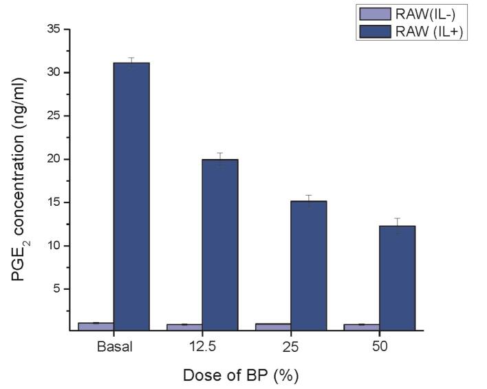
The activity of COX-1 and COX-2 is directly related to prostaglandin production. The production of prostaglandins induced by several cell types when a proinflammatory event occurs. 32 It is known that macrophages produce PGE 2 after interleukine-1β (IL-1β) activation due to this cytokine is a mediator of the proinflammatory response and is related to cell proliferation, differentiation, and apoptosis. It has been described that in the initial wound stage, the tumor necrosis factor (TNF)-α and IL-1β are produced, while the shutdown of TNF-α and IL-1β at such stage of the inflammation facilitates wound healing. In contrast, the prolonged blockade may negatively affect the healing process. 33 On the contrary, in an in vitro model, it has been previously reported that 5 ng/mL of IL-1β as the optimal dose for the production of pro-inflammatory cytokine. 34 In the present study, it was evaluated the effect of chitosan on cyclooxygenase pathway by PGE 2 expression in RAW 264.7 cells treated with IL-1β, which significantly enhanced the PGE 2 production and the addition of BP gel resulted in a statistically significant decrease of PGE 2 production in IL 1β-treated cells. Note that PGE 2 production by RAW 264.7 cells wasinversely dose-dependent to the dilutions of BP gel. However, in the absence of IL-1β activation did not reduce the basal PGE 2 formation. The results of the PGE 2 expression are shown in Table 3 Fig. 3 .
Table 3. Results of prostaglandin E2 production after interleukin-1β activation using enzyme-linked immunosorbent assay method.
| Dose of BP (%) | RAW 264.7 cells | |
|---|---|---|
| Without IL-1β Mean (ng/mL) ± SD |
With IL-1β Mean (ng/mL) ± SD |
|
| Abbreviations: BP, Bexident post topical gel; IL, interleukin; SD, standard deviation. Note: Same letters are not statistically significant different. Uppercase letters represent differences between the doses and, lowercase letters represent differences between the IL-1β (absent or present) stimulation using in both Mann–Whitney U test. p ≤ 0.05. | ||
| Basal | 1.09 A,a ± 0.08 | 31.12 A,b ± 0.59 |
| 12.5 | 0.92 A,a ± 0.08 | 19.93 B,b ± 0.82 |
| 25 | 0.98 A,a ± 0.07 | 15.14 C,b ± 0.77 |
| 50 | 0.93 A,a ± 0.09 | 12.29 D,b ± 0.88 |
Fig. 3.
Prostaglandin E 2 production by RAW 264.7 macrophage cell line in contact with Bexident post topical gel.
Regarding clinical use, it has been reported the results of different studies using BP showing good results. For instance, a clinical study for assessing the wound healing process and, level of trismus, facial swelling, and pain performed in 50 patients with bilaterally third molars indicated for oral surgery, which were randomly assigned to a placebo group or an experimental BP group; and it was found during 1 to 14 day that no differences in mouth opening, facial swelling, and pain were showed in comparison to the controls; however, the wound healing score was better in the BP group, both on day 7 and day 14 of follow-up. 35 Probably, the BP gel application after surgical injury resulted in a faster healing wound that could be attributed to the decrease in PGE 2 production due to chitosan anti-inflammatory action.
Some clinical indications on the safety of the BP gel remain unclear such as its use in children. On the other hand, the warning on the label instructs the patients allergic to shellfish to consult a dentist. Chitin is covalently linked to proteins—when came from crustaceous—due to that the warning about the potential allergy could be presented. It has been reported in a pilot study that glucosamine is probably safe for patients with shellfish allergy, 36 which is relevant since glucosamine and N-acetyl-D -glucosamine are copolymers that constituted the chitosan. 9 However, the isolated, when the chitosan has been purified as a plain polysaccharide, that process reduces its allergy potential 37 since the allergic to shellfish is detonated by tropomyosin protein—which causes IgE-mediated hypersensitivity after ingestion, 38 not by a polysaccharide. Even so, the chitosan, by itself, like any substance could trigger an allergic reaction even if the patient does not have any previous allergic reaction, so the patient should be instructed so that if he/her observes any adverse reaction when applying the BP gel, discontinue its application, and go to the dental practitioner office for an examination. Therefore, in future studies that lead to answering these questions should be conducted.
Recently, it has been developing materials like membranes, beads, hydrogels, and fibers from chitosan for potential use in dentistry. Cahyanto research team developed a chitosan-collagen membrane from Barramundi ( Latescalcarifer ) for guided tissue regeneration techniquefor periodontal therapy. 39 Further, periodontal ligament cells sheet and arginine-glycyl-aspartic acid-modified chitosan scaffold for periodontal tissue regeneration showed biocompatibility and high cell proliferation in a horizontal periodontal defect model. 40 On the other hand, chitosan hydrogels (chitosan-H-propolis, chitosan-H-propolis-nystatin, and chitosan-H-nystatin) treated dentine gave significantly high shear bond values by their swelling properties and antioxidant effect. 41 Besides, the anticarious mats of chitosan fibers containing Garcinia mangostana extract have shown promising results to prevent dental caries. 42 43
Also, chitosan has been widely used for diverse purposes as in solutions for regenerative medicine. This encompasses the inclusions of nanoparticles by electrospinning, which is exemplified by the fact that chitosan and silk fibroin composite, collagen chitosan, agarose chitosan, and chitosan with a fiber-forming agent have broad research for their use in neural tissue regeneration, bone regeneration, wound dressings, mucoadhesive mats, and other applications. 6
Conclusion
The limitations of this study were that, like all in vitro studies, the data of the results are not fully extrapolated to the clinical performance. However, the in vitro design of this study allowed us to determine the biological properties of BP gel. The results of the present study indicate that the BP gel has a great antibacterial effect, adequate biocompatibility, showing a decrease in the expression of PGE 2 on cells with previously induced proinflammatory activation. Due to the above, its use as a therapeutic agent after oral surgery seems to be adequate. In the future, genotoxic effects and other in vivo tests in animal models with appropriate follow-up time intervals are necessary to determine if prolonged use for other clinical conditions does not represent a risk.
Acknowledgments
L.A.F. thanks Cátedras-CONACYT program, Posgrade Division and Dentistry School, Universidad Autónoma Benito Juárez de Oaxaca for their support. R.T.R thanks Cuerpo académico “Investigación en Salud” UABJO-CA-63. R.G.C thanks Red de Farmacoquímicos. R.T.R. and A.M.R. thank the Strengthening Program in Educational Quality 2018 years 2019 from Secretary of Public Education.
Funding Statement
Funding This study received its financial support from CONACyT (project number CB-2016–01–284495) assigned to RTR and DGAPA-PAPIIT: IA205518 assigned to RGC.
Footnotes
Conflict of Interest None declared.
References
- 1.Guo S, Dipietro L A. Factors affecting wound healing. J Dent Res. 2010;89(03):219–229. doi: 10.1177/0022034509359125. [DOI] [PMC free article] [PubMed] [Google Scholar]
- 2.Hersh E V, Moore P A. Comment on controlling dental post-operative pain and the intraoral local delivery of drugs. Curr Med Res Opin. 2015;31(12):2185–2187. doi: 10.1185/03007995.2015.1109504. [DOI] [PubMed] [Google Scholar]
- 3.Lopez-Lopez J, Jan-Pallí E, lez-Navarro B G, Jané-Salas E, Estrugo-Devesa A, Milani M. Efficacy of chlorhexidine, dexpanthenol, allantoin and chitosan gel in comparison with bicarbonate oral rinse in controlling post-interventional inflammation, pain and cicatrization in subjects undergoing dental surgery. Curr Med Res Opin. 2015;31(12):2179–2183. doi: 10.1185/03007995.2015.1108909. [DOI] [PubMed] [Google Scholar]
- 4.Arbia W, Arbia L, Adour L, Amrane A. Chitin extraction from crustacean shells using biological methods: a review. Food Technol Biotechnol. 2013;51(01):12–25. [Google Scholar]
- 5.Sarwar M S, Huang Q, Ghaffar A et al. A smart drug delivery system based on biodegradable chitosan/poly(allylamine hydrochloride) blend films. Pharmaceutics. 2020;12(02):131. doi: 10.3390/pharmaceutics12020131. [DOI] [PMC free article] [PubMed] [Google Scholar]
- 6.Qasim S B, Zafar M S, Najeeb S et al. Electrospinning of chitosan-based solutions for tissue engineering and regenerative medicine. Int J Mol Sci. 2018;19(02):407. doi: 10.3390/ijms19020407. [DOI] [PMC free article] [PubMed] [Google Scholar]
- 7.Vargas Villanueva J G, Sarmiento Huertas P A, Galan F S, Esteban Rueda R J, Briceño Triana J C, Casas Rodriguez J P. Bio-adhesion evaluation of a chitosan-based bone bio-adhesive. Int J Adhes Adhes. 2019;92:80–88. [Google Scholar]
- 8.Tarsi R, Muzzarelli R A, Guzmán C A, Pruzzo C. Inhibition of Streptococcus mutans adsorption to hydroxyapatite by low-molecular-weight chitosans. J Dent Res. 1997;76(02):665–672. doi: 10.1177/00220345970760020701. [DOI] [PubMed] [Google Scholar]
- 9.Kong M, Chen X G, Xing K, Park H J. Antimicrobial properties of chitosan and mode of action: a state of the art review. Int J Food Microbiol. 2010;144(01):51–63. doi: 10.1016/j.ijfoodmicro.2010.09.012. [DOI] [PubMed] [Google Scholar]
- 10.Rahimi S, Janani M, Lotfi M et al. A review of antibacterial agents in endodontic treatment. Iran Endod J. 2014;9(03):161–168. [PMC free article] [PubMed] [Google Scholar]
- 11.Basrani B R, Manek S, Sodhi R N, Fillery E, Manzur A. Interaction between sodium hypochlorite and chlorhexidine gluconate. J Endod. 2007;33(08):966–969. doi: 10.1016/j.joen.2007.04.001. [DOI] [PubMed] [Google Scholar]
- 12.Hidalgo E, Dominguez C.Mechanisms underlying chlorhexidine-induced cytotoxicity Toxicol In Vitro 200115(4-5)271–276. [DOI] [PubMed] [Google Scholar]
- 13.Araújo L U, Grabe-Guimarães A, Mosqueira V C, Carneiro C M, Silva-Barcellos N M. Profile of wound healing process induced by allantoin. Acta Cir Bras. 2010;25(05):460–466. doi: 10.1590/s0102-86502010000500014. [DOI] [PubMed] [Google Scholar]
- 14.Argueta-Figueroa L, Delgado-García J J, García-Contreras R et al. Mineral trioxide aggregate enriched with iron disulfide nanostructures: an evaluation of their physical and biological properties. Eur J Oral Sci. 2018;126(03):234–243. doi: 10.1111/eos.12408. [DOI] [PubMed] [Google Scholar]
- 15.Kim K S, Anthony B F. Importance of bacterial growth phase in determining minimal bactericidal concentrations of penicillin and methicillin. Antimicrob Agents Chemother. 1981;19(06):1075–1077. doi: 10.1128/aac.19.6.1075. [DOI] [PMC free article] [PubMed] [Google Scholar]
- 16.Cuellar-Flores M, Acosta-Torres L S, Martínez-Alvarez O et al. Effects of alkaline treatment for fibroblastic adhesion on titanium. Dent Res J (Isfahan) 2016;13(06):473–477. doi: 10.4103/1735-3327.197043. [DOI] [PMC free article] [PubMed] [Google Scholar]
- 17.Torres-Gómez N, Nava O, Argueta-Figueroa L, García-Contreras R, Baeza-Barrera A, Vilchis-Nestor A R. Shape tuning of magnetite nanoparticles obtained by hydrothermal synthesis: effect of temperature. J Nanomater. 2019;2019:7.921273E6. [Google Scholar]
- 18.Li W, Zhou J, Xu Y. Study of the in vitro cytotoxicity testing of medical devices. Biomed Rep. 2015;3(05):617–620. doi: 10.3892/br.2015.481. [DOI] [PMC free article] [PubMed] [Google Scholar]
- 19.Garcia-Contreras R, Sugimoto M, Umemura N et al. Alteration of metabolomic profiles by titanium dioxide nanoparticles in human gingivitis model. Biomaterials. 2015;57:33–40. doi: 10.1016/j.biomaterials.2015.03.059. [DOI] [PubMed] [Google Scholar]
- 20.Bae K, Jun E J, Lee S M, Paik D I, Kim J B. Effect of water-soluble reduced chitosan on Streptococcus mutans, plaque regrowth and biofilm vitality. Clin Oral Investig. 2006;10(02):102–107. doi: 10.1007/s00784-006-0038-3. [DOI] [PubMed] [Google Scholar]
- 21.Savitha A, SriRekha A, Vijay R. Ashwija, Champa C, Jaykumar T. An in vivo comparative evaluation of antimicrobial efficacy of chitosan, chlorhexidine gluconate gel and their combination as an intracanal medicament against Enterococcus faecalis in failed endodontic cases using real time polymerase chain reaction (qPCR) Saudi Dent J. 2019;31(03):360–366. doi: 10.1016/j.sdentj.2019.03.003. [DOI] [PMC free article] [PubMed] [Google Scholar]
- 22.Nair G, Panchal A, Gandhi B, Shah S, Shah R. Evaluation and comparision of antimicrobial effects of chlorhexidine (CHX) and chitosan (CHT) mouthwash in chronic periodontitis (CGP) patients: a clinicomicrobiological study. IOSR J Dent Med Sci. 2017;16(10):26–32. [Google Scholar]
- 23.Decker E M, von O hle, C, Weiger R, Wiech I, Brecx M. A synergistic chlorhexidine/chitosan combination for improved antiplaque strategies. J Periodontal Res. 2005;40(05):373–377. doi: 10.1111/j.1600-0765.2005.00817.x. [DOI] [PubMed] [Google Scholar]
- 24.Chronopoulou L, Nocca G, Castagnola M et al. Chitosan based nanoparticles functionalized with peptidomimetic derivatives for oral drug delivery. N Biotechnol. 2016;33(01):23–31. doi: 10.1016/j.nbt.2015.07.005. [DOI] [PubMed] [Google Scholar]
- 25.Ambrogi V, Pietrella D, Nocchetti M et al. Montmorillonite-chitosan-chlorhexidine composite films with antibiofilm activity and improved cytotoxicity for wound dressing. J Colloid Interface Sci. 2017;491(01):265–272. doi: 10.1016/j.jcis.2016.12.058. [DOI] [PubMed] [Google Scholar]
- 26.Giunchedi P, Juliano C, Gavini E, Cossu M, Sorrenti M. Formulation and in vivo evaluation of chlorhexidine buccal tablets prepared using drug-loaded chitosan microspheres. Eur J Pharm Biopharm. 2002;53(02):233–239. doi: 10.1016/s0939-6411(01)00237-5. [DOI] [PubMed] [Google Scholar]
- 27.Kean T, Thanou M. Biodegradation, biodistribution and toxicity of chitosan. Adv Drug Deliv Rev. 2010;62(01):3–11. doi: 10.1016/j.addr.2009.09.004. [DOI] [PubMed] [Google Scholar]
- 28.Minami S, Oh-oka M, Okamoto Y et al. Chitosan-inducing hemorrhagic pneumonia in dogs. Carbohydr Polym. 1996;29(03):241–246. [Google Scholar]
- 29.Aranaz I, Acosta N, Civera C et al. Cosmetics and cosmeceutical applications of chitin, chitosan and their derivatives. Polymers (Basel) 2018;10(02):213. doi: 10.3390/polym10020213. [DOI] [PMC free article] [PubMed] [Google Scholar]
- 30.Arai K. Toxicity of chitosan. Bull Tokai Reg Fish Lab. 1968;56:86–94. [Google Scholar]
- 31.Thonemann B, Schmalz G, Hiller K A, Schweikl H. Responses of L929 mouse fibroblasts, primary and immortalized bovine dental papilla-derived cell lines to dental resin components. Dent Mater. 2002;18(04):318–323. doi: 10.1016/s0109-5641(01)00056-2. [DOI] [PubMed] [Google Scholar]
- 32.Ricciotti E, FitzGerald G A. Prostaglandins and inflammation. Arterioscler Thromb Vasc Biol. 2011;31(05):986–1000. doi: 10.1161/ATVBAHA.110.207449. [DOI] [PMC free article] [PubMed] [Google Scholar]
- 33.Morand D N, Davideau J L, Clauss F, Jessel N, Tenenbaum H, Huck O. Cytokines during periodontal wound healing: potential application for new therapeutic approach. Oral Dis. 2017;23(03):300–311. doi: 10.1111/odi.12469. [DOI] [PubMed] [Google Scholar]
- 34.Koh T, Murakami Y, Tanaka S, Machino M, Sakagami H. Re-evaluation of anti-inflammatory potential of eugenol in IL-1β-stimulated gingival fibroblast and pulp cells. In Vivo. 2013;27(02):269–273. [PubMed] [Google Scholar]
- 35.Madrazo-Jiménez M, Rodríguez-Caballero Á, Serrera-Figallo M Á et al. The effects of a topical gel containing chitosan, 0.2% chlorhexidine, allantoin and despanthenol on the wound healing process subsequent to impacted lower third molar extraction. Med Oral Patol Oral Cir Bucal. 2016;21(06):e696–e702. doi: 10.4317/medoral.21281. [DOI] [PMC free article] [PubMed] [Google Scholar]
- 36.Gray H C, Hutcheson P S, Slavin R G. Is glucosamine safe in patients with seafood allergy? J Allergy Clin Immunol. 2004;114(02):459–460. doi: 10.1016/j.jaci.2004.05.050. [DOI] [PubMed] [Google Scholar]
- 37.Waibel K H, Haney B, Moore M, Whisman B, Gomez R. Safety of chitosan bandages in shellfish allergic patients. Mil Med. 2011;176(10):1153–1156. doi: 10.7205/milmed-d-11-00150. [DOI] [PubMed] [Google Scholar]
- 38.Fuller H, Goodwin P, Morris G. An enzyme-linked immunosorbent assay (ELISA) for the major crustacean allergen, tropomyosin, in food. Food Agric Immunol. 2006;17(01):43–52. [Google Scholar]
- 39.Susanto A, Satari M H, Abbas B, Koesoemowidodo R SA, Cahyanto A. Fabrication and characterization of chitosan-collagen membrane from barramundi (lates calcarifer) scales for guided tissue regeneration. Eur J Dent. 2019;13(03):370–375. doi: 10.1055/s-0039-1698610. [DOI] [PMC free article] [PubMed] [Google Scholar]
- 40.Amir L R, Soeroso Y, Fatma D et al. Periodontal ligament cell sheets and RGD-modified chitosan improved regeneration in the horizontal periodontal defect model. Eur J Dent. 2020;14(02):306–314. doi: 10.1055/s-0040-1709955. [DOI] [PMC free article] [PubMed] [Google Scholar]
- 41.Perchyonok V T, Zhang S, Grobler S R, Oberholzer T G. Insights into and relative effect of chitosan-H, chitosan-H-propolis, chitosan-H-propolis-nystatin and chitosan-H-nystatin on dentine bond strength. Eur J Dent. 2013;7(04):412–418. doi: 10.4103/1305-7456.120666. [DOI] [PMC free article] [PubMed] [Google Scholar]
- 42.Samprasit W, Kaomongkolgit R, Sukma M, Rojanarata T, Ngawhirunpat T, Opanasopit P. Mucoadhesive electrospun chitosan-based nanofibre mats for dental caries prevention. Carbohydr Polym. 2015;117(01):933–940. doi: 10.1016/j.carbpol.2014.10.026. [DOI] [PubMed] [Google Scholar]
- 43.Samprasit W, Rojanarata T, Akkaramongkolporn P, Ngawhirunpat T, Kaomongkolgit R, Opanasopit P. Fabrication and in vitro/in vivo performance of mucoadhesive electrospun nanofiber mats containingα-mangostin. AAPS PharmSciTech. 2015;16(05):1140–1152. doi: 10.1208/s12249-015-0300-6. [DOI] [PMC free article] [PubMed] [Google Scholar]


