Abstract
We present a case of ectopic insertion of the whole tendon of the pectoralis minor muscle associated with impingement syndrome. Based on this case, we performed a review of the literature focused on the association between this ectopic tendon insertion and anterior shoulder pain.
Keywords: Pectoralis minor muscle, Ultrasonography, Shoulder
Introduction
The pectoralis minor muscle (PMi) generally originates from the third to fifth ribs and usually inserts on the medial and superior margins of the anterior portion of the coracoid process. Unusual insertions of the PMi are more frequent than previously thought and have been described in 15–37.8% of cadaveric studies [1–3]. Imaging techniques seem not to be highly sensitive in detecting these anomalous insertions, ranging from 1.5 to 13.4% in MRI [4] and 9.5% in ultrasound [5]. The abnormal insertion may affect the whole tendon or, more frequently, part of it. These anomalous insertions include the joint capsule, greater and lesser tuberosities, coracoacromial ligament, supraspinatus tendon, glenoid labrum, coracobrachialis muscle, anatomical neck of the humerus, and clavicle [2]. Some authors consider that tendon attachment to the joint capsule can cause shoulder pain or discomfort related to coracoid impingement syndrome, shoulder stiffness, or a superior labral lesion [6]. When the whole tendon overpasses the coracoid process, tendinopathy (thickening) and a snapping sensation may be produced due to sliding of the tendon over the coracoid process [7]. Here we present an unusual case, in which the whole PMi tendon overpasses the coracoid process to insert into the greater tuberosity, causing anterosuperior shoulder pain.
Case report
A 72-year-old female patient with 6-month ongoing right shoulder pain and anterosuperior clicking was sent to our center for ultrasound-guided injection of the acromioclavicular joint (ACJ). Clinical records and previous medical images of the patient were reviewed before ultrasound. On radiography, small calcifications adjacent to the coracoid process and mild degenerative glenohumeral joint osteoarthritis (GHJ OA) were observed. GHJ injection performed by a general practitioner had led to symptomatic relief, but pain recurred after 2 weeks. Next, MRI was requested to assess the integrity of rotator cuffs and to quantify GHJ OA. The reported findings included moderate-to-advanced supraspinatus and infraspinatus tendinopathies, fraying of cranial subscapularis fibers without definite tear, a moderate volume of fluid that distended the long head of the biceps tendon and the subdeltoid bursa, and moderate ACJ OA. No abnormalities regarding the PMi insertion were reported.
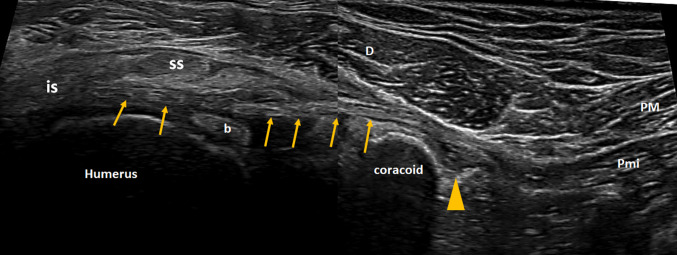
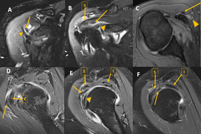
Based on these data, an ultrasound-guided ACJ injection was performed. The injection led to partial long-term pain relief (> 50%, 6 months follow up). Ultrasound exam confirmed most of the findings reported in radiography and MRI. In addition, the anomalous PMi tendon was clearly depicted overlying the coracoid process, over an intertwining bursa, and passing at the rotator interval over the biceps tendon and under the supraspinatus and infraspinatus tendons toward the greater tuberosity, into which it inserted (Fig. 1). These findings were retrospectively identified in MRI images (Fig. 2).
Fig. 1.
Composition of two ultrasound images depicting the PMi tendon (arrows) running over the coracoid and entering the rotator interval over the biceps tendon (b) and under the supraspinatus, (SS) and infraspinatus (IS) muscle and tendons. Arrowhead: adventitial bursa. PM pectoralis major, PMi pectoralis minor, D deltoid
Fig. 2.
a, b Coronal T2 fat sat images showing the PMi tendon (arrow) medially to the coracoacromial ligament (arrowhead in a) under the supraspinatus muscle (S) and over the biceps tendon (arrowhead in b). c Axial T2* showing the PMi tendon (arrow) over the coracoid, separated by an adventitial bursa (arrowhead). d, f Sagittal DP fat sat images showing the PMi tendon (arrow) over the coracoid process and bursa (D) between the supraspinatus (S) and the long head of the biceps tendon (arrowhead) (E) and under the supraspinatus (S)–infraspinatus (I) interval. PMi pectoralis minor
Discussion
Little has been published in the medical literature about the prevalence of anomalous insertions of the PMi tendon. Anatomical dissections report a higher prevalence of PMi tendon variants than do imaging techniques, possibly indicating that ultrasound and MRI are relatively insensitive to these variants, even when the studies specifically aim to find them [4, 5]. Several factors may account for this lack of sensitivity.
First of all, this abnormality may involve only a part of the tendon and the small size of this anomalous structure running over the coracoid process may prevent proper visualization. Among the three types described by Le Double, type II involves only part of the tendon and is considered the most frequent [2, 3]. Our case, on the contrary, was a type I variant, i.e., the whole tendon runs without attachment over the coracoid process and the musculotendinous junction of the PMi lies inferior to the coracoid process. This is different than the rarest type III variant, in which the whole muscle (not merely the tendon) runs over the coracoid process, because the musculotendinous junction is superior to the coracoid process (Fig. 3).
Fig. 3.
Schematic representation of PMi variants according to Le Double. a Normal insertion. b Type I. The complete tendon runs over the coracoid process and be inserted at different places. c Type II. Only the aberrant part of the tendon runs over the coracoid process. d Type III. The whole muscle runs over the coracoid process
Second, due to the variety of insertion sites for the anomalous tendon, it may be confused with other anatomical structures such as the coracoacromial, coracoglenoid, or coracohumeral ligaments. In fact, comparison of MRI with arthroscopy resulted in a sensitivity of 64% and specificity of 80% in detecting anomalous insertions of the PMi tendon. The presence of an anomalous PMi tendon is associated with increased variability of insertion, size, and shape of normal shoulder ligaments, inducing a potential misinterpretation of the ectopic PMi tendon as one of these structures [2].
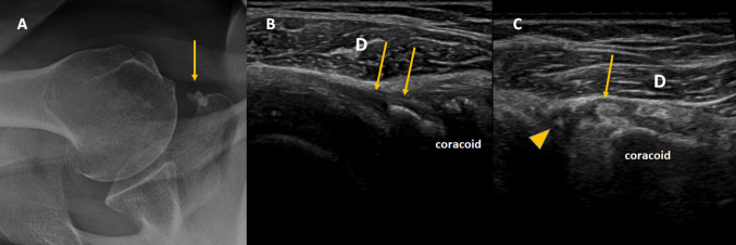
Regarding ultrasound, routine scanning of the shoulder for impingement syndrome focuses mainly on the study of the muscles and tendons of the rotator cuff, ACJ, subdeltoid bursa, and the long head of the biceps tendon, rather than ligaments. Nevertheless, in this case, the coracohumeral ligament was identified as a thickened, calcified structure over the subscapularis muscle (Fig. 4). It was clearly separated from the anomalous PMi tendon, running over the coracoid and the coracoacromial ligament, lateral and perpendicular to the anomalous tendon.
Fig. 4.
a Axial radiography showing calcifications medially to the coracoid process. b Axial ultrasound demonstrated that they were located at the coracoacromial ligament insertion (arrows). c Short axis ultrasound view of the PMi tendon (arrow) over the coracoid and medially to the coracoacromial ligament (arrowhead). d Deltoid
The ectopic PMi tendon may have clinical and surgical implications. It has been considered as a rare cause of impingement; thus, it should be sought only after more common etiologies of impingement have been ruled out. If surgery is required, tenotomy of the ectopic tendon has been recommended to release the cuff completely and relieve tension on the repair [6].
Abnormalities in the PMi muscle and tendons have also been implicated in scapular dyskinesia, because the tightened tendon tilts the coracoid inferiorly [8]. In our case, the adventitial bursa over the coracoid and the calcification of the coracoid insertion of the coracohumeral ligament might be the result of the tension caused by the anomalous tendon. It might have also led to stress on the coracoacromial ligament, thus promoting the development of ACJ OA.
In summary, we have presented a case of ectopic insertion of the whole PMi tendon associated with impingement syndrome. In our opinion, more attention should be paid to the PMi muscle and tendon in shoulder ultrasound in the presence of anterior shoulder pain and clicking, especially when more common causes of shoulder pain do not explain the symptoms after diagnostic ultrasound or guided injection tests.
Compliance with ethical standards
Conflict of interest
The authors declare that they have no conflict of interest to disclose.
Ethical approval
All procedures performed in studies involving human participants were in accordance with the ethical standards of the institutional and/or national research committee and with the 1964 Helsinki Declaration and its later amendments or comparable ethical standards.
Informed consent
Informed consent was obtained from all individual participants included in the study.
Footnotes
Publisher's Note
Springer Nature remains neutral with regard to jurisdictional claims in published maps and institutional affiliations.
References
- 1.Lee KW, Choi YJ, Lee HJ, Gil YC, Kim HJ, Tansatit T, Hu KS. Classification of unusual insertion of the pectoralis minor muscle. Surg Radiol Anat. 2018;40(12):1357–1361. doi: 10.1007/s00276-018-2107-0. [DOI] [PubMed] [Google Scholar]
- 2.Schwarz GM, Hirtler L. Ectopic tendons of the pectoralis minor muscle as cause for shoulder pain and motion inhibition—explaining clinically important variabilities through phylogenesis. PLoS One. 2019;14(6):e0218715. doi: 10.1371/journal.pone.0218715. [DOI] [PMC free article] [PubMed] [Google Scholar]
- 3.Le Double A. Traité des variations du système musculaire de l’homme et de leur signification au point de vue de l’anthropologie zoologique. Paris: Schleicher frères; 1987. [Google Scholar]
- 4.Lee CB, Choi SJ, Ahn JH, Ryu DS, Park MS, Jung SM, et al. Ectopic insertion of the pectoralis minor tendon: inter-reader agreement and findings in the rotator interval on MRI. Korean J Radiol. 2014;15(6):764–770. doi: 10.3348/kjr.2014.15.6.764. [DOI] [PMC free article] [PubMed] [Google Scholar]
- 5.Homsi C, Rodrigues MB, Silva JJ, Stump X, Morvan G. Anomalous insertion of the pectoralis minor muscle: ultrasound findings. J Radiol. 2003;84(9):1007–1011. [PubMed] [Google Scholar]
- 6.Moineau G, Cikes A, Trojani C, Boileau P. Ectopic insertion of the pectoralis minor: implication in the arthroscopic treatment of shoulder stiffness. Knee Surg Sports Traumatol Arthrosc. 2008;16(9):869–871. doi: 10.1007/s00167-008-0535-9. [DOI] [PubMed] [Google Scholar]
- 7.Low S, Tan S. Ectopic insertion of the pectoralis minor muscle with tendinosis as a cause of shoulder pain and clicking. Clin Radiol. 2010;65(3):254–256. doi: 10.1016/j.crad.2009.11.004. [DOI] [PubMed] [Google Scholar]
- 8.Burkhart SS, Morgan CD, Kibler WB. The disabled throwing shoulder: spectrum of pathology Part III: The SICK scapula, scapular dyskinesis, the kinetic chain, and rehabilitation. Arthroscopy. 2003;19(4):404–420. doi: 10.1053/jars.2003.50128. [DOI] [PubMed] [Google Scholar]


