Abstract
Objective
This study was performed to obtain normative data of the distal femoral cartilage thickness in healthy adults by ultrasound.
Methods
This cross-sectional study included 72 healthy adults. The demographic characteristics of the participants were recorded, and the thickness of the femoral articular cartilage was measured using a 5- to 18-MHz linear probe.
Results
Significant statistical difference towards the male side at left medial condyle (P = 0.001) and left lateral condyle (P = 0.009). Weakly positive statistical difference was noted towards the male side at right medial condyle (P = 0.06) and right lateral condyle (P = 0.07). The femoral cartilage thickness in the study participants did not correlate with weight, body mass index, and age (P >0.05). Positive statistical correlation with height noted in right medial condyle, right lateral condyle, right intercondylar area, and left medial condyle.
Conclusion
This study increases the pool of normative data of femoral cartilage thickness measurements. Additionally, the findings of this study emphasize the fact that women have thinner cartilage than men in four of the studied parameters.
Keywords: Femoral cartilage thickness, ultrasound, sex-related differences, normative data, osteoarthritis, knee
Introduction
Articular cartilage is a special type of connective tissue consisting of solid and fluid phases. Its main function is to provide a low-friction surface with a reasonable degree of lubrication and to promote transmission of shear forces to the underlying subchondral bone.1,2 Osteoarthritis of the knee joint is a worldwide health problem associated with irreversible damage to the articular cartilage.3,4 Other causes of cartilage degeneration include trauma and rheumatoid arthritis. Measurement of the femoral cartilage thickness is considered an important tool for the diagnosis and follow-up of osteoarthritis.4 Magnetic resonance imaging is a trusted diagnostic tool in the assessment of femoral cartilage thickness. However, the high cost, limited availability, and relatively long examination time of magnetic resonance imaging limit its use. Ultrasonography is a low-cost, widely available, and dynamic diagnostic imaging tool that is rapidly emerging as an aid in assessment of the femoral cartilage.5,6 Some research articles have described the use of high-resolution ultrasound for evaluation of the femoral cartilage thickness; however, the pool of normative data for the adult population must be enriched to increase the confidence in this modality.
The present study was performed to investigate the femoral cartilage thickness using ultrasound in healthy adults.
Methods
Participants
The participants in this cross-sectional study were recruited from August to October 2019. The inclusion criteria were a clinically healthy status, male or female sex, and age of 18 to 65 years. The exclusion criteria were a history of trauma to the knee joint, surgery involving the lower limb, osteoarthritis, or inflammatory arthritis. Each participant’s sex, age, weight, body mass index (BMI), and height were recorded.
Technique
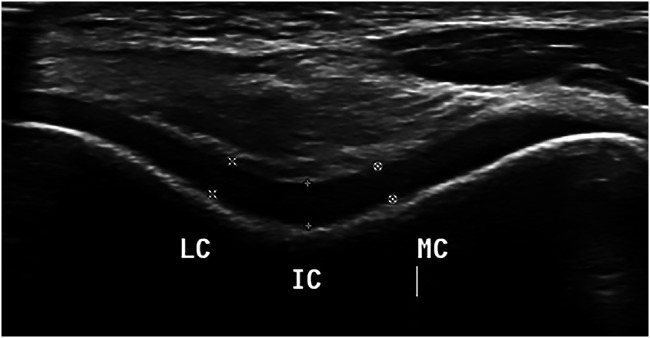
Two radiologists with 10 years of experience performed the ultrasound scans using a linear 5- to 18-MHz linear transducer (Epiq 7 version 1.5 Ultrasound System; Philips, Amsterdam, Netherlands). Each patient was scanned three times. The transducer was positioned in the axial plane on the suprapatellar region. All participants were placed in the supine position with maximum knee flexion. Midpoint measurements were taken from each of three locations in both knees: left medial condyle (LMC), left lateral condyle (LLC), left intercondylar area (LIC), right medial condyle (RMC), right lateral condyle (RLC), and right intercondylar area (RIC) (Figure 1).
Figure 1.
Axial scan of the suprapatellar region with femoral cartilage thickness measurements. IC, intercondylar area; MC, medial condyle; LC, lateral condyle.
Statistical analysis
Statistical analysis was performed using Statistical Package for the Social Sciences (SPSS) version 21 software (IBM Corp., Armonk, NY, USA). A sample size of ≥50 patients was required, with 25 patients per group. Considering a dropout rate of 20%, 72 participants were enrolled in the study. All data are presented as mean ± standard deviation and range. Differences in the measured values were compared between the right and left sides using Wilcoxon’s signed rank test. Correlations between age, weight, height, and BMI were evaluated using Pearson’s correlation coefficient (r). A P value of <0.05 was considered statistically significant.
Ethics
This study was approved by the institutional review board of the College of Medicine, Prince Sattam Bin Abdulaziz University (September 2019, Alkharj). All patients were informed of the study protocol and provided written consent.
Results
Measurements were taken from 144 knees of 72 healthy adult volunteers (36 men, 36 women). The demographic features of the study participants are shown in Table 1. The intra-observer reliability calculations resulted in an overall intra-class correlation coefficient of 0.86. The inter-rater reliability calculations showed an overall intraclass correlation coefficient of 0.79. The femoral cartilage thickness in the study population is shown in Table 2. No difference in the cartilage thickness in the intercondylar region, lateral condyle, or medial condyle was found between the right and left knees. However, the cartilage of the LMC and LLC was significantly thicker in men than in women (P = 0.001 and 0.009, respectively). The cartilage of the RMC and RLC was also thicker in men than in women (P = 0.06 and 0.07, respectively) (Table 3). The femoral cartilage thickness was not correlated with weight, BMI, or age. However, the cartilage thickness of the RMC, RLC, RIC, and LMC was significantly correlated with height (P < 0.05).
Table 1.
Demographic characteristics of healthy adult volunteers.
| Patients(n = 72) | |
|---|---|
| Age, years | 30.60 ± 6.13 |
| Sex | |
| Female | 36 (50) |
| Male | 36 (50) |
| Weight, kg | 64.61 ± 15.48 |
| Height, cm | 161.07 ± 9.73 |
| BMI, kg/m2 | 24.70 ± 4.14 |
Data are presented as mean ± standard deviation or n (%).
BMI, body mass index.
Table 2.
Femoral cartilage thickness in the study population.
| Patients(n = 72) | |
|---|---|
| RIC, cm | 0.21 ± 0.04 |
| RMC, cm | 0.20 ± 0.04 |
| RLC, cm | 0.20 ± 0.03 |
| LIC, cm | 0.21 ± 0.05 |
| LMC, cm | 0.19 ± 0.04 |
| LLC, cm | 0.23 ± 0.03 |
Data are presented as mean ± standard deviation.
LMC, left medial condyle; LLC, left lateral condyle; LIC, left intercondylar area; RMC, right medial condyle; RLC, right lateral condyle; RIC, right intercondylar area.
Table 3.
Independent-samples t test comparing femoral cartilage thickness measurements between men and women.
| Sex | n | Mean ± standard deviation | T | P value | |
|---|---|---|---|---|---|
| RIC, cm | Female | 36 | 0.20 ± 0.04 | 1.567 | 0.119 |
| Male | 36 | 0.21 ± 0.04 | |||
| RMC, cm | Female | 36 | 0.19 ± 0.03 | 1.911 | 0.06 |
| Male | 36 | 0.21 ± 0.03 | |||
| RLC, cm | Female | 36 | 0.19 ± 0.03 | 1.805 | 0.07 |
| Male | 36 | 0.20 ± 0.03 | |||
| LIC, cm | Female | 36 | 0.20 ± 0.04 | 1.503 | 0.135 |
| Male | 36 | 0.21 ± 0.04 | |||
| LMC, cm | Female | 36 | 0.18 ± 0.03 | 3.408 | 0.001 |
| Male | 36 | 0.20 ± 0.03 | |||
| LLC, cm | Female | 36 | 0.19 ± 0.3 | 2.661 | 0.009 |
| Male | 36 | 0.21 ± 0.03 |
LMC, left medial condyle; LLC, left lateral condyle; LIC, left intercondylar area; RMC, right medial condyle; RLC, right lateral condyle; RIC, right intercondylar area.
Discussion
In this study, we used high-resolution ultrasound to measure the femoral cartilage thickness in healthy adult volunteers. We evaluated both the medial and lateral condyles together with the intercondylar region bilaterally. Cartilage degeneration is a main component of knee osteoarthritis. In addition to conventional ultrasound, sonoelastography has been used in recent studies to assess the stiffness of the articular cartilage, hypothesizing that the pathological cartilage is softer than normal cartilage.7,8 In the present study, height was correlated with femoral cartilage thickness in four of the six parameters. However, no other demographic factors were correlated with the cartilage thickness. Additionally, women tended to have thinner cartilage than men in four of the six parameters.
Measurements of the femoral cartilage thickness in our study were comparable with those in other studies involving healthy adults.2,4,5,9 Our results showed no difference in the cartilage thickness among the medial condyle, lateral condyle, and intercondylar region, which is consistent with the findings reported by Özçakar et al.9 and Malas et al.10 but not with those reported by Roberts et al.5 Additionally, our study showed that women had thinner cartilage than men, which is consistent with other studies.5,9
The present study has some limitations. The sample size was relatively small and heterogeneous, limiting generalization of our results. Further studies with larger sample sizes and wider age ranges are recommended. Multicenter studies with more variation in age groups and different populations are advised.
In conclusion, this study increases the pool of normative data of femoral cartilage thickness measurements. The findings of this study also emphasize the fact that women have thinner cartilage than men in four of the studied parameters.
Acknowledgement
The authors thank the Deanship of Scientific Research at Prince Sattam Bin Abdulaziz University.
Authors’ contributions
MA Bedewi designed the study and conducted the data search and was the major contributor in drafting, writing, and editing of the manuscript. AA Elsifey assisted in interpretation of the data. AK Saleh assisted in interpretation of the data. MF Naguib co-designed the study. NB Nwihadh co-designed the study. AA Abd-Elghany assisted in interpretation of the data. SM Swify assisted in the design of the study. All authors read and approved the final manuscript.
Availability of data and materials
All data generated or analyzed during the study are available from the first (corresponding) author.
Declaration of conflicting interest
The authors declare that there is no conflict of interest.
Funding
This research received no specific grant from any funding agency in the public, commercial, or not-for-profit sectors.
ORCID iDs
Mohamed A Bedewi https://orcid.org/0000-0001-6723-0749
Ayman A. Elsifey https://orcid.org/0000-0002-3834-4461
References
- 1.Kazam JK, Nazarian LN, Miller TT, et al. Sonographic evaluation of femoral trochlear cartilage in patients with knee pain. J Ultrasound Med 2011; 30: 797–802. [DOI] [PubMed] [Google Scholar]
- 2.Kilic G, Kilic E, Akgul O, et al. Ultrasonographic assessment of diurnal variation in the femoral condylar cartilage thickness in healthy young adults. Am J Phys Med Rehabil 2015; 94: 297–303. [DOI] [PubMed] [Google Scholar]
- 3.Naredo E, Acebes C, Möller I, et al. Ultrasound validity in the measurement of knee cartilage thickness. Ann Rheum Dis 2009; 68: 1322–1327. [DOI] [PubMed] [Google Scholar]
- 4.Schmitz RJ, Wang HM, Polprasert DR, et al. Evaluation of knee cartilage thickness: a comparison between ultrasound and magnetic resonance imaging methods. Knee 2017; 24: 217–223. [DOI] [PubMed] [Google Scholar]
- 5.Roberts HM, Moore JP, Thom JM. The reliability of suprapatellar transverse sonographic assessment of femoral trochlear cartilage thickness in healthy adults. J Ultrasound Med 2019; 38: 935–946. [DOI] [PubMed] [Google Scholar]
- 6.Mesci N, Mesci E, Külcü DG. Association of neuropathic pain with ultrasonographic measurements of femoral cartilage thickness and clinical parameters in patients with knee osteoarthritis. J Phys Ther Sci 2016; 28: 2190–2195. [DOI] [PMC free article] [PubMed] [Google Scholar]
- 7.Cay N, Ipek A, Isik C, et al. Strain ratio measurement of femoral cartilage by real-time elastosonography: preliminary results. Eur Radiol 2015; 25: 987–993. [DOI] [PubMed] [Google Scholar]
- 8.Akkaya M, Cay N, Gursoy S, et al. Sonoelastography of the knee joint. Clin Anat 2019; 32: 99–104. [DOI] [PubMed] [Google Scholar]
- 9.Özçakar L, Tunç H, Öken Ö, et al. Femoral cartilage thickness measurements in healthy individuals: learning, practicing and publishing with TURK-MUSCULUS. J Back Musculoskelet Rehabil 2014; 27: 117–124. [DOI] [PubMed] [Google Scholar]
- 10.Malas FU, Kara M, Kaymak B, et al. Ultrasonographic evaluation in symptomatic knee osteoarthritis: clinical and radiological correlation. Int J Rheum Dis 2014; 17: 536–540. [DOI] [PubMed] [Google Scholar]
Associated Data
This section collects any data citations, data availability statements, or supplementary materials included in this article.
Data Availability Statement
All data generated or analyzed during the study are available from the first (corresponding) author.


