Abstract
Background
Claudin-1 plays an important part in maintaining the mucosal structures and physiological functions. Several studies showed a relationship between claudin-1 and colorectal cancer (CRC), but its prognostic significance is inconsistent. This meta-analysis assessed the prognostic value and clinical significance of claudin-1 in CRC.
Materials and Methods
We retrieved eligible studies from PubMed, Cochrane Library, Embase, and Web of Science databases before February 10, 2020. The hazard ratio (HR) with 95% confidence interval (CI) was applied to assess the correlation between claudin-1 and prognosis and clinical features. Heterogeneity was assessed by the Cochran Q test and I-square (I2), while publication bias was evaluated by the Begg test and Egger test. Test sequence analysis (TSA) was used to estimate whether the included studies' number is sufficient. The stability of the results was judged by sensitivity analysis. Metaregression was utilized to explore the possible covariance which may impact on heterogeneity among studies.
Results
Eight studies incorporating 1704 patients met the inclusion criteria. Meta-analysis showed that the high expression of claudin-1 was associated with better overall survival (HR, 0.46; 95% CI, 0.28–0.76; P = 0.002) and disease-free survival (HR, 0.44; 95% CI, 0.29–0.65; P = 0.003) in CRC. In addition, we found that claudin-1 was related to the better tumor type (n = 6; RR, 0.60; 95% CI, 0.49–0.73; P < 0.00001), negative venous invasion (n = 4; RR, 0.81; 95% CI, 0.70–0.95; P = 0.001), and negative lymphatic invasion (n = 4; RR, 0.83; 95% CI, 0.74–0.92; P = 0.0009).
Conclusion
The increased claudin-1 expression in CRC is associated with better prognosis. In addition, claudin-1 was related to the better tumor type and the less venous invasion and lymphatic invasion.
1. Introduction
Colorectal cancer (CRC) is the third most common malignant tumor all of the world. There were 1.4 million new CRC cases every year [1]. It is expected to increase by 60% to 2.2 million new CRC cases and 1.1 million deaths in ten years [2]. The treatments include surgery, chemotherapy, and radiotherapy. Over the past 30 years, effective screening measures and multimodal therapies had depressed the incidence and the mortality rate and improved long-term survival rate. The incidence of CRC had decreased approximately 3% per year between 2003 and 2012 [3, 4]. In high-income countries, 5-year relative survival has reached almost 65%, but in low-income countries, it remained less than 50% [5–7]. Tumor stage is the most important criterion for judging prognosis and guiding treatment. At present, the domestic and internationally recognized standards for CRC staging are the TNM and the improved Dukes staging method developed by the International Union Against Cancer (UICC) and the American Cancer Society (AJCC). However, the current Dukes or TNM staging cannot monitor tumor progression dynamically and reflect the metastasis accurately. Recently, new prognostications were identified and played an important role in CRC, like biologic, genetic, and other molecular information [8–11].
Claudins are the major components of tight junctions (TJs), a kind of transmembrane proteins, and localize at the apex of epithelial cells in the colon [12, 13, 14]. In normal colon tissue, claudin-1 participates in maintaining the mucosal barrier structure and normal physiological functions [15, 16], regulating the permeability of the intestinal mucosal barrier, and preventing harmful macromolecular substances from entering the intestine. In recent years, the complex function of claudin-1 in tumors was unraveled by analyzing the expression of claudin-1 in colorectal adenocarcinoma and normal mucosa. Many studies have shown that the abnormal expression of claudins is related to the tumor development and prognosis, such as prostate cancer [17], gallbladder cancer [18], breast cancer [19, 20], esophageal adenocarcinomas [21], gastric adenocarcinoma [22], laryngeal carcinoma [23], lung cancer [24–26], and glioblastoma [27].
However, the prognostic value and clinical significance of claudin-1 are controversial [28]. Some studies have shown that the decreased expression of claudin-1 indicates worse prognostic and aggressive tumor behaviors [29] and linked with higher histological grade, invasion depth, and lymph invasion in CRC [30, 31]. However, other studies showed that there is no relation between them [32]. Therefore, we performed this meta-analysis to investigate the prognostic and clinical significance of claudin-1 expression in CRC.
2. Materials and Methods
2.1. Data Sources and Search Strategy
This study was based on Preferred Reporting Items for Systematic Reviews and Meta-analyses (PRISMA) guideline (File S1). We retrieved articles published before February 18, 2020, in PubMed, Embase, the Cochrane Library, and Web of Science databases using medical subject headings (MeSH) and their free-text words. Search terms include “Colorectal Neoplasm”/“Colorectal Tumor”/“Colorectal Carcinoma”/“Colorectal Cancer”/“colonic cancer”/“rectal cancer”/“crc”/“colon cancer”/“rectum cancer” and “claudin-1”/“claudin 1”/“CLDN-1”/“CLDN 1”(File S2). No ethical approval or patient consent was required in this study because this meta-analysis was based on previous studies and does not contain any studies with human or animal subjects.
2.2. Inclusion and Exclusion Criteria
Inclusion criteria were as follows: (1) the study belonged to a cohort study; (2) the study object were patients with colorectal cancer; (3) the study content was the relationship between claudin-1 expression and CRC survival rate; and (4) the outcome of the study is the survival rate of colorectal cancer. These studies were excluded if they (1) were duplicate publications or overlapping studies; (2) exclusively used animals or cell lines; (3) were case reports, reviews, conference reports, abstracts, books, or letters; or (4) do not have enough data to assess the correlation between claudin-1 and survival outcome.
2.3. Data Extraction and Quality Assessment
There were two independent authors who extracted and summarized data. Any disagreement was settled by the adjudicating senior authors until consensus was reached. We extracted the following information: first author, publication year, country, number of patients, tumor site, mean age, TNM stage, follow-up time, claudin-1 expression, detection method, antibody, cutoff value for claudin-1, claudin-1 expression rate, survival outcome, Newcastle Ottawa Scale (NOS), and method for extracting survival data. If there were hazard ratios (HRs) in the article, we extract it directly. If HRs were not provided directly, we used the software Engauge Digitizer Version 4.1 to extract Kaplan-Meier curve data and calculated HRs. We assessed the quality of eligible studies by NOS [33].
2.4. Statistical Analysis
We used Preferred Reporting Items for Systematic Reviews and Meta-analyses (PRISMA) guidelines in this meta-analysis [34]. Hazard ratio (HR) and relative risk (RR) with corresponding 95% confidence interval (CI) were applied to evaluate the correlation between claudin-1 expression levels and prognosis (OS/DFS) and clinical characteristics of CRC [35]. The Cochran Q test and I2 test were used to evaluate the impact of study heterogeneity on the results of the meta-analysis [36]. Based on the Cochrane review guidelines, I2 > 50% indicates severe heterogeneity and the analysis should use a random effects model. Otherwise, the fixed effect models were utilized [37]. In addition, we performed metaregression analysis to explore the source of heterogeneity, Begg's and Egger's tests to detect publication bias [38, 39], trial sequential analysis (TSA) to estimate whether the sample sizes required for the meta-analysis were sufficient [40, 41], and sensitivity analysis by excluding one study at a time to confirm the robustness of the results. All statistical analyses were carried out by STATA (version 14.0, Stata Corporation, College Station, TX, USA) and Review Manager (version 5.3, Cochrane Collaboration, Copenhagen, Denmark).
3. Results
3.1. Characteristics of Enrolled Studies
Figure 1 summarizes the flow diagram of the literature searching, and Table 1 shows the detailed characteristics of the eligible studies. Among the 192 articles that were retrieved, 171 records were excluded after screening the titles and abstracts. Among the other 21 articles, 13 articles were excluded, including reviews and conference (5) and lack of study endpoint (8). Thus, a total of eight studies were eventually included in this study [42–49]. The sample size was 1704 totally and ranged from 119 to 344 patients. The included studies were conducted in the USA, Japan, Korea, Turkey, and Canada and were published between 2005 and 2019. The NOS assessment for all studies is shown in Table S1, indicating the studies were of high quality.
Figure 1.
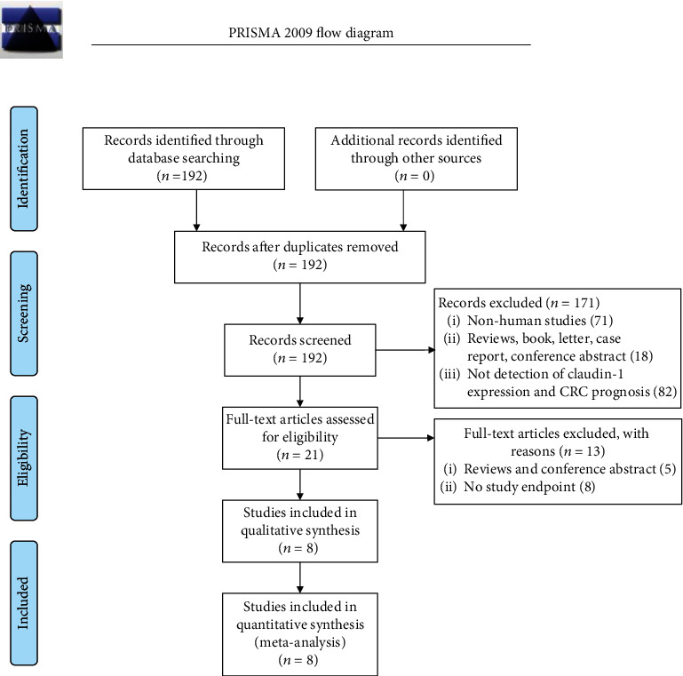
Table 1.
Main characteristics of the included publications.
| First author | Year | Country | No. of patients | Site | Mean age (years) | TNM stage | Follow-up (months) | Claudin-1 expression | Detection method | Antibody | Cutoff value | High expression | Outcome | NOS (score) | Data extraction method |
|---|---|---|---|---|---|---|---|---|---|---|---|---|---|---|---|
| Karabulut | 2015 | Turkey | 140 | Colorectal | 60.0 | I-IV | 14.0 | Serum | ELISA | YHB0737HU | >8.4 ng/ml | 50.0% | OS/DFS | 7 | K-M method |
| Matsuoka | 2011 | Japan | 156 | Colorectal | 65.0 | II-IV | 79.0 | Protein | IHC | E3411 | PP > 33% | 20.5% | OS/DFS | 8 | Univariate |
| Nakagawa | 2011 | Japan | 119 | Colorectal | NR | I-IV | 46.8 | mRNA | RT-PCR | NR | PP > median | 50.0% | OS/DFS | 8 | K-M method |
| Resnick | 2005 | USA | 129 | Colon | 72.5 | II | 96.0 | Protein | IHC | Polyclonal rabbit | SI ≥ 2 | 24.8% | OS/DFS | 9 | K-M method |
| Shibutani | 2013 | Japan | 344 | Colorectal | 66.8 | II-III | 51.7 | Protein | IHC | Polyclonal rabbit | PP > 25% | 68.0% | OS/DFS | 9 | K-M method |
| Singh | 2011 | USA | 250 | Colon | 64.6 | I-IV | 46.1 | Protein | IHC | Anti-claudin1 | >median | 50.0% | OS | 6 | K-M method |
| Yoshida | 2011 | Japan | 306 | Rectum | 64.0 | II-III | 38.0 | Protein | IHC | Monoclonal antibodies | PP > 30% | 46.7% | OS/DFS | 8 | K-M method |
| Kim | 2019 | Korea | 260 | Colon | 63.5 | I-IV | NR | Protein | IHC | Anti-claudin-1 | ISS ≥ 6 | 57.3% | OS/DFS | 8 | K-M method |
NR: not reported; IHC: immunohistochemistry; RT-PCR: reverse transcriptase polymerase chain reaction; SI: staining intensity; PP: positive cell percentage; immunostaining score (ISS) = PP∗SI; OS: overall survival; DFS: disease-free survival; K-M: Kaplan-Meier; NOS: Newcastle Ottawa Scale.
3.2. Claudin-1 Expression and Survival Rate
A total of eight studies explored the relationship between claudin-1 expression and OS. Because of the significant heterogeneity (I2 = 73%, P = 0.0005), random effects model was employed for evaluation. Our data indicated that the high expression of claudin-1 was associated with better OS (HR, 0.46; 95% CI, 0.28–0.76; P = 0.002; Figure 2).
Figure 2.

There were seven studies that reported the relationship between claudin-1 expression and DFS. Due to the significant heterogeneity (I2 = 65%, P = 0.009) between these studies, a random effects model was applied for meta-analysis. Our data indicated that the high expression of claudin-1 was associated with better DFS (HR, 0.44; 95% CI, 0.29–0.65; P < 0.0001; Figure 3).
Figure 3.

3.3. Claudin-1 Expression and Clinical Features
Table 2 summarizes the relationship between claudin-1 expression and clinicopathological characteristics. The high expression of claudin-1 was significantly related to the better tumor type (n = 6; RR, 0.60; 95% CI, 0.49–0.73; P < 0.00001), negative venous invasion (n = 4; RR, 0.81; 95% CI, 0.70–0.95; P = 0.001), and negative lymphatic invasion (n = 4; RR, 0.83; 95% CI, 0.74–0.92; P = 0.0009). In addition, meta-analysis showed that claudin-1 was associated with early tumor stage (n = 3; RR, 0.75; 95% CI, 0.54–1.04; P = 0.09) and negative lymph node metastasis(n = 4; RR, 0.91; 95% CI, 0.82–1.02; P = 0.10), although it was not statistically significant.
Table 2.
Meta-analysis of the correlation between claudin-1 expression and clinicopathological factors of colorectal cancer.
| Clinicopathological parameter | No. of studies | Participants | RR (95% CI) | Analysis model | Heterogeneity | Test for overall effect | ||
|---|---|---|---|---|---|---|---|---|
| I 2 (%) | P value | Z test | P value | |||||
| Tumor type (poorly vs. well) | 6 | 1312 | 0.60 (0.49, 0.73) | Fixed | 33 | 0.19 | 4.94 | <0.00001 |
| Venous invasion (+ vs. -) | 4 | 1029 | 0.81 (0.70, 0.95) | Fixed | 0 | 0.40 | 1.59 | 0.0010 |
| Lymphatic invasion (+ vs. -) | 4 | 1028 | 0.83 (0.74, 0.92) | Fixed | 0 | 0.42 | 3.32 | 0.0009 |
| Perineural invasion (+ vs. -) | 2 | 566 | 0.57 (0.25, 1.31) | Random | 80 | 0.03 | 1.33 | 0.18 |
| Depth of invasion (T3, 4 vs. T1, 2) | 4 | 1029 | 0.94 (0.72, 1.22) | Random | 70 | 0.02 | 0.47 | 0.64 |
| Lymph node metastasis (+ vs. -) | 4 | 1029 | 0.91 (0.82, 1.02) | Fixed | 0 | 0.60 | 1.66 | 0.10 |
| Distant metastasis (+ vs. -) | 2 | 379 | 0.88 (0.67, 1.15) | Fixed | 0 | 0.50 | 0.94 | 0.35 |
| TNM stage (III, IV vs. I, II) | 3 | 722 | 0.75 (0.54, 1.04) | Random | 69 | 0.04 | 1.71 | 0.09 |
| Size (larger vs. smaller) | 2 | 275 | 0.88 (0.60, 1.29) | Fixed | 0 | 0.68 | 0.65 | 0.52 |
| Gender (male vs. female) | 5 | 1185 | 1.02 (0.92, 1.14) | Fixed | 0 | 0.44 | 0.43 | 0.66 |
| Age (older vs. younger) | 2 | 275 | 1.15 (0.83, 1.60) | Fixed | 0 | 0.50 | 0.97 | 0.33 |
| Tumor site (colon vs. rectum) | 2 | 500 | 1.01 (0.87, 1.17) | Fixed | 0 | 0.81 | 0.10 | 0.92 |
RR: risk ratio; Random, random effects model; Fixed: fixed effect model.
Besides, we did not observe correlations between claudin-1 expression and other clinicopathological features, including perineural invasion (RR, 0.57; 95% CI, 0.25–1.31; P = 0.18; n = 2), depth of invasion (RR, 0.94; 95% CI, 0.72–1.22; P = 0.64; n = 4), lymph node metastasis (RR, 0.91; 95% CI, 0.82–1.02; P = 0.1; n = 4), distant metastasis (RR, 0.88; 95% CI, 0.67–1.15; P = 0.35; n = 2), size (RR, 0.88; 95% CI, 0.60–1.29; P = 0.52; n = 2), tumor site (RR, 1.01; 95% CI, 0.87–1.17; P = 0.92; n = 2), gender (RR, 1.02; 95% CI, 0.92–1.14; P = 0.66; n = 5), or age (RR, 1.15; 95% CI, 0.83–1.60; P = 0.33; n = 2).
3.4. Sensitivity Analysis and Publication Bias
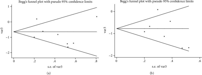
We performed sensitivity analysis by excluding one study at a time to confirm the robustness of the results for OS (Figure 4(a)) and DFS (Figure 4(b)). In addition, Egger's linear regression (OS, P = 0.480, Figure 5(a); DFS, P = 0.470, Figure 5(b)) and Begg's rank correlation test (OS, P = 1.000; Figure 6(a); DFS, P = 0.368, Figure 6(b)) showed that there was no publication bias in this study.
Figure 4.
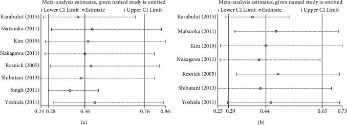
Figure 5.
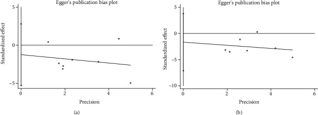
Figure 6.
3.5. Trial Sequential Analysis and Metaregression Analysis
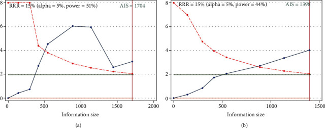
The cumulative Z-curve (blue line) reached the required information size (RIS) indicating that the number of cases included in this meta-analysis is sufficient. The blue line crosses the traditional threshold (horizontal line) and the TSA threshold (red line) indicating that the high expression level of claudin-1 was statistically significant with better OS (Figure 7(a)) and DFS (Figure 7(b)) of colorectal cancer.
Figure 7.
We performed a metaregression analysis to explore possible sources of heterogeneity. The results showed that covariates (year, country, site, TNM stage, detection method, NOS score, and survival analysis) did not significantly affect OS and DFS of colorectal cancer (Table 3).
Table 3.
Metaregression analysis for OS and DFS.
| Covariates | Multivariate analysis (OS) | Multivariate analysis (DFS) | ||||
|---|---|---|---|---|---|---|
| Coefficient | SE | P value | Coefficient | SE | P value | |
| Site | -0.206 | 0.518 | 0.717 | -0.522 | 0.181 | 0.213 |
| TNM stage | 0.039 | 0.435 | 0.934 | 0.121 | 0.133 | 0.531 |
| Detection method | 0.167 | 0.758 | 0.840 | 0.066 | 0.315 | 0.868 |
| NOS score | -0.573 | 0.809 | 0.530 | -0.638 | 0.335 | 0.308 |
| Data extraction method | 0.344 | 0.513 | 0.550 | -1.313 | 0.550 | 0.253 |
4. Discussion
CRC is one of the most common gastrointestinal tumors. There are approximately 1.2 million new CRC cases, and 600 000 die from the disease every year [50]. The prognosis of CRC has improved slowly but steadily during the past decades in the world. Early diagnosis and prognostic prediction are becoming more and more important for patients. This meta-analysis assessed the association between claudin-1 and prognosis, and the results showed that the high-expressed claudin-1 is correlated with lower aggressive tumor behavior and better prognosis (OS: HR, 0.46; 95% CI, 0.28–0.76; P = 0.002; DFS: HR, 0.44; 95% CI, 0.29–0.65; P < 0.0001).
This is an updated meta-analysis to clarify claudin-1 expression and prognosis in CRC. Previous meta-analysis by Jiang et al. showed that the claudin-1expression is associated with one, three, and five years of OS [51]. In comparison, the present analysis not only added an additional three studies [43, 44, 48] but also examined the HR for OS and DFS of CRC. We also included all claudin-1 detection methods, including ELISA and RT-PCR, which were excluded in the analysis by Jiang et al. In addition, new studies have emerged reporting on claudin-1 and CRC since the previous similar meta-analysis was published in 2017 [48, 52].
Since claudins were discovered, literature about the claudins' status and mechanisms during tumorigenesis is constantly expanding. Claudins in intestinal cytomembrane maintain the intestine's homeostasis; thus, abnormal expression of claudins may result in various pathophysiological conditions, and the loss of claudin-1 expression leads to tumor invasion and metastasis. In addition, NF-κB was frequently activated in CRC tissue with low-expressed claudin-1.Some studies have shown that claudin-1 is upregulated in CRC, but this overexpression has a good prognostic value, overall survival, disease-free survival, less metastases, and less aggressive disease [45, 47]. Matsuoka et al. suggested that claudin-1 plays a part in tumorigenesis as a promoter, and the expression of this protein will be lost in tumor cells once the tumor is established and started the invasion process [45].
Since the clinical and pathological characteristics were often used to predict prognosis of CRC, the relationship between claudin-1 and the clinical and pathological characteristics of CRC was also discussed. The results indicate that claudin-1 expression is related to better tumor type, negative venous invasion, and negative lymphatic invasion. In addition, claudin-1 was associated with early tumor stage and negative lymph node metastasis, although not statistically significant (P = 0.09 and P = 0.10). It is well known that better tumor type, negative venous invasion, negative lymphatic invasion, early tumor stage, and negative lymph node metastasis were good indicators for predicting the prognosis of colorectal cancer. These data also confirm that high expression of claudin-1 can be used as a good prognostic biomarker for colorectal cancer. However, whether the relationship of high expression of claudin-1 with better prognosis is due to its relationship with better tumor type, negative venous invasion, and negative lymphatic invasion is not clear, which needs more studies to confirm it. In addition, we did not find any correlation between the expression of claudin-1 and nerve infiltration, depth of infiltration, lymph node metastasis, distant metastasis, tumor size, location, gender, and age due to the insufficient number of included studies.
Claudin-1 can be detected by immunohistochemistry in blood samples, which is a simple, easy, and feasible method. We can know well the progress, prognosis, and treatment effect of CRC by continuously monitoring the claudin-1 level. In addition, claudin-1 could guide us to formulate a reasonable treatment strategy. For example, we could choose different treatment strategies for resectable, unresectable, and potentially resectable tumors in advanced colorectal cancer patients. So, claudin-1 is of great significance in guiding the clinical treatment decisions and predicting prognosis.
There are some limitations in this study. Firstly, significant heterogeneity could be induced by the different detection methods and cutoff level. Secondly, all of the included studies were in English and observational studies, which contributed to selection bias and recall bias. Third, there may be some errors in the extraction of survival data from the Kaplan-Meier curve, which may affect the accuracy of the results. Finally, the robustness of the statistical results could be impacted by the sufficient number of eligible articles and patients.
5. Conclusion
This meta-analysis identified eight studies assessing the association between claudin-1 and prognosis and clinical characteristics of CRC. These studies suggest that the high expression of claudin-1 was associated with better overall survival and disease-free survival in CRC. Moreover, claudin-1 was related to the better tumor type, negative venous invasion, and negative lymphatic invasion. Overall, this meta-analysis showed that claudin-1 may be a valuable indicator for predicting prognosis and helping us accurately intervene in the progress of CRC. However, whether the relationship of high expression of claudin-1 with better prognosis is due to its relationship with better tumor type, negative venous invasion, and negative lymphatic invasion is not clear, which needs more randomized controlled trials (RCTs) to confirm the conclusion.
Contributor Information
Chao Li, Email: lichao37521@163.com.
Guang Ning, Email: gning_sibs@163.com.
Conflicts of Interest
The authors declare that there is no conflict of interest regarding the publication of this paper.
Authors' Contributions
Didi Zuo was responsible for investigation, data curation, formal analysis, and writing of original draft. Jiantao Zhang was responsible for data curation, formal analysis, and validation. Tao Liu was responsible for data curation and formal analysis. Chao Li and Guang Ning were responsible for funding acquisition, project administration, and supervision.
Supplementary Materials
The supplementary material showed the quality assessment of the included studies by Newcastle Ottawa Scale (NOS). The NOS scores range from 0 to 9, and studies scoring out of 6 were considered high quality.
References
- 1.Siegel R. L., Miller K. D., Jemal A. Cancer statistics, 2019. CA: a Cancer Journal for Clinicians. 2018;69(1):7–34. doi: 10.3322/caac.21551. [DOI] [PubMed] [Google Scholar]
- 2.Arnold M., Sierra M. S., Laversanne M., Soerjomataram I., Jemal A., Bray F. Global patterns and trends in colorectal cancer incidence and mortality. Gut. 2017;66(4):683–691. doi: 10.1136/gutjnl-2015-310912. [DOI] [PubMed] [Google Scholar]
- 3.Cheng L., Eng C., Nieman L. Z., Kapadia A. S., Du X. L. Trends in colorectal cancer incidence by anatomic site and disease stage in the United States from 1976 to 2005. American Journal of Clinical Oncology. 2011;34(6):573–580. doi: 10.1097/COC.0b013e3181fe41ed. [DOI] [PubMed] [Google Scholar]
- 4.Siegel R. L., Miller K. D., Jemal A. Cancer statistics, 2017. CA: a Cancer Journal for Clinicians. 2017;67(1):7–30. doi: 10.3322/caac.21387. [DOI] [PubMed] [Google Scholar]
- 5.Sankaranarayanan R., Swaminathan R., Brenner H., et al. Cancer survival in Africa, Asia, and Central America: a population-based study. The Lancet Oncology. 2010;11(2):165–173. doi: 10.1016/S1470-2045(09)70335-3. [DOI] [PubMed] [Google Scholar]
- 6.Brenner H., Bouvier A. M., Foschi R., et al. Progress in colorectal cancer survival in Europe from the late 1980s to the early 21st century: the EUROCARE study. International Journal of Cancer. 2012;131(7):1649–1658. doi: 10.1002/ijc.26192. [DOI] [PubMed] [Google Scholar]
- 7.Siegel R., DeSantis C., Virgo K., et al. Cancer treatment and survivorship statistics, 2012. CA: a Cancer Journal for Clinicians. 2012;62(4):220–241. doi: 10.3322/caac.21149. [DOI] [PubMed] [Google Scholar]
- 8.Allegra C. J., Jessup J. M., Somerfield M. R., et al. American Society of Clinical Oncology provisional clinical opinion: testing for KRAS gene mutations in patients with metastatic colorectal carcinoma to predict response to anti-epidermal growth factor receptor monoclonal antibody therapy. Journal of Clinical Oncology. 2009;27(12):2091–2096. doi: 10.1200/JCO.2009.21.9170. [DOI] [PubMed] [Google Scholar]
- 9.De Roock W., De Vriendt V., Normanno N., Ciardiello F., Tejpar S. _KRAS_ , _BRAF_ , _PIK3CA_ , and _PTEN_ mutations: implications for targeted therapies in metastatic colorectal cancer. The Lancet Oncology. 2011;12(6):594–603. doi: 10.1016/S1470-2045(10)70209-6. [DOI] [PubMed] [Google Scholar]
- 10.Febbo P. G., Ladanyi M., Aldape K. D., et al. NCCN Task Force Report: Evaluating the Clinical Utility of Tumor Markers in Oncology. Journal of the National Comprehensive Cancer Network. 2011;9(Suppl_5):S-1–S-32. doi: 10.6004/jnccn.2011.0137. [DOI] [PubMed] [Google Scholar]
- 11.Locker G. Y., Hamilton S., Harris J., et al. ASCO 2006 update of recommendations for the use of tumor markers in gastrointestinal cancer. Journal of Clinical Oncology. 2006;24(33):5313–5327. doi: 10.1200/JCO.2006.08.2644. [DOI] [PubMed] [Google Scholar]
- 12.Van Itallie C. M., Anderson J. M. The Molecular Physiology of Tight Junction Pores. Physiology. 2004;19(6):331–338. doi: 10.1152/physiol.00027.2004. [DOI] [PubMed] [Google Scholar]
- 13.Bertiaux-Vandaële N., Youmba S. B., Belmonte L., et al. The Expression and the Cellular Distribution of the Tight Junction Proteins Are Altered in Irritable Bowel Syndrome Patients With Differences According to the Disease Subtype. American Journal of Gastroenterology. 2011;106(12):2165–2173. doi: 10.1038/ajg.2011.257. [DOI] [PubMed] [Google Scholar]
- 14.Yang J. J., Ma Y. L., Zhang P., Chen H. Q., Liu Z. H., Qin H. L. Histidine decarboxylase is identified as a potential biomarker of intestinal mucosal injury in patients with acute intestinal obstruction. Molecular Medicine. 2011;17(11-12):1323–1337. doi: 10.2119/molmed.2011.00107. [DOI] [PMC free article] [PubMed] [Google Scholar]
- 15.Singh A. B., Sharma A., Dhawan P. Claudin Family of Proteins and Cancer: An Overview. Journal of Oncology. 2010;2010:11. doi: 10.1155/2010/541957.e24978 [DOI] [PMC free article] [PubMed] [Google Scholar]
- 16.Lu Z., Ding L., Lu Q., Chen Y.-H. Claudins in intestines. Tissue Barriers. 2014;1(3):p. e24978. doi: 10.4161/tisb.24978. [DOI] [PMC free article] [PubMed] [Google Scholar]
- 17.Kind S., Büscheck F., Höflmayer D., et al. Claudin-1 upregulation is associated with favorable tumor features and a reduced risk for biochemical recurrence in ERG-positive prostate cancer. World Journal of Urology. 2019 doi: 10.1007/s00345-019-03017-w. [DOI] [PubMed] [Google Scholar]
- 18.Jin H., Zhang Q., Zhang S., Liu H., Man Z., Wang Y. Effects of claudin-1 downregulation on the physiological processes of gallbladder cancer SGC996 cells. Oncology Letters. 2019;17(2):1688–1694. doi: 10.3892/ol.2018.9740. [DOI] [PMC free article] [PubMed] [Google Scholar]
- 19.Danzinger S., Tan Y. Y., Rudas M., et al. Association of Cytokeratin 5 and Claudin 3 expression with BRCA1 and BRCA2 germline mutations in women with early breast cancer. BMC Cancer. 2019;19(1):p. 695. doi: 10.1186/s12885-019-5908-6. [DOI] [PMC free article] [PubMed] [Google Scholar]
- 20.Tabariès S., McNulty A., Ouellet V., et al. Afadin cooperates with Claudin-2 to promote breast cancer metastasis. Genes & Development. 2019;33(3-4):180–193. doi: 10.1101/gad.319194.118. [DOI] [PMC free article] [PubMed] [Google Scholar]
- 21.Coati I., Lotz G., Fanelli G. N., et al. Claudin-18 expression in oesophagogastric adenocarcinomas: a tissue microarray study of 523 molecularly profiled cases. British Journal of Cancer. 2019;121(3):257–263. doi: 10.1038/s41416-019-0508-4. [DOI] [PMC free article] [PubMed] [Google Scholar]
- 22.Rohde C., Yamaguchi R., Mukhina S., Sahin U., Itoh K., Türeci Ö. Comparison of Claudin 18.2 expression in primary tumors and lymph node metastases in Japanese patients with gastric adenocarcinoma. Japanese Journal of Clinical Oncology. 2019;49(9):870–876. doi: 10.1093/jjco/hyz068. [DOI] [PMC free article] [PubMed] [Google Scholar]
- 23.Zhou S., Piao X., Wang C., Wang R., Song Z. Identification of claudin‑1, ‑3, ‑7 and ‑8 as prognostic markers in human laryngeal carcinoma. Molecular Medicine Reports. 2019;20(1):393–400. doi: 10.3892/mmr.2019.10265. [DOI] [PMC free article] [PubMed] [Google Scholar]
- 24.Wei D. F., Tang M. K., Liu Y., Zhang C. Y., Qin L. J. Effect of hypoxia inducible factor-1 alpha on brain metastasis from lung cancer and its mechanism. Sichuan Da Xue Xue Bao. Yi Xue Ban. 2019;50(2):188–192. [PubMed] [Google Scholar]
- 25.Sun X., Cui S., Fu X., Liu C., Wang Z., Liu Y. MicroRNA-146-5p promotes proliferation, migration and invasion in lung cancer cells by targeting claudin-12. Cancer Biomarkers. 2019;25(1):89–99. doi: 10.3233/CBM-182374. [DOI] [PubMed] [Google Scholar]
- 26.Akizuki R., Eguchi H., Endo S., Matsunaga T., Ikari A. ZO-2 suppresses cell migration mediated by a reduction in matrix metalloproteinase 2 in claudin-18-expressing lung adenocarcinoma A549 cells. Biological & Pharmaceutical Bulletin. 2019;42(2):247–254. doi: 10.1248/bpb.b18-00670. [DOI] [PubMed] [Google Scholar]
- 27.Chen Q. BMP4 inhibits glioblastoma invasion by promoting E-cadherin and claudin expression. Frontiers in Bioscience. 2019;24(6):1060–1070. doi: 10.2741/4768. [DOI] [PubMed] [Google Scholar]
- 28.Georges R., Bergmann F., Hamdi H., et al. Sequential biphasic changes in claudin1 and claudin4 expression are correlated to colorectal cancer progression and liver metastasis. Journal of Cellular and Molecular Medicine. 2012;16(2):260–272. doi: 10.1111/j.1582-4934.2011.01289.x. [DOI] [PMC free article] [PubMed] [Google Scholar]
- 29.Morita K., Furuse M., Fujimoto K., Tsukita S. Claudin multigene family encoding four-transmembrane domain protein components of tight junction strands. Proceedings of the National Academy of Sciences of the United States of America. 1999;96(2):511–516. doi: 10.1073/pnas.96.2.511. [DOI] [PMC free article] [PubMed] [Google Scholar]
- 30.Abdelzaher E., Rizk A. M., Bessa S. S., Omer K. M. Predictive value of immunohistochemical expression of claudin-1 in colonic carcinoma. Journal of the Egyptian National Cancer Institute. 2011;23(4):123–131. doi: 10.1016/j.jnci.2011.10.002. [DOI] [PubMed] [Google Scholar]
- 31.Sezer C. Loss of tight junction proteins (Claudin 1, 4, and 7) correlates with aggressive behavior in colorectal carcinoma. Medical Science Monitor. 2014;20:1255–1262. doi: 10.12659/MSM.890598. [DOI] [PMC free article] [PubMed] [Google Scholar]
- 32.Morata-Tarifa C., Jiménez G., García M. A., et al. Low adherent cancer cell subpopulations are enriched in tumorigenic and metastatic epithelial-to-mesenchymal transition-induced cancer stem-like cells. Scientific Reports. 2016;6(1) doi: 10.1038/srep18772. [DOI] [PMC free article] [PubMed] [Google Scholar]
- 33.Stang A. Critical evaluation of the Newcastle-Ottawa scale for the assessment of the quality of nonrandomized studies in meta-analyses. European Journal of Epidemiology. 2010;25(9):603–605. doi: 10.1007/s10654-010-9491-z. [DOI] [PubMed] [Google Scholar]
- 34.Moher D., Liberati A., Tetzlaff J., Altman D. G., PRISMA Group Preferred Reporting Items for Systematic Reviews and Meta-Analyses: The PRISMA Statement. Annals of Internal Medicine. 2009;151(4):264–9, W64. doi: 10.7326/0003-4819-151-4-200908180-00135. [DOI] [PubMed] [Google Scholar]
- 35.Tierney J. F., Stewart L. A., Ghersi D., Burdett S., Sydes M. R. Practical methods for incorporating summary time-to-event data into meta-analysis. Trials. 2007;8(1) doi: 10.1186/1745-6215-8-16. [DOI] [PMC free article] [PubMed] [Google Scholar]
- 36.Higgins J. P., Thompson S. G., Deeks J. J., Altman D. G. Measuring inconsistency in meta-analyses. BMJ. 2003;327(7414):557–560. doi: 10.1136/bmj.327.7414.557. [DOI] [PMC free article] [PubMed] [Google Scholar]
- 37.DerSimonian R., Laird N. Meta-analysis in clinical trials revisited. Contemporary Clinical Trials. 2015;45(Part A):139–145. doi: 10.1016/j.cct.2015.09.002. [DOI] [PMC free article] [PubMed] [Google Scholar]
- 38.Begg C. B., Mazumdar M. Operating characteristics of a rank correlation test for publication bias. Biometrics. 1994;50(4):1088–1101. doi: 10.2307/2533446. [DOI] [PubMed] [Google Scholar]
- 39.Egger M., Smith G. D., Schneider M., Minder C. Bias in meta-analysis detected by a simple, graphical test. BMJ. 1997;315(7109):629–634. doi: 10.1136/bmj.315.7109.629. [DOI] [PMC free article] [PubMed] [Google Scholar]
- 40.Wacholder S., Chanock S., Garcia-Closas M., el ghormli L., Rothman N. Assessing the probability that a positive report is false: an approach for molecular epidemiology studies. Journal of the National Cancer Institute. 2004;96(6):434–442. doi: 10.1093/jnci/djh075. [DOI] [PMC free article] [PubMed] [Google Scholar]
- 41.Wetterslev J., Thorlund K., Brok J., Gluud C. Trial sequential analysis may establish when firm evidence is reached in cumulative meta-analysis. Journal of Clinical Epidemiology. 2008;61(1):64–75. doi: 10.1016/j.jclinepi.2007.03.013. [DOI] [PubMed] [Google Scholar]
- 42.Resnick M. B., Konkin T., Routhier J., Sabo E., Pricolo V. E. Claudin-1 is a strong prognostic indicator in stage II colonic cancer: a tissue microarray study. Modern Pathology. 2005;18(4):511–518. doi: 10.1038/modpathol.3800301. [DOI] [PubMed] [Google Scholar]
- 43.Singh A. B., Sharma A., Smith J. J., et al. Claudin-1 up-regulates the repressor ZEB-1 to inhibit E-cadherin expression in colon cancer cells. Gastroenterology. 2011;141(6):2140–2153. doi: 10.1053/j.gastro.2011.08.038. [DOI] [PMC free article] [PubMed] [Google Scholar]
- 44.Karabulut M., Alis H., Bas K., et al. Clinical significance of serum claudin-1 and claudin-7 levels in patients with colorectal cancer. Molecular and Clinical Oncology. 2015;3(6):1255–1267. doi: 10.3892/mco.2015.626. [DOI] [PMC free article] [PubMed] [Google Scholar]
- 45.Matsuoka T., Mitomi H., Fukui N., et al. Cluster analysis of claudin-1 and -4, E-cadherin, and β-catenin expression in colorectal cancers. Journal of Surgical Oncology. 2011;103(7):674–686. doi: 10.1002/jso.21854. [DOI] [PubMed] [Google Scholar]
- 46.Yoshida T., Kinugasa T., Akagi Y., et al. Decreased expression of claudin-1 in rectal cancer: a factor for recurrence and poor prognosis. Anticancer Research. 2011;31(7):2517–2525. [PubMed] [Google Scholar]
- 47.Nakagawa S., Miyoshi N., Ishii H., et al. Expression of CLDN1 in colorectal cancer: a novel marker for prognosis. International Journal of Oncology. 2011;39(4):791–796. doi: 10.3892/ijo.2011.1102. [DOI] [PubMed] [Google Scholar]
- 48.Kim N. Y., Pyo J. S., Kang D. W., Yoo S. M. Loss of claudin-1 expression induces epithelial-mesenchymal transition through nuclear factor-κB activation in colorectal cancer. Pathology, Research and Practice. 2019;215(3):580–585. doi: 10.1016/j.prp.2019.01.015. [DOI] [PubMed] [Google Scholar]
- 49.Shibutani M., Noda E., Maeda K., Nagahara H., Ohtani H., Hirakawa K. Low expression of claudin-1 and presence of poorly-differentiated tumor clusters correlate with poor prognosis in colorectal cancer. Anticancer Research. 2013;33(8):3301–3306. [PubMed] [Google Scholar]
- 50.Brenner H., Kloor M., Pox C. P. Colorectal cancer. Lancet. 2014;383(9927):1490–1502. doi: 10.1016/S0140-6736(13)61649-9. [DOI] [PubMed] [Google Scholar]
- 51.Jiang L., Yang L., Huang H., Liu B. Y., Zu G. Prognostic and clinical significance of claudin-1 in colorectal cancer: a systemic review and meta-analysis. International Journal of Surgery. 2017;39:214–220. doi: 10.1016/j.ijsu.2017.02.005. [DOI] [PubMed] [Google Scholar]
- 52.Gowrikumar S., Ahmad R., Uppada S. B., et al. Upregulated claudin-1 expression promotes colitis-associated cancer by promoting β-catenin phosphorylation and activation in Notch/p-AKT-dependent manner. Oncogene. 2019;38(26):5321–5337. doi: 10.1038/s41388-019-0795-5. [DOI] [PMC free article] [PubMed] [Google Scholar]
Associated Data
This section collects any data citations, data availability statements, or supplementary materials included in this article.
Supplementary Materials
The supplementary material showed the quality assessment of the included studies by Newcastle Ottawa Scale (NOS). The NOS scores range from 0 to 9, and studies scoring out of 6 were considered high quality.


