

Abstract
Background:
In the setting of complete distal biceps tendon rupture, surgical repair has become the standard of care to restore optimal elbow function, but the optimal approach and method of tendon fixation are still subjects of debate and have remained controversial for more than half a century.
Purpose:
To evaluate patient-reported long-term outcomes after distal biceps tendon repair using a modified double-incision technique.
Study Design:
Case series; Level of evidence, 4.
Methods:
We reviewed primary distal biceps tendon repairs after isolated tendon rupture using the modified muscle-splitting double-incision approach and transosseous suture fixation technique described by Morrey et al (1985), which had been performed at our level 1 trauma center between January 2000 and December 2013. Outcome measures included the subjective elbow value (SEV), the Oxford Elbow Score (OES) with its 3 domains (function, pain, and social-psychological), a self-performed hook test, and the 3-level version of the EuroQoL 5-dimensional instrument (EQ-5D-3L) as a measure of health status. Levels of overall satisfaction were determined by asking whether the patient would consent to the operation again. In addition, patients were asked to report any complications.
Results:
A total of 30 patients met the inclusion criteria, and 25 patients were available for the survey. Mean age at the time of rupture was 47 years. All patients were male. Mean follow-up was 120 months (range, 57-207 months). The follow-up rate was 83.34%. The following outcome results were obtained: SEV, 88.16% ± 25.18%; OES, 43.80 ± 10.56 out of 48 points; OES Pain, 92.50% ± 23.03%; OES Function, 92.25% ± 22.19%; OES Social-Psychological, 89% ± 23.68%; EQ-5D-3L, 0.93 ± 0.21. All patients described a negative hook test. Patient-reported complications included painless limitation in forearm rotation in 8% of patients (n = 2); reduced flexion and forearm rotation strength with and without pain in 8% (n = 2) and 4% (n = 1), respectively; synostosis after 1 year requiring revision surgery in 4% (n = 1); and transient wrist drop in 4% (n = 1). The overall complication rate was 28% (7/25), and 96% (n = 24) would consent to the operation again.
Conclusion:
Despite the cited approach-related morbidity, we report an excellent patient-reported long-term outcome for the double-incision distal biceps repair technique.
Keywords: distal biceps tendon repair, modified double-incision, transosseous suture fixation, distal biceps tendon rupture
Although relatively uncommon, distal biceps tendon rupture is the most common acute tendinous injury around the elbow. This injury predominantly occurs in male patients between the fourth and sixth decades of life, with an estimated incidence of surgically repaired tendon ruptures of 2.55 per 100,000 patients per year.15
The injury usually results from an eccentric force on the biceps brachii with the elbow in a flexed position. In the setting of complete tendon avulsion from the radial tuberosity (Figure 1), which is the most common presentation, surgical repair has become the standard of care to optimally restore elbow and forearm function. However, for more than half a century, the optimal approach and method of tendon fixation have continued to be subjects of debate and remain controversial without any clear consensus.20
Figure 1.

A 40-year-old male patient from this study with acute distal biceps tendon rupture of the left elbow in the (A) frontal and (B) sagittal views with a typical Popeye sign.
Distal biceps tendon repair can be performed through a single-incision or double-incision approach. Because the original single-incision technique developed by Dobbie5 resulted in an unacceptably high incidence of neurologic injury, the double-incision technique described by Boyd and Anderson,3 later modified by Kelly et al14 and Morrey et al,17 became the procedure of choice for several decades. Although this technique may have reduced the risk of nerve injury, it requires dissection through the radioulnar syndesmosis and has been reported to increase the risk for heterotopic ossification (HO).1,6,7
To date, no surgical technique has been proven superior, and results with sufficient long-term follow-up are lacking. The purpose of this study was to evaluate patient-reported long-term outcomes 10 years after distal biceps tendon repair using the modified double-incision technique described by Morrey et al.17
Methods
This study was authorized by the local ethical committee and was carried out in accordance with the ethical standards of the 1964 Declaration of Helsinki as updated in 2004. We reviewed primary distal biceps tendon repairs after isolated tendon rupture using the modified muscle-splitting double-incision approach and transosseous suture fixation, performed at our institution over a 13-year period between January 2000 and December 2013. Patients were identified by searching in our institutional database for coded diagnosis (S46.2, M66.32) according to the International Classification of Diseases–10th Revision as well as related procedure codes (5-855.02, 5-855.12, 5-855.x2) according to the German version of the International Classification of Procedures in Medicine. Exclusion criteria included the presence of an associated fracture, joint dislocation, any concomitant soft tissue injury, and use of other approaches or fixation methods. These criteria were verified by checking operation reports and discharge letters.
Eligible patients were contacted and asked to complete a survey using patient-reported outcome measures (PROMs), including the subjective elbow value (SEV)21; the Oxford Elbow Score (OES)4 as a 12-item PROM with its 3 unidimensional domains (Function, Pain, and Social-Psychological); a self-performed hook test; and the 3-level version of the EuroQoL 5-dimensional instrument (EQ-5D-3L).2,23 Level of overall satisfaction was determined by asking whether the patient would consent to the operation again. In addition, patients were asked to report any complications that occurred postoperatively, without mention of any examples or answer options by the assessor.
Modified Double-Incision Technique
All patients underwent the same surgical approach and fixation method, which has been the standard in our trauma department for nearly 3 decades. The muscle-splitting double-incision approach modified by Morrey and others14,17 uses a transverse incision in the antecubital fossa. After identification of the distal portion of the biceps tendon, the degenerated part was resected. Two Ethibond Excel No. 2 nonabsorbable sutures (Ethicon, Inc) were passed through the distal part of the tendon in a Krakow pattern, leaving 2 free ends distally (Figure 2). In maximal supination, a blunt clamp was inserted into the interosseous space along the medial aspect of the radial tuberosity, without any subperiosteal exposure of the ulna, until it appeared on the dorsolateral aspect of the proximal forearm. A second longitudinal incision was made over the clamp (Figure 3, A and B). With the forearm in full pronation, the radial tuberosity was exposed through a muscle-splitting approach in line with the fibers of the common extensors and the supinator. A 10-15 mm × 7-8 mm cavity, deep enough to accommodate the prepared tendon, was created within the radial tuberosity with a high-speed bur while carefully preserving the protuberance of the tuberosity that acts as a cam effect (Figure 3, C and D). Bone debris that emerged during burring was removed meticulously. A curved hemostat was inserted in the anterior incision and used to pass the prepared tendon through the interosseous space out the second incision (Figure 3E). Three 2.0-mm drill holes were placed approximately at 1-cm intervals through the dorsal cortical margin of the tuberosity. The tendon sutures were then passed through the drill holes. With the elbow at 90° of flexion and the forearm pronated, the tendon was pulled into the bicipital tuberosity, and the sutures were pulled tight and tied (Figure 3F).14,17
Figure 2.

The distal biceps tendon is exposed through a transverse incision in the antecubital fossa and prepared with two No. 2 nonabsorbable sutures in a Krakow pattern (same patient from Figure 1).
Figure 3.

While the arm is in maximal supination, (A) a blunt clamp is inserted into the interosseous space until the clamp appears on the dorsolateral aspect of the proximal forearm, and (B) a second longitudinal incision is made over the clamp. (C and D) After exposure of the radial tuberosity with a muscle-splitting technique in full pronation, a cavity is created with a high-speed bur. (E) The prepared tendon is pulled out through the second incision into the bicipital tuberosity and (F) repaired through transosseous drill holes (same patient from Figure 1).
Postoperative rehabilitation in a functional brace for 6 weeks allowed early gradual increase of range of motion. In the first week, the elbow was immobilized at 90° of flexion. From the second week, the brace was unlocked to allow 30° to full flexion with active extension and full pronation as well as passive flexion and full supination. From the fourth week, the brace was unlocked and full active extension pursued. From the sixth week, the brace was discontinued and gentle active flexion and pronation against gravity were permitted. Progressive strengthening was allowed around 3 months after surgery, with unrestricted activity permitted at 6 months.
Results
A total of 30 patients met the inclusion criteria, and 25 patients were available for the survey, for a follow-up rate of 83.34%. Follow-up occurred at an average of 120 months (range, 57-207 months). All 25 patients were men, with a mean age of 47 years (range, 31-76 years) at the time of injury. The dominant arm was involved in 84% of cases (n = 21), and the right side was involved in 68% of cases (n = 17). In 96% of cases (n = 24), surgery was performed within the first 6 weeks after injury.
The mean SEV was 88.16% ± 25.18%, and the OES was 43.80 ± 10.56 out of 48 points. The means of the 3 subdomains of the OES were 92.50% ± 23.03% for OES Pain, 92.25% ± 22.19% for OES Function, and 89% ± 23.68% for OES Social-Psychological. The mean EQ-5D-3L was 0.93 ± 0.21. All patients described a negative hook test. We noted that 96% of patients (n = 24) would consent to the operation again (Table 1).
Table 1.
Results of Outcome Measurementsa
| Outcome Measurement | Result |
|---|---|
| OES (of a possible 0-48 points) | 43.80 ± 10.56 |
| OES domains, % | |
| Pain | 92.50 ± 23.03 |
| Function | 92.25 ± 22.19 |
| Social-Psychological | 89 ± 23.68 |
| SEV, % | 88.16 ± 25.18 |
| EQ-5D-3L score (of a possible score of 0-1.00) | 0.93 ± 0.21 |
| Negative hook test, n (%) | 25 (100) |
| Patients who would consent to the operation again, n (%) | 24 (96) |
aValues are expressed as mean ± SD unless otherwise noted. EQ-5D-3L, 3-level version of the EuroQoL 5-dimensional instrument; OES, Oxford Elbow Score; SEV, subjective elbow value.
Patient-reported complications included painless limitation in forearm rotation in 8% of patients (n = 2) after 6 and 12 weeks; reduced flexion and forearm rotation strength with and without pain in 8% (n = 2) and 4% (n = 1), respectively, after more than 12 weeks; synostosis after 1 year requiring revision surgery in 4% (n = 1); and transient wrist drop in 4% (n = 1) immediately after surgery with complete remission after 6 months (Table 2). Figures 4 and 5 show 15-year clinical and radiological results of the patient described in Figures 1 through 3.
Table 2.
Patient-Reported Complicationsa
| Patient-Reported Complications | n | Scores | Consent to Operation Again? |
|---|---|---|---|
| Minor | |||
| Painless limitation in active and passive forearm rotation | 2 | OES, 41; SEV, 80% OES, 39; SEV, 30% Pain, 100% Function, 100% Social-Psychological, 43.75% |
Yes Yes |
| Painful decreased strength of elbow flexion and forearm supination | 2 | OES, 40; SEV, 80% OES, 24; SEV, 50% Pain, 43.75% Function, 50% Social-Psychological, 56.25% |
Yes Yes |
| Painless decreased strength of elbow flexion and forearm supination | 1 | OES, 44; SEV, 80% | Yes |
| Major | |||
| Synostosis after 1 year requiring revision surgery | 1 | OES, 0; SEV, 0% | No |
| Transient wrist drop | 1 | OES, 100; SEV, 100% | Yes |
aA total of 7 patients reported complications, for a 28% overall complication rate. OES, Oxford Elbow Score; SEV, subjective elbow value.
Figure 4.

Same patient 15 years postoperatively with negative self-performed hook test and full range of motion. Oxford Elbow Score, 48/48 points. Subjective elbow value, 100%. EuroQoL 5-dimensional instrument,3-level version, 1. No patient-reported complications.
Figure 5.
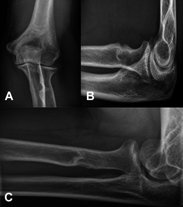
Radiographs of the left elbow in (A) anteroposterior, (B) lateral, and (C) Coyle views 15 years postoperatively without any heterotopic ossification.
Discussion
In response to a high rate of HO and synostosis observed after the double-incision technique described by Boyd and Anderson,3 a modification of this approach was described by Morrey et al17 in 1985, who proposed a muscle-splitting approach through the extensors in an attempt to limit subperiosteal dissection and thereby reduce the likelihood of HO and synostosis. Despite the successful modification, invention of new fixation devices and remaining concerns about HO and synostosis revived the anterior limited single-incision approach. The only existing prospective, randomized clinical trial, that by Grewal et al,9 did not find any difference in final outcomes at 2 years, clinically significant occurrence of HO formation, or strength recovery between the limited single-incision approach with the use of 2 suture anchors and the modified double-incision approach with transosseous suture fixation, except for a 10% advantage in final isometric flexion strength in the latter. In addition, a significantly lower overall complication rate was seen due to a lower number of early transient neurapraxias involving the lateral antebrachial cutaneous nerve (19/47 vs 3/43 in the double-incision group; P < .001).9,20
Although the approach does not dictate the type of fixation, transosseous suture fixation is typically combined with the modified double-incision approach, whereas alternative fixation methods such as suture anchors, intraosseous screws, and cortical buttons are associated with the single-incision approach. To date, clinical evidence reported in the literature does not support a clear advantage of one technique over the other.20 Good to excellent outcomes are described regardless of approach and fixation method.16
However, numerous studies have demonstrated that the posterior muscle-splitting approach provides the required exposure for a more closely anatomic footprint repair than the anterior single-incision approach.11 Forthman et al8 described in their cadaveric study an average angular orientation of the bicipital tuberosity of 65° with a wide range between 15° and 120° of pronation with respect to the coronal plane of the radius and an average site of tendon insertion ranging from –5° to 105° of pronation. They concluded that 35% of bicipital tuberosities were pronated greater than 60° and, therefore, anatomic reinsertion of the tendon could not be achieved through an anterior approach under the assumption that the maximum angle of orientation for operative instruments would be about 60° of pronation from an anterior single-incision approach (Figure 6).8 These significant individual variations in local anatomic features were highlighted in a cadaveric study by Hasan et al,12 demonstrating that a virtually generated bone tunnel was within the original tendon footprint in only 9.7% of the cases from the anterior approach and in 73.4% of cases from the posterior approach. Schmidt et al19 gave these findings clinical significance in a retrospective study of single-incision repairs, quantifying the biceps reattachment site with postoperative magnetic resonance imaging as 73° more anterior than the original footprint and positing that this nonanatomic position might be responsible for the measured postoperative decrease in supination strength of 10% in neutral and 33% in 60° of supination compared with the uninjured side.
Figure 6.

Exposure of the anatomic attachment site of the distal biceps tendon through a posterior muscle-splitting approach, described by Morrey et al,17 in a right elbow in 90° of flexion and full pronation (left). For comparison, nonanatomic exposure through an anterior single-incision approach in extension and full supination (right).
In elbow surgery, a wide range of outcome tools could potentially be used to measure clinical outcome. Currently, the only elbow-specific outcome tool that has been validated with a high-quality method, whether patient-reported or physician-administered, is the OES,22 used herein. It was developed in response to the lack of an appropriate standardized method of outcome assessment in elbow surgery and was designed specifically to be a patient-focused outcome measure, independent from the operating surgeon, thereby minimizing the risk of bias.4,10 The SEV, also known as Single Assessment Numeric Evaluation (SANE), was viewed as worthwhile by a subcommittee of the American Shoulder and Elbow Surgeons Value Committee, established to make recommendations regarding evaluation of more than 25 different shoulder and elbow PROMs to compare surgeon performance independent from the operating surgeon.13 Furthermore, a previous study demonstrated a correlation between the SEV and the physician-administered Mayo Elbow Performance Score and thus established a convergent validity.21 Moreover, the SEV was able to detect changes in outcome after treatment.21 The EQ-5D-3L as a measure of health status has been validated in previous studies for use among adults in a general population and has been recommended as a postinjury assessment tool.2,23
These PROMs were used for the above reasons as the primary outcome measurement tools in the current study. This is a strength of the study, as other nonspecific outcome measures such as the Disabilities of the Arm, Shoulder and Hand (DASH) could underestimate disability after biceps rupture and treatment. For example, a single-incision repair study reported an average postoperative DASH score of 10 (10.1 is normal for the US population), but 47% of the patients complained of symptoms when performing physical activities.11,20 Nevertheless, it should be considered that neither the OES nor the SEV is currently valid in determining a minimal clinically important difference across a heterogeneous study population, defined as the smallest change in the measure that patients believe is significant and that would cause clinicians to consider reevaluating a patient’s management.21
This is the first study describing long-term outcomes with an average 10-year follow-up for distal biceps tendon repair using the modified double-incision technique described by Morrey and others.17 The study makes use of patient-reported outcome measurements, and the results could serve as a reference for future long-term benchmarking studies.
Nevertheless, this study had several limitations. The hook test is useful in differentiating between a complete avulsion and an incomplete injury. The test is reported to be 100% sensitive and 100% specific in detecting complete distal ruptures.18 All patients reported a negative hook test, suggesting at least no case of complete rerupture in this study. However, the reliability of the hook test in a self-performed setting, as was done in this study, is unclear, and it should be considered that experienced clinicians can at times have difficulties with this test. The fact that this test serves as the only evidence for adequate integrity of the repaired tendon and that imaging methods such as magnetic resonance imaging or ultrasonography are lacking should be considered a major limitation.
The modified double-incision technique has been the current standard of treatment at our institution for nearly 3 decades, since it was first described by Morrey et al.17 All surgeries in this study were performed by experienced senior physicians, which could be seen as a further limitation, because some authors have suggested that repair of the distal biceps tendon has a steep learning curve, so that more experienced surgeons may have a lower complication rate, although Kelly et al14 demonstrated the contrary.
Even though an overall complication rate of 28% seems high, it remains within the range of reported complication rates and is almost identical with the overall rate reported by Kelly et al.14 A weakness of the current study is certainly the inability to interpret individual complications, which results from our patient-reported study design. Only 1 complication, a radioulnar synostosis (1/25; 4%), was attested to objectively by an external physician and required revision surgery. This occurrence suggests that the modified approach has decreased but not eliminated the risk of radioulnar synostosis, as previously reported by some authors describing a similar revision surgery rate of 4.1% due to HO or synostosis.6 The role of routine HO prophylaxis is also missing in this study; because indomethacin was not routinely prescribed, the rate of compliance with this treatment was not documented retrospectively, and, owing to the long follow-up intervals, it was not feasible to ask the patients about compliance. Follow-up radiographs were not routinely performed to assess for the presence of asymptomatic HO. In addition, objective assessment of range of motion and strength was not performed either pre- or postoperatively to assess amount of improvement, and we did not collect data on rates of return to work. Moreover, as a case series without any comparison or control group, this study cannot contribute any clarification regarding the superiority of one technique over another.
Asking patients about complications without mentioning any examples or answer options offers the opportunity to assess only those complications that are relevant from a patient’s point of view, without physician influence on the definition of a complication. However, reports about limited forearm rotation and reduced strength in this study remain unclear in their origin and extent. They suggest HO formation, lack of adequate preservation of the radial tuberosity’s protuberance, and/or iatrogenic approach–related supinator damage as potential causes. However, these aspects appear to be clinically nonsignificant in view of the excellent overall outcomes and the fact that only 1 patient had regrets about consenting to the surgery.
Conclusion
Despite the cited approach-related morbidity, we report excellent long-term patient-reported outcomes for the double-incision distal biceps repair technique.
Footnotes
Final revision submitted May 21, 2020; accepted June 8, 2020.
The authors declared that there are no conflicts of interest in the authorship and publication of this contribution. AOSSM checks author disclosures against the Open Payments Database (OPD). AOSSM has not conducted an independent investigation on the OPD and disclaims any liability or responsibility relating thereto.
Ethical approval for this study was obtained from Hannover Medical School.
References
- 1. Amin NH, Volpi A, Lynch TS, et al. Complications of distal biceps tendon repair: a meta-analysis of single-incision versus double-incision surgical technique. Orthop J Sports Med. 2016;4(10):2325967116668137. [DOI] [PMC free article] [PubMed] [Google Scholar]
- 2. Black JA, Herbison GP, Lyons RA, Polinder S, Derrett S. Recovery after injury: an individual patient data meta-analysis of general health status using the EQ-5D. J Trauma. 2011;71:1003–1010. [DOI] [PubMed] [Google Scholar]
- 3. Boyd AD, Anderson LD. A method for reinsertion of the distal biceps brachii tendon. J Bone Joint Surg Am. 1961;43:1041–1043. [Google Scholar]
- 4. Dawson J, Doll H, Boller I, et al. The development and validation of a patient-reported questionnaire to assess outcomes of elbow surgery. J Bone Joint Surg Br. 2008;90:466–473. [DOI] [PubMed] [Google Scholar]
- 5. Dobbie R. Avulsion of the lower biceps brachii tendon: analysis of fifty-one previously unreported cases. Am J Surg. 1941;51:662–683. [Google Scholar]
- 6. Dunphy TR, Hudson J, Batech M, Acevedo DC, Mirzayan R. Surgical treatment of distal biceps tendon ruptures: an analysis of complications in 784 surgical repairs. Am J Sports Med. 2017;45:3020–3029. [DOI] [PubMed] [Google Scholar]
- 7. Ford SE, Andersen JS, Macknet DM, Connor PM, Loeffler BJ, Gaston RG. Major complications after distal biceps tendon repairs: retrospective cohort analysis of 970 cases. J Shoulder Elbow Surg. 2018;27(10):1898–1906. [DOI] [PubMed] [Google Scholar]
- 8. Forthman CL, Zimmerman RM, Sullivan MJ, Gabel GT. Cross-sectional anatomy of the bicipital tuberosity and biceps brachii tendon insertion: relevance to anatomic tendon repair. J Shoulder Elbow Surg. 2008;17:522–526. [DOI] [PubMed] [Google Scholar]
- 9. Grewal R, Athwal GS, MacDermid JC, et al. Single versus double-incision technique for the repair of acute distal biceps tendon ruptures: a randomized clinical trial. J Bone Joint Surg Am. 2012;94:1166–1174. [DOI] [PubMed] [Google Scholar]
- 10. Guyver PM, Cattell AE, Hall MJ, Brinsden MD. Oxford elbow scores in an asymptomatic population. Ann R Coll Surg Engl. 2013;95:415–417. [DOI] [PMC free article] [PubMed] [Google Scholar]
- 11. Hansen G, Smith A, Pollock JW, et al. Anatomic repair of the distal biceps tendon cannot be consistently performed through a classic single-incision suture anchor technique. J Shoulder Elbow Surg. 2014;23:1898–1904. [DOI] [PubMed] [Google Scholar]
- 12. Hasan SA, Cordell CL, Rauls RB, Bailey MS, Sahu D, Suva LJ. Two-incision versus one-incision repair for distal biceps tendon rupture: a cadaveric study. J Shoulder Elbow Surg. 2012;21:935–941. [DOI] [PubMed] [Google Scholar]
- 13. Hawkins RJ, Thigpen CA. Selection, implementation, and interpretation of patient-centered shoulder and elbow outcomes. J Shoulder Elbow Surg. 2018;27:357–362. [DOI] [PubMed] [Google Scholar]
- 14. Kelly EW, Morrey BF, O’Driscoll SW. Complications of repair of the distal biceps tendon with the modified two-incision technique. J Bone Joint Surg Am. 2000;82:1575–1581. [DOI] [PubMed] [Google Scholar]
- 15. Kelly MP, Perkinson SG, Ablove RH, Tueting JL. Distal biceps tendon ruptures: an epidemiological analysis using a large population database. Am J Sports Med. 2015;43:2012–2017. [DOI] [PubMed] [Google Scholar]
- 16. Kodde IF, Baerveldt RC, Mulder PG, Eygendaal D, van den Bekerom MP. Refixation techniques and approaches for distal biceps tendon ruptures: a systematic review of clinical studies. J Shoulder Elbow Surg. 2016;25:e29–e37. [DOI] [PubMed] [Google Scholar]
- 17. Morrey BF, Askew LJ, An KN, Dobyns JH. Rupture of the distal tendon of the biceps brachii: a biomechanical study. J Bone Joint Surg Am. 1985;67:418–421. [PubMed] [Google Scholar]
- 18. O’Driscoll SW, Goncalves LB, Dietz P. The hook test for distal biceps tendon avulsion. Am J Sports Med. 2007;35:1865–1869. [DOI] [PubMed] [Google Scholar]
- 19. Schmidt CC, Diaz VA, Weir DM, Latona CR, Miller MC. Repaired distal biceps magnetic resonance imaging anatomy compared with outcome. J Shoulder Elbow Surg. 2012;21:1623–1631. [DOI] [PubMed] [Google Scholar]
- 20. Schmidt CC, Savoie FH III, Steinmann SP, et al. Distal biceps tendon history, updates, and controversies: from the closed American Shoulder and Elbow Surgeons meeting—2015. J Shoulder Elbow Surg. 2016;25:1717–1730. [DOI] [PubMed] [Google Scholar]
- 21. Schneeberger AG, Kosters MC, Steens W. Comparison of the subjective elbow value and the Mayo elbow performance score. J Shoulder Elbow Surg. 2014;23:308–312. [DOI] [PubMed] [Google Scholar]
- 22. The B, Reininga IH, El Moumni M, Eygendaal D. Elbow-specific clinical rating systems: extent of established validity, reliability, and responsiveness. J Shoulder Elbow Surg. 2013;22:1380–1394. [DOI] [PubMed] [Google Scholar]
- 23. Van Beeck EF, Larsen CF, Lyons RA, Meerding WJ, Mulder S, Essink-Bot ML. Guidelines for the conduction of follow-up studies measuring injury-related disability. J Trauma. 2007;62:534–550. [DOI] [PubMed] [Google Scholar]