Abstract
A 51-year-old woman visited our hospital with a chief complaint of an abnormal chest shadow in the right lung detected during a routine annual check-up. Chest computed tomography showed a 14-mm ground-glass opacity in the right upper lobe, suspicious for lung cancer. At the same time, a tracheal bronchus originating directly from the trachea was observed. In addition to the tracheal bronchus, a pulmonary vein variation running dorsal to the pulmonary artery was detected. She underwent thoracoscopic apical segmentectomy and mediastinal lymph node sampling. Her postoperative course was uneventful. Tracheal bronchus is a rare anomaly, with an incidence of 0.1% to 2%. However, tracheal bronchus is often accompanied by pulmonary vessel variations, and care should thus be taken when performing thoracoscopic lung resection.
Keywords: Thoracoscopy, segmentectomy, lung cancer, tracheal bronchus, pulmonary vein, venous malformation
Introduction
Tracheal bronchus (TB) is a congenital anomaly originating from the trachea or main bronchus directed to the upper lobe, with an incidence of 0.1% to 2%.1 TB is usually diagnosed incidentally during bronchoscopy or bronchography, and can be classified into four types: displaced, rudimentary, supernumerary, and anomalous right upper lobe bronchus.2 Here, we report the case of a patient with TB and abnormal pulmonary vein anatomy who underwent thoracoscopic right apical segmentectomy for lung cancer, and also review the relevant literature.
Case presentation
A 51-year-old woman with no history of smoking was admitted to our hospital because of a chest shadow in the right lung detected during a routine annual check-up. Computed tomography (CT) scan suggested a 14 × 10 mm pure ground-glass nodule in the apical segmental (S1) bronchus of the right upper lobe (Figure 1). Three-dimensional CT demonstrated that the apical (S1) and posterior segmental (S2) bronchus originated directly from the main trachea, and the pulmonary vein of the apical segmental (V1) bronchus was seen running dorsal to the pulmonary artery, instead of in front of it (Figure 2). The bronchus anatomy was confirmed by tracheoscopy (Figure 3). A reconstructed three-dimensional CT image demonstrated the variable bronchus and pulmonary vein (Figure 4).
Figure 1.
Pure ground-glass nodule in the apical segmental (S1) bronchus of the right upper lobe on computed tomography imaging (arrow).
Figure 2.
(a) Apical segmental (S1) and posterior segmental (S2) bronchus originating directly from the main trachea. (b) Pulmonary vein of the apical and posterior segmental (V1+V2) bronchus running dorsal to the pulmonary artery, instead of in front of it.
Figure 3.
Tracheal bronchus (TB) on computed tomography imaging (arrow).
Figure 4.
Tracheal bronchus (TB) by tracheoscopy (arrow).
The patient was asymptomatic according to normal physical examination and blood test analysis. No distant metastases were detected by bone radioisotopic scanning. The patient was scheduled for right apical segmentectomy with mediastinal lymph node sampling for suspected adenocarcinoma.
Intraoperative scanning showed that the apical and posterior segmental bronchus originated directly from the right wall of the trachea, as indicated preoperatively. First, the pulmonary artery was exposed through the front of the pulmonary hilum and separated by careful dissection. The artery of the apical segment (A1) was then ligated and cut, and the apical segmental bronchus (B1) was isolated and cut. The pulmonary vein (V1) was ligated and cut at the rear of the apical segmental bronchus (Figure 5). The lymph nodes around the TB were resected, and right apical segmentectomy was completed using end staplers. The patient’s postoperative course was uneventful. The pathological diagnosis was minimally invasive adenocarcinoma with negative margins and no evidence of lymph node metastasis. The patient recovered well and was discharged 5 days post-operation. No problems were observed 13 months after surgery (Figure 6).
Figure 5.
Variable bronchus on reconstructed three-dimensional image.
Figure 6.
Relationships among the variable bronchus (green), abnormal pulmonary vein (blue), and pulmonary artery (red) in reconstructed three-dimensional image.
The patient provided informed consent for publication of this case report.
Discussion
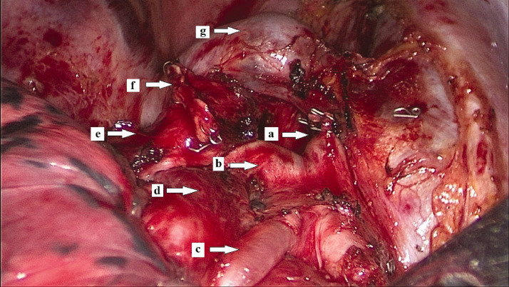
TB is a rare anomaly in which a bronchus arises directly from the trachea. Inada and Kishimoto,3 Kubo et al.,4 and Xu et al.5 all reported tracheal bronchi originating from the main trachea and ventilating the apical and anterior segmental bronchus, and posterior segmental bronchi originating from the right main bronchus. However, to the best of our knowledge, the current case provides the first report of a tracheal bronchus ventilating the apical and posterior segmental bronchus, with an anterior segmental bronchus originating from the right main bronchus. This is also the first reported case of an abnormal tracheal bronchus to the target segment in a patient with successful thoracoscopic right apical segmentectomy (Figure 7).
Figure 7.
Structures of lung segmental hilum: (a) pulmonary artery of the apical segmental (A1) (stump) bronchus; (b) pulmonary artery of the posterior segmental (A2) bronchus; (c) pulmonary artery of the anterior segmental (A3) bronchus; (d) pulmonary vein of the apical and posterior segmental (V1+V2) (before dealing with) bronchus; (e) bronchus of the posterior segment (B2); (f) bronchus of the apical segment (B1) (stump); and (g) azygous vein.
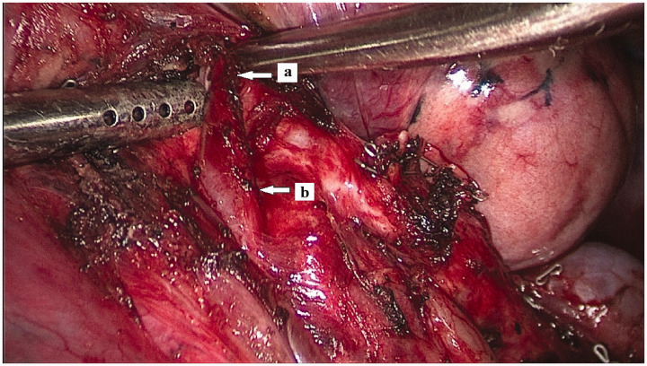
We believed that thoracoscopic segmentectomy can be performed safely by skilled surgeons, but it is important to recognize the presence of TB before surgery. Reconstructed three-dimensional imaging is especially useful, and is carried out before most thoracoscopic segmentectomy procedures in our institute. In addition, because TB is likely to be associated with pulmonary vessel variations,4 being aware of the course of the pulmonary blood vessels and bronchi before surgery, using CT bronchography or 3D-CT angiography, can help to ensure the safety of the procedure. In the current case, we noted a variable V1 pulmonary vein running dorsal to the pulmonary artery trunk and directly into the central vein before surgery (Figure 8).
Figure 8.
Pulmonary vein of the apical segmental (V1) bronchus: (a) pulmonary vein of the apical segmental (V1) bronchus and (b) pulmonary vein of the apical and posterior segmental (V1+V2) bronchus.
In conclusion, TB is a rare anomaly frequently associated with pulmonary vessel variations. Preoperative chest CT, including reconstructed 3-D imaging, and flexible bronchoscopy are useful for preoperative planning and can facilitate successful surgery.
Declaration of conflicting interest
The authors declare that there is no conflict of interest.
Funding
The authors disclosed receipt of the following financial support for the research, authorship, and/or publication of this article: This study was supported by the program of Jiaxing Science and Technology Bureau [grant number 2017AY33007] and 2019 Jiaxing Key Discipline of Medicine—Thoracic Surgery (Supporting Subject) [grant number 2019-zc-09].
ORCID iD
References
- 1.Ritsema GH. Ectopic right bronchus: indication for bronchography. AJR Am J Roentgenol 1983; 140: 671–674. [DOI] [PubMed] [Google Scholar]
- 2.Wooten C, Patel S, Cassidy L, et al. Variations of the tracheobronchial tree: anatomical and clinical significance. Clin Anat 2014; 27: 1223–1233. [DOI] [PubMed] [Google Scholar]
- 3.Inada K, Kishimoto S. An anomalous tracheal bronchus to the right upper lobe; report of two cases. Dis Chest 1957; 31: 109–112. [DOI] [PubMed] [Google Scholar]
- 4.Okubo K, Ueno Y, Isobe J. Upper sleeve lobectomy for lung cancer with tracheal bronchus. J Thorac Cardiovasc Surg 2000; 120: 1011–1012. [DOI] [PubMed] [Google Scholar]
- 5.Xu XF, Chen L, Wu WB, et al. Thoracoscopic right posterior segmentectomy of a patient with anomalous bronchus and pulmonary vein. Ann Thorac Surg 2014; 98: e127–e129. [DOI] [PubMed] [Google Scholar]


