Abstract
Correct regulation of cell contractility is critical for the function of many biological systems. The reproductive system of the hermaphroditic nematode C. elegans contains a contractile tube of myoepithelial cells known as the spermatheca, which stores sperm and is the site of oocyte fertilization. Regulated contraction of the spermatheca pushes the embryo into the uterus. Cell contractility in the spermatheca is dependent on actin and myosin and is regulated, in part, by Ca2+ signaling through the phospholipase PLC-1, which mediates Ca2+ release from the endoplasmic reticulum. Here, we describe a novel role for GSA-1/Gαs, and protein kinase A, composed of the catalytic subunit KIN-1/PKA-C and the regulatory subunit KIN-2/PKA-R, in the regulation of Ca2+ release and contractility in the C. elegans spermatheca. Without GSA-1/Gαs or KIN-1/PKA-C, Ca2+ is not released, and oocytes become trapped in the spermatheca. Conversely, when PKA is activated through either a gain of function allele in GSA-1 (GSA-1(GF)) or by depletion of KIN-2/PKA-R, the transit times and total numbers, although not frequencies, of Ca2+ pulses are increased, and Ca2+ propagates across the spermatheca even in the absence of oocyte entry. In the spermathecal-uterine valve, loss of GSA-1/Gαs or KIN-1/PKA-C results in sustained, high levels of Ca2+ and a loss of coordination between the spermathecal bag and sp-ut valve. Additionally, we show that depleting phosphodiesterase PDE-6 levels alters contractility and Ca2+ dynamics in the spermatheca, and that the GPB-1 and GPB-2 Gβ subunits play a central role in regulating spermathecal contractility and Ca2+ signaling. This work identifies a signaling network in which Ca2+ and cAMP pathways work together to coordinate spermathecal contractions for successful ovulations.
Author summary
Organisms are full of biological tubes that transport substances such as food, liquids, and air through the body. Moving these substances in a coordinated manner, with the correct directionality, timing, and rate is critical for organism health. In this study we used Caenorhabditis elegans, a small transparent worm, to study how cells in biological tubes coordinate how and when they squeeze and relax. The C. elegans spermatheca is part of the reproductive system, which uses calcium signaling to drive the coordinated contractions that push fertilized eggs out into the uterus. Using genetic analysis and a calcium-sensitive fluorescent protein, we show that the G-protein GSA-1 functions with protein kinase A to regulate calcium release, and contraction of the spermatheca. These findings establish a link between G-protein and cAMP signaling that may apply to similar signaling pathways in other systems.
Introduction
Regulation of cellular contractility and relaxation is essential for the function of epithelial and endothelial tubes, which are subjected to changing flow, strain, and pressure as they transport liquid, gases, and other cells throughout the body [1,2]. The Caenorhabditis elegans spermatheca, part of the hermaphroditic reproductive system, is an excellent model for the study of coordinated cell contractility [3–8]. The hermaphrodite reproductive system is composed of two symmetrical gonad arms surrounded by smooth muscle-like sheath cells, the spermathecae, and a common uterus [9,10]. The spermatheca, the site of fertilization, consists of an 8-cell distal neck, a 16-cell central bag, and a syncytial 4-cell spermatheca-uterine valve (sp-ut valve). During ovulation, gonadal sheath cells contract and the distal neck of the spermatheca is pulled open to allow entry of the oocyte. After a regulated period of time the sp-ut valve opens and the distal spermathecal bag constricts to expel the embryo, [11,12]. Failure to coordinate these distinct contraction events during ovulation leads to embryos that fail to reach the uterus, become misshapen, or have decreased viability [4,7].
The signaling pathways that regulate acto-myosin contractility in the spermatheca are similar to those found in smooth muscle and non-muscle cells [13–15]. Two pathways, a Ca2+ dependent and a Rho-dependent pathway are both necessary for spermathecal contractility. These pathways culminate in the phosphorylation and activation of non-muscle myosin. The Ca2+ dependent pathway relies on the activation of PLC-1/phospholipase C-ε, which cleaves phosphatidyl inositol (PIP2) into the second messengers diacylglycerol (DAG) and inositol 1,4,5 triphosphate (IP3). IP3 stimulates the release of Ca2+ from the endoplasmic reticulum (ER) through the ITR-1/IP3 receptor [3,5]. Cytosolic Ca2+ activates the MLCK-1/myosin light chain kinase, which phosphorylates and activates myosin [16]. The Rho-dependent pathway is activated by oocyte entry, which displaces a mechanosensitive Rho GAP, SPV-1, from the membrane, leading to the activation of RHO-1 [7]. GTP-bound RHO-1 then activates LET-502/ROCK, which in turn phosphorylates myosin and inhibits the myosin phosphatase, subsequently increasing the levels of phosphorylated myosin [5,7]. The sp-ut valve opens as the bag constricts to push the embryo into the uterus. Although Ca2+ signaling and actin-based contractility are critical [4,5,8,16–19], little is known about the mechanisms regulating the timing and spatial coordination of Ca2+ release during embryo transit through the spermatheca.
In order to better understand the regulation of Ca2+ signaling in the spermatheca, we performed a candidate RNA interference (RNAi) screen and identified the heterotrimeric G-protein alpha subunit GSA-1/Gαs as an important regulator of spermathecal Ca2+ release. Gαs is a GTPase that regulates diverse biochemical responses in multiple tissue types, including the contractility of airway and arteriole smooth muscle cells [20,21]. In C. elegans, GSA-1 regulates an array of behaviors including larval viability, egg laying, oocyte maturation and locomotion [22–25]. GSA-1 is expressed in neurons, muscle cells, pharynx and the male tail [25–27] and throughout the somatic gonad, including the sheath cells, the spermatheca, uterus and the vulval muscles [23,25], however, the role of GSA-1 in the spermatheca has not been studied.
Heterotrimeric G proteins are composed of α, β, and γ subunits and are typically found downstream of the 7-pass transmembrane class of G-protein coupled receptors (GPCR). Upon ligand binding, the GPCR acts as a guanine nucleotide exchange factor (GEF), exchanging GDP for GTP on the α subunit, and activating the heterotrimeric G protein. Heterotrimeric G proteins can also be activated via a receptor-independent mechanism facilitated by G protein regulator proteins (GPRs) [20,28]. Upon activation, the GTP-bound α subunit disassociates from the βγ subunit and both the α and βγ subunits can initiate downstream signaling pathways [29]. C. elegans express two Gβ subunits, encoded by GPB-1 and GPB-2. GPB-1 shares 86% homology with mammalian β subunits and interacts with all Gα subunits in C. elegans [30,31]. There are two γ subunits, GPC-1 and GPC-2, in the C. elegans genome. GPC-1/γ is expressed in sensory neurons, while GPC-2/γ is expressed more broadly [32]. Gβγ subunits can regulate ion channels [33,34] including Ca2+ channels [35], as well as activate or inhibit adenylyl cyclase [36]. GPB-2 acts downstream of the Gα subunit GOA-1 in pharyngeal pumping [37], and is required for egg-laying and locomotion [38,39].
In smooth muscle, activation of Gαs inhibits acto-myosin contractility through the activation of adenylyl cyclase, which converts adenosine triphosphate (ATP) to 3’-5’-cyclic adenosine monophosphate (cAMP). cAMP levels are in part regulated by phosphodiesterases (PDEs), which convert cAMP into AMP. PKA is composed of two catalytic (PKA-C) and two regulatory (PKA-R) subunits, and is activated when cAMP binds to the PKA-R subunits, causing the release of PKA-C. Therefore, PKA-R acts as an inhibitor of PKA-C in the absence of cAMP. Once released, PKA-C interacts with multiple downstream effectors and regulates lipid metabolism [40], cell migration [41], and vasodilation [42], among many other functions [43]. In airway smooth muscle, PKA inhibits contraction by antagonizing Gαq mediated increases in Ca2+, as well as by phosphorylating and inhibiting Rho [44] and other regulators of contractility. In C. elegans, PKA is encoded by catalytic subunit kin-1/PKA-C and regulatory subunit kin-2/PKA-R. KIN-1 has been implicated in a variety of functions, such as cold tolerance [45], oocyte meiotic maturation [46], locomotion [47], immune responses [48], and Ca2+ influx into motor neurons [49].
Here we describe a role for GSA-1/Gαs and its downstream effectors, KIN-1/PKA-C and KIN-2/PKA-R, in the regulation of Ca2+ release and contractility in the C. elegans spermatheca. Dysregulation of these proteins results in abnormal Ca2+ signaling and loss of coordinated contractility between the sp-ut valve and the spermathecal bag. Additionally, we describe a role for Gβ subunits GPB-1 and GPB-2 and phosphodiesterase PDE-6 in spermathecal contractility and Ca2+ signaling.
Results
GSA-1/Gαs and KIN-1/PKA-C are necessary for proper oocyte transit through the spermatheca
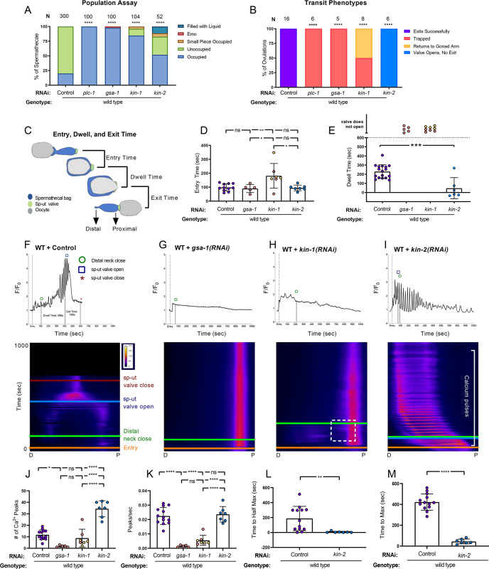
To determine if GSA-1/Gαs is required for oocyte transit, we depleted it using RNA interference (RNAi) in animals expressing GCaMP3 (GFP-calmodulin-M13 Peptide, version 3), a genetically encoded Ca2+ sensor, in the spermatheca [5,50]. Expression of GCaMP3 in the spermatheca provided both a visible GFP outline for scoring and a sensor for Ca2+ levels. The percentage of spermathecae occupied by an embryo was scored in adult animals. Depletion of factors that play a role in embryo transit through the spermatheca results in an increase in the percentage of occupied spermathecae compared to empty vector, or control RNAi, treated animals (18% occupied, n = 300). We used plc-1/PLC-ε, a gene essential for embryo transit, as a positive control (100% occupied, n = 214) [3–5] (Fig 1A).
Fig 1. GSA-1/Gαs and KIN-1/PKA-C, and KIN-2/PKA-R regulate contractility and Ca2+ dynamics in the spermatheca.
(A) Population assay of wild type animals grown on control RNAi, plc-1(RNAi), gsa-1(RNAi), kin-1(RNAi), and kin-2(RNAi). Spermathecae were scored for the presence or absence of an embryo (occupied or unoccupied), presence of a fragment of an embryo or presence of liquid (small piece occupied, filled with liquid), or the presence of endomitotic oocytes in the gonad arm (emo). The total number of unoccupied spermatheca were compared to the sum of all other phenotypes using the Fisher’s exact t-test. N is the total number of spermathecae counted. (B) Transit phenotypes of the ovulations of wild type animals treated with control RNAi, plc-1(RNAi), gsa-1(RNAi), kin-1(RNAi), and kin-2(RNAi) and scored for successful embryo transits through the spermatheca (exits successfully), failure to exit (trapped), reflux into the gonad arm (returns to gonad arm), and the situation in which the sp-ut valve opens, but the embryo does not exit (valve opens, no exit). For transit phenotype analysis, statistics were performed using the total number of oocytes that exited the spermatheca successfully compared to the sum of all other phenotypes. Fisher’s exact t-tests were used for both population assays and transit phenotype analysis, and control RNAi was compared between all other RNAi treatments. (C) Schematic representation of a spermatheca undergoing an ovulation, with entry, dwell, and exit times indicated. Entry time (D) and dwell time (E) analysis of animals treated with control, gsa-1(RNAi), kin-1(RNAi), and kin-2(RNAi). Color coding of the data points corresponds to the transit phenotypes in B. One-way ANOVA with a multiple comparison Tukey’s test was conducted to compare dwell times, and Fishers exact t-test (two dimensional x2 analysis) was performed on the exit times. Representative normalized Ca2+ traces and kymograms of movies in B (F-I) are shown with time of entry, distal neck closure, and time the sp-ut valve opens and closes indicated. Levels of Ca2+ signal were normalized to 30 frames before oocyte entry. Kymograms generated by averaging over the columns of each movie frame (see methods). Refer to S3A–S3D Fig for additional Ca2+ traces and S4A–S4D Fig for additional kymograms. Refer to S3H Fig for Ca2+ traces of wild type animals treated with plc-1(RNAi). (J-M) Color coding of the data points corresponds to the transit phenotypes in B. (J) The number of Ca2+ peaks and (K) the peaks per second was determined for animals treated with control, gsa-1(RNAi), kin-1(RNAi), and kin-2(RNAi), and were compared using one-way ANOVA with a multiple comparison Tukey’s test. The amount of time after oocyte entry required to reach either the (L) half maximum or (M) maximum Ca2+ signal was quantified for animals treated with control and kin-2(RNAi) and compared using Fishers exact t-test. Stars designate statistical significance (**** p<0.0001, *** p<0.005, ** p<0.01, * p<0.05).
Depletion of gsa-1 by RNAi in GCaMP3 expressing animals resulted in almost complete spermathecal occupancy (98% occupied, n = 61) with a low penetrance entry defect, in which gonad arms were filled with endomitotically duplicating oocytes (EMO) [51] (2% EMO, n = 61;Fig 1A). The EMO phenotype occurs when mature oocytes fail to enter the spermatheca and re-enter the mitotic cycle, accumulating DNA [51]. Depletion of GSA-1 resulted in 90% spermathecal occupancy (Fig 1B), with the other 10% exhibiting entry defects (n = 87). The null allele gsa-1(pk75) results in larval arrest, and therefore cannot be used to study the role of GSA-1 in the spermatheca [24]. These results reveal a novel role for GSA-1 in the regulation of spermathecal contractility.
Because GSA-1/Gαs often stimulates the production of cAMP and activation of PKA, we next explored a possible role for PKA in regulation of spermathecal contractility. KIN-1/PKA-C is expressed in the spermathecal bag and the sp-ut valve [40]. Depletion of KIN-1/PKA-C resulted in significantly increased spermathecal occupancy (85% occupied, n = 104; Fig 1A). Depletion of the regulatory subunit KIN-2/PKA-R also resulted in increased spermathecal occupancy (52% occupied, n = 52; Fig 1A,). These results suggest that regulation of PKA activity is critical for successful transit of fertilized embryos through the spermatheca.
GSA-1/Gαs KIN-1/PKA-C, and KIN-2/PKA-R regulate spermathecal contractility and Ca2+ dynamics
To better understand how GSA-1/Gαs, KIN-1/PKA-C, and KIN-2/PKA-R regulate transit of oocytes through the spermatheca, we scored time-lapse movies of animals expressing GCaMP3 in the spermatheca for the presence of four transit phenotypes: successful exit of the embryo (Exited Successfully), retention of the embryo in the spermatheca (Trapped), reflux of the embryo into the oviduct (Returned to Gonad Arm), and failure of the embryo to exit, despite an open sp-ut valve (Valve Opens, No Exit). As expected, all control RNAi embryos exited the spermatheca (100% Exited Successfully, n = 16; Fig 1B,) and all gsa-1(RNAi) embryos failed to exit the spermatheca (100% Trapped, n = 5). Unlike wild type ovulations (S1 Movie), in gsa-1(RNAi) animals, the embryo remained in the spermatheca with the distal neck and sp-ut valve fully closed (S2 Movie). In kin-1(RNAi) animals, 50% of the embryos remained in the spermatheca, while the others returned to the gonad arm (n = 8). We observed embryos that entered, returned to the gonad arm, and re-entered the spermatheca multiple times during imaging, while the sp-ut valve remained closed (S3 Movie). In 100% of the kin-2(RNAi) ovulations, the sp-ut valve opened, often prematurely, but the spermatheca failed to produce the contractions necessary for the embryo to be pushed into the uterus (n = 6).
To further quantify the transit dynamics, we measured the entry time, dwell time, and exit time [16,52] in the time lapse movies. The entry time is the time from the beginning of oocyte entry to when the distal neck closes, fully enclosing the oocyte in the spermatheca. The dwell time is measured from time of distal neck closure to the point the sp-ut valve begins to open. The exit time is measured from sp-ut valve opening, through expulsion of the fertilized embryo into the uterus and full closure of the sp-ut valve (Fig 1C). Entry times in gsa-1(RNAi) and kin-2(RNAi) did not differ significantly from wild type. However, because of the defects in distal neck closure discussed above, kin-1(RNAi) animals had a significantly increased entry time (Fig 1D). Because the sp-ut valve never opens in gsa-1(RNAi) and kin-1(RNAi) animals, neither dwell time nor exit time could be measured. This is reflected by the color-coded values annotated “valve does not open” in Fig 1D. In contrast, in kin-2(RNAi) animals, the sp-ut valve opened significantly faster than wild type, resulting in low, and sometimes negative, dwell times (Fig 1E). Despite these short dwell times, the oocytes consistently failed to exit the spermatheca during the 30-minute imaging period, resulting in no exit times for kin-2(RNAi) animals.
Taken together, these results are consistent with the hypothesis that PKA negatively regulates contractility in the spermatheca and sp-ut valve. We propose that when KIN-1/PKA-C is depleted, increased contractility in the proximal bag and sp-ut valve prevent exit of the fertilized embryo. In this case, the embryo is trapped or pushed back into the oviduct. Conversely, when KIN-2/PKA-R is depleted, activating KIN-1/PKA-C, neither the spermathecal bag nor sp-ut valve is contractile, resulting in reduced exit of the fertilized embryos. In this case, the embryo is not pushed out.
Because Ca2+ signaling plays a key role in spermatheca contractility [5], we next explored the role of GSA-1 and PKA in spermathecal Ca2+ dynamics. We collected time-lapse fluorescent images of animals expressing GCaMP3 and plotted the data both as 1D traces of total Ca2+ levels, and as 2D kymographs with time on the y-axis and space in the x-axis, which allows visualization of the spatial and temporal aspects of Ca2+ signaling. In wild type animals, the sp-ut valve exhibits a bright pulse of Ca2+ immediately upon oocyte entry, which is followed by a quiet period. Once the oocyte is completely enclosed, Ca2+ oscillates across the spermatheca, increasing in intensity until peaking concomitantly with distal spermathecal constriction and embryo exit [5,52] (Fig 1F, S1 Movie). Depletion of gsa-1(RNAi) resulted in low levels of Ca2+ signal in the spermathecal bag. In contrast, Ca2+ signal in the sp-ut valve remained high throughout the entire observation period (n = 5; Fig 1G; S2 Movie). We observed similarly low levels of Ca2+ signal in kin-1(RNAi) animals, however, some Ca2+ signal was observed on the proximal side of the spermathecal bag (n = 8; Fig 1H, indicated by box; S2 Movie). This increased proximal Ca2+ signal (Fig 1H) coincided with retrograde motion of the embryo back into the gonad arm (see Fig 1B; S3 Movie). These results suggest GSA-1 and KIN-1 are required for proper levels of Ca2+ signaling in the spermathecal bag and for the correct inhibition of Ca2+ in the sp-ut valve.
We next investigated the effect of hyperactivation of the catalytic subunit KIN-1/PKA-C by depleting the regulatory subunit, KIN-2/PKA-R. Because depletion of KIN-1 results in a loss of Ca2+ signaling in the spermatheca bag, we expected that the loss of the regulatory subunit, KIN-2/PKA-R, might increase Ca2+ signaling by allowing the catalytic subunit to remain uninhibited. In kin-2(RNAi) animals, Ca2+ signaling increased immediately upon oocyte entry (Fig 1I). Ca2+ transients propagated from the distal to the proximal side of the spermatheca in rhythmic succession (S4 Movie, S1 Fig). These Ca2+ transients appeared as pulses in the Ca2+ trace data and as horizontal lines in the kymogram (Fig 1I). Although kin-2(RNAi) stimulated Ca2+ release, embryos failed to exit the spermatheca (n = 7, Fig 1B). These results suggest unregulated activity of KIN-1/PKA-C, through loss of KIN-2/PKA-R, results in abnormal Ca2+ signaling in the spermatheca, which is insufficient to stimulate embryo exit.
To quantify the differences in Ca2+ signal and enable statistical analysis over many embryo transits, we used Matlab scripts to identify peaks in the GCaMP time series. To capture the rate of increase in calcium signal during embryo transit, we quantified the amount of time after oocyte entry required to reach half the maximum Ca2+ signal [52]. To determine the length of time required for the strongest Ca2+ pulses to occur, we calculated the time to the maximum peak value. To quantitatively define ‘flat’ traces, we calculated the variance of the first derivative, which expresses how quickly the intensity values in the data series are changing with respect to time [53]. As described above, when signal through GSA-1 or PKA was reduced by gsa-1(RNAi) and kin-1(RNAi), few transients were seen and the Ca2+ traces were significantly flatter than the wild type control (Compare Fig 1F to Fig 1G and 1H; S2A Fig). Because of these flat profiles, both gsa-1(RNAi) and kin-1(RNAi) exhibited significantly reduced Ca2+ peaks (Fig 1J), and Ca2+ peaks per second (Fig IK). Because the gsa-1(RNAi) and kin-1(RNAi) traces did not increase in intensity, the time to half max (Fig 1L) and time to max metrics are not biologically informative metrics. In contrast, when PKA activity was increased through the depletion of KIN-2/PKA-R, the number of Ca2+ peaks increased significantly (Fig 1J), however, when we accounted for the duration of the transit, peak frequency per se was not significantly different than wild type (Fig 1K). In kin-2(RNAi) animals, onset of Ca2+ transients occurred almost immediately after oocyte entry, resulting in a significantly reduced time to both half maximum (Fig 1L) and maximum Ca2+ signal (Fig 1M). These results support the conclusion that GSA-1 and KIN-1/PKA stimulate the production of Ca2+ transients in the C. elegans spermatheca.
GSA-1(GF) induction of Ca2+ signaling in the spermatheca is dependent on KIN-1/PKA-C and PLC-1
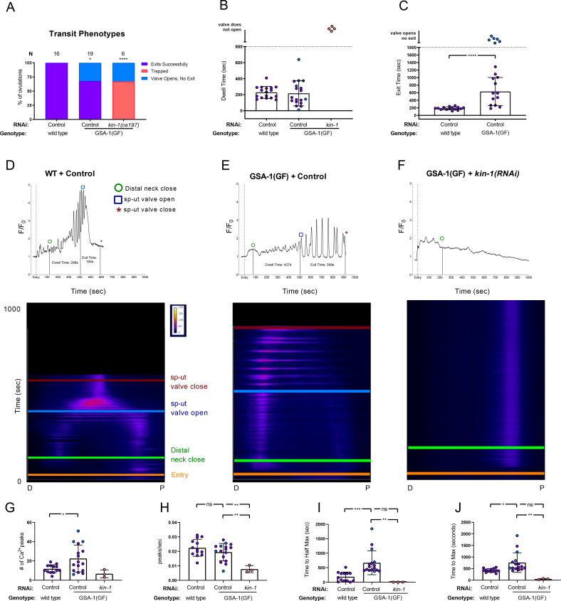
We next explored the effects of hyperactivating GSA-1 signaling on spermathecal transits. The allele gsa-1(ce94) is a gain of function allele (referred to here as GSA-1(GF)) in which the GTP-bound form of GSA-1 is stabilized, resulting in an elevated level of GSA-1 activity [54]. Most fertilized embryos in GSA-1(GF) animals passed successfully through the spermatheca, however, in 31% of transits, the sp-ut valve opened, but the embryo was unable to exit (n = 13; Fig 2A), suggesting that the bag may not contract with the timing or force needed to efficiently expel the embryo into the uterus (Fig 2C). We reasoned that if PKA is downstream of GSA-1, depletion of KIN-1/PKA-C in the GSA-1(GF) background would result in increased trapping of embryos in the spermatheca. Indeed, GSA-1(GF) animals treated with kin-1(RNAi) resulted in a complete failure of embryos to exit the spermatheca. We observed 67% of the embryos remained entirely enclosed by the spermatheca, while in the remaining 33% the valve opened but the embryo was unable to exit (n = 6; Fig 2A).
Fig 2. GSA-1(GF) induction of Ca2+ signaling in the spermatheca is dependent on KIN-1/PKA-C.
(A) Transit phenotypes of the ovulations of wild type animals treated with control RNAi, and GSA-1(GF) animals treated with control RNAi and kin-1(RNAi), were scored for successful embryo transits through the spermatheca (exits successfully), failure to exit (trapped), reflux into the gonad arm (returns to gonad arm), and the situation in which the sp-ut valve opens, but the embryo does not exit (valve opens, no exit). The total number of oocytes that exited the spermatheca successfully was compared to the sum of all other phenotypes using the Fisher’s exact t-test. Control RNAi was compared to all other RNAi treatments. Dwell (B) and exit (C) times of movies in A were analyzed using Fishers exact t-test (two dimensional x2 analysis). Color coding of the data points corresponds to the transit phenotypes in A. Representative normalized Ca2+ traces and kymograms of movies in A (D-F) are shown with time of entry, distal neck closure, and time the sp-ut valve opens and closes indicated. Levels of Ca2+ signal were normalized to 30 frames before oocyte entry. Kymograms generated by averaging over the columns of each movie frame (see methods). Refer to S3A Fig and S5A and S5B Fig for additional Ca2+ traces, and S4A Fig and S6A and S6B Fig for additional kymograms. (G-J) Color coding of the data points corresponds to the transit phenotypes in A. (G) The number of Ca2+ peaks, (H) the peaks per second, and the amount of time after oocyte entry required to reach either (I) half the maximum or (J) maximum Ca2+ signal were quantified for wild type animals treated with control RNAi, and GSA-1(GF) animals treated with control RNAi and kin-1(RNAi). These were compared using one-way ANOVA with a multiple comparison Tukey’s test. Stars designate statistical significance (*** p<0.005, ** p<0.01, * p<0.05).
Because gsa-1(RNAi) reduced Ca2+ signaling in the spermathecal bag but increased signaling in the sp-ut valve, we predicted increasing GSA-1 activity would produce the opposite effect. However, rather than exhibiting prematurely elevated Ca2+, expression of GSA-1(GF) resulted in low amplitude peaks for the first few hundred seconds, after which Ca2+ was released in pulses that continued until embryo exit. In the sp-ut valve, Ca2+ signal was depressed, as expected (Compare Fig 2D and 2F). The number of Ca2+ peaks increased significantly in GSA-1(GF) animals (Fig 2G), however peak frequency was not significantly different than wild type (Fig 2H). This suggests that, as with depletion of KIN-2/PKA-R (see Fig 1) the observed increase in overall number of peaks in GSA-1(GF) is due to the increased transit lengths.
If PKA is downstream of GSA-1(GF), depletion of KIN-1/PKA-C should suppress the Ca2+ phenotypes seen in the GSA-1(GF) animals. Indeed, depletion of KIN-1 in GSA-1(GF) prevented the Ca2+ pulses observed in GSA-1(GF) animals (Fig 2G, S3B Fig), significantly decreasing the number of Ca2+ peaks (Fig 2H) and peaks per second (Fig 2I). Because the Ca2+ signal in kin-1(RNAi) animals did not ramp up over time, the highest amplitude peaks are reached early in the time series, which is reflected in the reduced time to half maximum (Fig 2I) and maximum Ca2+ signal (Fig 2J) in GSA-1(GF) animals treated with kin-1(RNAi). These metrics are similar to those seen when wild type animals were treated with kin-1(RNAi) (See Fig 1G–1J). These results are consistent with the hypothesis that KIN-1 is acting downstream of GSA-1 in the spermatheca to regulate Ca2+ release.
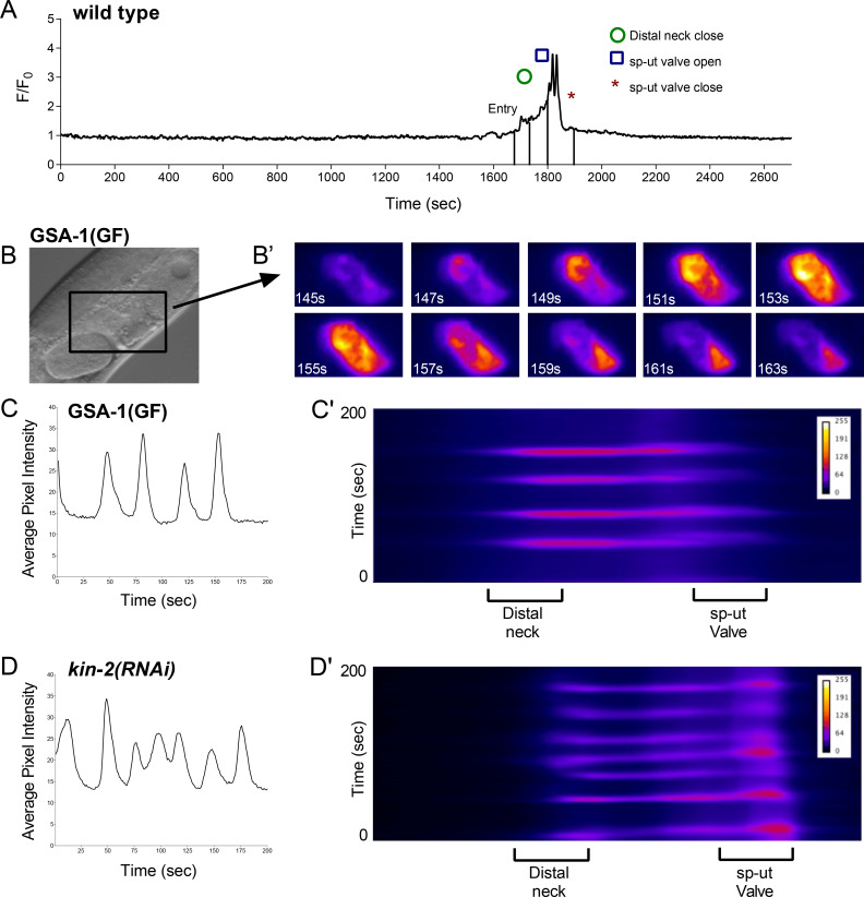
In wild type animals, oocyte entry stimulates Ca2+ release [5]. The spermathecal Ca2+ remains at baseline until ovulation and returns to baseline following embryo exit (Fig 3A). However, while observing GSA-1(GF) animals, we noticed Ca2+ pulses in unoccupied spermathecae, which traveled from the distal spermatheca through the bag to the sp-ut valve (Fig 3B and 3C, S5 Movie). Similarly, in kin-2(RNAi) animals, Ca2+ repeatedly increased, peaked, and then dropped to baseline levels in empty spermathecae (Fig 3D). These results suggest that activating GSA-1 or PKA can bypass the need for oocyte entry as a trigger for Ca2+ release. In addition, these similarities between GSA-1(GF) and kin-2(RNAi) support the hypothesis that GSA-1 triggers Ca2+ signaling through KIN-1/KIN-2. The GSA-1(GF) and kin-2(RNAi) Ca2+ traces share features, such as triggering Ca2+ signaling in empty spermathecae, and reducing the time to half max and max signal in occupied spermathecae (see Fig 1I and 1J and Fig 2I and 2J), however, activating PKA through kin-2(RNAi) produces high amplitude peaks sooner after oocyte entry. This difference could be a result of either the degree of activation of PKA, or differences in downstream effectors.
Fig 3. GSA-1(GF) and knockdown of KIN-2/PKA-R induce Ca2+ signaling in an empty spermatheca.
(A) Normalized Ca2+ trace of a wild type ovulation before, during, and after transit. Ca2+ signal does not rise above basal levels before or after the transit. Time of entry, distal neck closure, and time the sp-ut valve opens and closes are indicated. (B) DIC image of a GSA-1(GF) animal; the spermatheca is indicated by a box. (B’) Ca2+ signaling in the unoccupied GSA-1(GF) spermatheca shown in B at an arbitrary timepoint before an ovulation event. Ca2+ repeatedly increases, peaks, and then drops to baseline levels. These pulses travel from the distal spermatheca through the bag to the sp-ut valve (S5 Movie). Average pixel intensity trace of the Ca2+ signal (C) and kymogram (C’) of the GSA-1(GF) spermatheca are shown. Knockdown of KIN-2 with RNAi results in a similarly pulsing unoccupied spermatheca, as shown by the average pixel intensity trace of the Ca2+ signal (D) and kymogram (D’) of a kin-2(RNAi) animal.
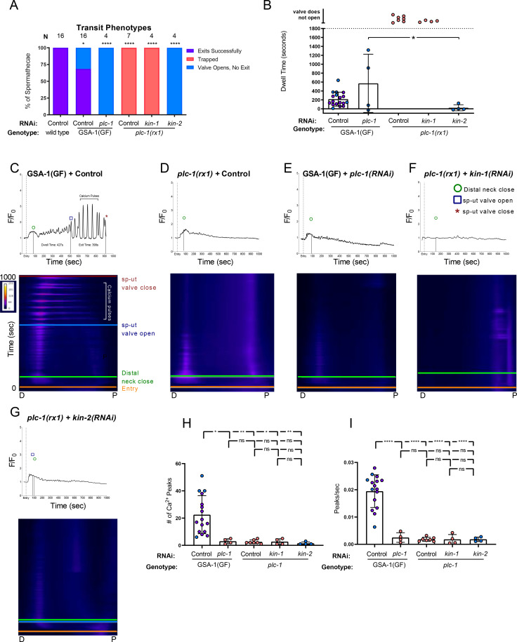
Previous work has shown that the phospholipase PLC-1 is required for spermathecal contractility and Ca2+ release [5]. Therefore, we next asked whether the phospholipase PLC-1 was required for transits in GSA-1(GF) animals. As expected, the null allele plc-1(rx1) resulted in 100% spermathecal occupancy (n = 7; Fig 4A). Similarly, in GSA-1(GF) animals treated with plc-1(RNAi), embryos did not exit the spermatheca, however the sp-ut valve did open (n = 4; Fig 4A). Occasionally, the valve opened before the distal neck closed, resulting in negative dwell times (Fig 4B). We next asked if the Ca2+ signal observed in the GSA-1(GF) animals (Fig 4C) required phospholipase-stimulated Ca2+ release. In plc-1 null animals, Ca2+ signaling remained at baseline levels in the spermathecal bag even after oocyte entry (Fig 6D). When GSA-1(GF) animals were treated with plc-1(RNAi), the Ca2+ signal remained low in the spermathecal bag, and we no longer observed the Ca2+ pulses characteristic of GSA-1(GF) (Fig 4E). Similarly, loss of plc-1 prevented both the excess proximal Ca2+ release observed in kin-1(RNAi) spermathecae (Fig 4F), and the Ca2+ pulses observed in kin-2(RNAi) spermathecae (Fig 4G). In GSA-1(GF) and kin-2(RNAi) animals, the number of Ca2+ peaks (Fig 4H) and Ca2+ peaks per second (Fig 4I), were reduced to the level seen in plc-1(RNAi) animals. Because the intensity of the Ca2+ did not increase over time in these low variance traces (S2C Fig, S6E Fig, S8A–S8C Fig) the time to half max and time to max are not biologically informative metrics. These results suggest the Ca2+ pulses observed when either GSA-1 or PKA signaling is activated require PLC-1, and indicate GSA-1 and PKA act upstream or in parallel to PLC-1 to stimulate Ca2+ release in the spermatheca.
Fig 4. Ca2+ signal is dependent on PLC-1.
(A) Transit phenotypes of the ovulations of wild type animals treated with control RNAi, GSA-1(GF) animals treated with control RNAi and plc-1(RNAi), and plc-1 null animals treated with control RNAi, kin-1(RNAi), and kin-2(RNAi) were scored for successful embryo transits through the spermatheca (exits successfully), failure to exit (trapped), and the situation in which the sp-ut valve opens, but the embryo does not exit (valve opens, no exit). The total number of oocytes that exited the spermatheca successfully was compared to the sum of all other phenotypes using the Fisher’s exact t-test. Control RNAi was compared to all other RNAi treatments. (B) Dwell times derived from movies in A were compared using One-way ANOVA with a multiple comparison Tukey’s test. Color coding of the data points corresponds to the transit phenotypes in A. Representative normalized Ca2+ traces and kymograms of movies in A (C-G) are shown with time of entry, distal neck closure, and time the sp-ut valve opens and closes indicated. Levels of Ca2+ signal were normalized to 30 frames before oocyte entry. Kymograms generated by averaging over the columns of each movie frame (see methods). Refer to S5A and S5E Fig and S7A–S7C Fig for additional Ca2+ traces, and S5A Fig and S6A, S6C, and S8A–S8C Fig for additional kymograms. (H-I) Color coding of the data points corresponds to the transit phenotypes in A. (H) The number of Ca2+ peaks (see methods) and (I) the peaks per second was determined for GSA-1(GF) animals treated with control RNAi and plc-1(RNAi), and plc-1 null animals treated with control RNAi, kin-1(RNAi), and kin-2(RNAi). Both were compared using one-way ANOVA with a multiple comparison Tukey’s test. Stars designate statistical significance (**** p<0.0001, *** p<0.005, ** p<0.01, * p<0.05).
Fig 6. Heterotrimeric G-protein beta subunits GPB-1 and GPB-2 regulate Ca2+ signaling and spermatheca contractility.
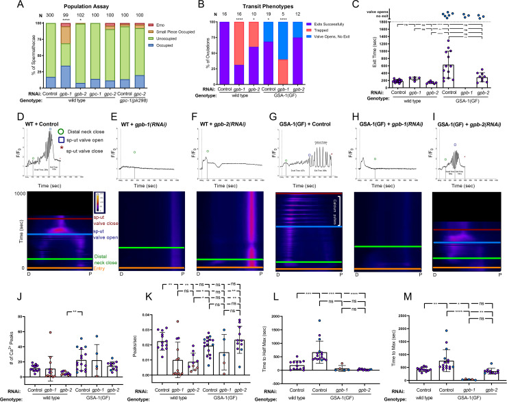
(A) Population assay of wild type young adults grown on control RNAi, gpb-1(RNAi), gpb-2(RNAi), gpc-1(RNAi), and gpc-2(RNAi) and gpc-1(pk298) grown on control RNAi and gpc-2(RNAi). Spermathecae were scored for the presence or absence of an embryo (occupied or unoccupied), presence of a fragment of an embryo (small piece occupied), or the presence of endomitotic oocytes in the gonad arm (emo) phenotypes. The total number of unoccupied spermatheca was compared to the sum of all other phenotypes using the Fisher’s exact t-test. N is the total number of spermathecae counted. (B) Transit phenotypes of the ovulations in wild type animals treated with control RNAi, gpb-1(RNAi) and gpb-2(RNAi), and GSA-1(GF) animals treated with control RNAi, gpb-1(RNAi), and gpb-2(RNAi) were scored for successful embryo transits through the spermatheca (exits successfully), failure to exit (trapped), and the situation in which the sp-ut valve opens, but the embryo does not exit (valve opens, no exit). Color coding of the data points corresponds to the transit phenotypes in B. For transit phenotype analysis, the total number of oocytes that exited the spermatheca successfully was compared to the sum of all other phenotypes. Fisher’s exact t-tests were used for both population assays and transit phenotype analysis. (C) Exit time of movies in B were compared using One-way ANOVA with a multiple comparison Tukey’s test. Representative normalized Ca2+ traces and kymograms of movies in B (D-I) are shown with time of entry, distal neck closure, and time the sp-ut valve opens and closes indicated. Levels of Ca2+ signal were normalized to 30 frames before oocyte entry. Kymograms generated by averaging over the columns of each movie frame (see methods). Refer to S3A, S3E, S3F Fig and S5A, S4C, S4D Figs for additional Ca2+ traces, and S6D and S6E Fig and S9B and S9C Fig for additional kymograms. (J-M) Color coding of the data points corresponds to the transit phenotypes in A. (J) The number of Ca2+ peaks and (K) the peaks per second was determined for wild type animals treated with control RNAi, gpb-1(RNAi) and gpb-2(RNAi), and GSA-1(GF) animals treated with control RNAi, gpb-1(RNAi), and gpb-2(RNAi). The amount of time after oocyte entry required to reach either the (L) half maximum or (M) maximum Ca2+ signal was quantified for wild type animals treated with control RNAi, GSA-1(GF) animals treated with control RNAi, gpb-1(RNAi), and gpb-2(RNAi). These values were compared using one-way ANOVA with a multiple comparison Tukey’s test. and compared using Fishers exact t-test. Stars designate statistical significance (**** p<0.0001, *** p<0.005, ** p<0.01, * p<0.05).
Phosphodiesterase PDE-6 is necessary for normal transit times and Ca2+ dynamics
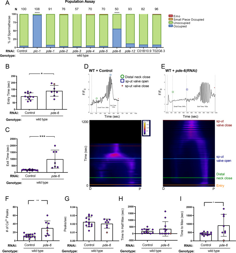
Because PKA is activated by cAMP, we anticipated that altering cAMP levels in the spermatheca would alter the contractility and Ca2+ dynamics during ovulation. Phosphodiesterases (PDEs) are enzymes that regulate the concentration of cAMP by catalyzing its hydrolysis into AMP [55]. To determine whether increasing cAMP levels by depleting PDEs would affect spermathecal transits, we depleted each of the known C. elegans PDEs and identified PDE-6 as an important regulator of spermathecal contractility. Depletion of pde-6 resulted in 56% (n = 50) of animals with embryos retained in the spermatheca (Fig 5A). To study the effects of PDE-6 on ovulations and Ca2+ dynamics, we collected time-lapse images of GCaMP3 animals fed pde-6(RNAi). Both the entry time and the exit time in pde-6(RNAi) animals were significantly longer than wild type (Fig 5B and Fig 5C. In one case, the sp-ut valve never opened, and the embryo remained trapped in the spermatheca (Fig 5D). The pde-6(RNAi) ovulations resulted in delayed Ca2+ release in the bag, after which the Ca2+ was released in pulses until embryo exit (n = 8; Fig 5E). The increased number of peaks observed in pde-6(RNAi) (Fig 5F), correspond to the increased transit time, rather than an increase in overall peak frequency (Fig 5G). While the time to half maximum Ca2+ value is not significantly different from wild type (Fig 5H), the time to maximum is significantly increased (Fig 5I), due to the delay before the onset of high amplitude Ca2+ pulses. The pde-6(RNAi) Ca2+ metrics are similar to the GSA-1(GF) metrics (S10 Fig). These results suggest PDE-6 regulates Ca2+ signaling and spermathecal contractility, likely through regulation of cAMP levels.
Fig 5. Phosphodiesterase PDE-6 is necessary for normal transit times and Ca2+ dynamics.
(A) population assay of wild type animals grown on control RNAi, plc-1(RNAi), pde-1(RNAi), pde-2(RNAi), pde-3(RNAi), pde-4(RNAi), pde-5(RNAi), pde-6(RNAi), pde-12(RNAi), C01B10.9 and T02G6.3. Spermathecae were scored for the presence or absence of an embryo (occupied or unoccupied), presence of a fragment of an embryo (small piece occupied), or the presence of endomitotic oocytes in the gonad arm (emo) phenotypes. The total number of unoccupied spermatheca was compared to the sum of all other phenotypes using the Fisher’s exact t-test. N is the total number of spermathecae counted. Control RNAi was compared to all other RNAi treatments. Entry time (B) and exit time (C) were compared using Fisher’s exact t-test (two dimensional x2 analysis). (B,C) Values are color coded corresponding to transit phenotype (purple: successful exit, orange: traps. Representative normalized Ca2+ traces and kymograms of control RNAi (D) and pde-6(RNAi) (E) are shown with time of entry, distal neck closure, and time the sp-ut valve opens and closes indicated. Levels of Ca2+ signal were normalized to 30 frames before oocyte entry. Kymograms were generated by averaging over the columns of each movie frame (see methods). Refer to S3A Fig and S3G Fig for additional Ca2+ traces, and S4A Fig and S9A Fig for additional kymograms. (F-I) Values are color coded corresponding to transit phenotype (purple: successful exit, orange: traps). (F) The number of Ca2+ peaks, (G) the peaks per second, and the amount of time after oocyte entry required to reach either (H) half the maximum or (I) maximum Ca2+ signal were quantified for wild type animals treated with control and pde-6(RNAi). Values were compared using Fisher’s exact t-test. Stars designate statistical significance (**** p<0.0001, *** p<0.005, ** p<0.01, * p<0.05).
Heterotrimeric G-protein beta subunit GPB-1 and GPB-2 are required to regulate Ca2+ signaling and spermatheca contractility
Heterotrimeric G proteins consist of an α, a β and a γ subunit. The β and γ subunits are closely associated and can be considered one functional unit. Activation of the Gα subunit causes Gα to dissociate from Gβγ, and both can then initiate downstream signaling pathways [56]. Therefore, we next explored potential roles for βγ in the spermatheca. Depletion of GPB-1/β resulted in a significant increase in the percent of occupied spermathecae (34%, n = 99) compared to control RNAi treated animals (18% occupied, n = 300; Fig 6A). GPB-1 depletion also resulted in a significant number of spermathecae occupied by a small piece of an embryo (Fig 6A). Surprisingly, while GPB-2 depletion showed no significant trapping in freely moving animals (Fig 6A), when the animals were immobilized for imaging, embryos failed to exit the spermatheca in 60% of gpb-2(RNAi) movies (n = 10) (Fig 6B), suggesting animals may be able to compensate somewhat for the loss of GPB-2 with body wall muscle contraction. Only 69% of gpb-1(RNAi) ovulations trapped, (Fig 6B). Of the ovulations that were able to exit successfully, neither gpb-1(RNAi) nor gpb-2(RNAi) on their own affected the time required for the embryo to exit the spermatheca (Fig 6C). Depletion of neither GPC-1/γ nor GPC-2/γ significantly affected ovulation when depleted via RNAi (Fig 6A). Similarly, neither the gpc-1(pk298) null allele, nor treating gpc-1(pk298) with gpc-2 RNAi resulted in ovulation defects. These data suggest GPB-1 may be associated with GSA-1 in the spermatheca (Fig 6A), but identification of the relevant γ subunits requires further investigation. Insufficient knockdown of GPC-1/γ nor GPC-2/γ may explain the lack of phenotype.
In order to explore a role for GPB-1 and GPB-2 in Ca2+ signaling, we observed ovulations in GCaMP3 expressing animals treated with gpb-1(RNAi) and gpb-2(RNAi). Compared to wild type movies, in which oocyte entry triggers a pulse of Ca2+ in the sp-ut valve, followed by Ca2+ oscillations in the spermathecal bag and sp-ut valve that increase in intensity until the embryo exits (Fig 6D), both gpb-1(RNAi) and gpb-2(RNAi) resulted in elevated and/or prolonged Ca2+ signal in the sp-ut valve and very little Ca2+ signal in the spermathecal bag (Fig 6E, Fig 6F, S2E Fig). While loss of GPB-1 or GPB-2 did not significantly reduce the overall number of peaks (Fig 6J), the longer duration of the transits resulted in reduced peak frequencies (Fig 6K). Because little rise in Ca2+ is observed, the time to half max (Fig 6L) and time to max (Fig 6M) are short for both gpb-1 and gpb-2(RNAi). Loss of the β subunits produces similar phenotypes to those observed for gsa-1(RNAi), suggesting GPB-1 and GPB-2 are required for the activation of Ca2+ signaling by GSA-1.
Activation of Gα liberates the Gβγ subunits, which can independently initiate signaling pathways [56]. In order to determine if activated signaling by Gβγ could partially explain the GSA-1(GF) phenotypes, we depleted GPB-1 and GPB-2 in the GSA-1(GF) background. First, we assessed the effect on GSA-1(GF) transit phenotypes. In GSA-1(GF) animals, even though the sp-ut valve opens, ~30% of embryos remained inside the spermatheca (n = 19; Fig 6B). Depletion of GPB-1 on its own resulted in significant trapping, with most spermathecae occupied with an embryo (36%) or an embryo fragment (26%) (n = 99; see Fig 6A). When GPB-1 was depleted in the GSA-1(GF) background, no successful transits were observed, with 40% of ovulations resulting in trapping, and 60% of ovulations in which the valve opened but the embryo was unable to exit (n = 5; Fig 6B). In contrast, in GSA-1(GF) gpb-2(RNAi) animals, the trapping phenotype did not differ significantly from GSA-1(GF) alone (Fig 6B). However, the significantly longer exit time observed in GSA-1(GF) animals was suppressed by gpb-2(RNAi) to wild type levels (Fig 6C).
As previously described, GSA-1(GF) results in strong Ca2+ pulses that propagate across the tissue (Fig 6G). When GPB-1 was depleted in GSA-1(GF) expressing animals, other than a brief pulse upon entry, only low amplitude peaks were observed in either the sp-ut valve or in the spermatheca bag (Fig 6H and S2E Fig). This result suggests GSA-1 requires GPB-1 to stimulate Ca2+ release, and is consistent with the model that binding to the Gβγ subunit GPB-1 is required for activation of GSA-1. In contrast, depletion of GPB-2 in GSA-1(GF) animals resulted in Ca2+ dynamics more similar to those observed in wild type animals (Fig 6I). Depletion of GPB-1 or GPB-2 did not reduce the number of peaks (Fig 6J) or peak frequency (Fig 6K) in GSA-1(GF) animals, although the peaks in the GSA-1(GF); gpb-1(RNAi) animals are much lower amplitude than in GSA-1(GF). This feature results in minimal increase in Ca2+ levels, reflected in the time to half max (Fig 6L) and time to max (Fig 6M), which were reached shortly after entry in GSA-1(GF) animals treated with gpb-1(RNAi). In contrast, when GSA-1(GF) animals were treated with gpb-2(RNAi), the time to max Ca2+ signal was reduced to wild type levels (Fig 6M). These results, in combination with the rescued exit times (Fig 6C), suggest loss of GPB-2 might suppress GSA-1(GF) activity to more wild type levels. Alternatively, the Gβs could be activating parallel signaling pathways needed for Ca2+ release, potentially including other Gα subunits active in the spermatheca.
Discussion
Ca2+ and cAMP are important second messengers with key roles in a diverse set of biological processes. Here, we show that GSA-1/Gαs and KIN-1/PKA-C function to regulate Ca2+ release and coordinated contractility in the C. elegans spermatheca. The Gβ subunits GPB-1 and GPB-2 are required for activation of GSA-1/Gαs, and phosphodiesterase PDE-6 functions in the spermatheca to regulate Ca2+ levels and overall tissue contractility.
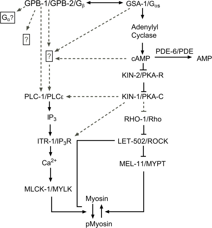
In the spermatheca, oocyte entry triggers Ca2+ waves, which propagate across the spermatheca and trigger spermathecal contractility. We found that loss of either GSA-1/Gαs or KIN-1/PKA-C leads to low levels of Ca2+ signal in the spermathecal bag, while increasing GSA-1 activity with a gain of function allele or activating PKA through loss of the regulatory subunit, KIN-2/PKA-R, leads to strong Ca2+ pulses, even in the absence of oocyte entry. This suggests GSA-1/Gαs and KIN-1/PKA-C stimulate Ca2+ release in the spermatheca. Previous work in our lab has shown that PLC-1/phospholipase C-ε is necessary to stimulate Ca2+ release in the spermatheca [5]. Here, we show that loss of PLC-1 can block the Ca2+ release observed in GSA-1(GF) and kin-2(RNAi) animals, suggesting PLC-1 acts either downstream or in parallel with Gαs and PKA (Fig 7). Mechanisms by which PKA could stimulate Ca2+ release include activation of the ITR-1/IP3 receptor, or activation of plasma membrane channels such as stretch-sensitive TRPV channels [57]. For example, in mouse cardiomyocytes, Gα activation can stimulate Ca2+ release through exchange protein directly activated by cAMP (EPAC) and Rap1 [58,59].
Fig 7. A working model of the spermathecal Ca2+ signaling network.
We propose KIN-1 and KIN-2 are working downstream of GSA-1, while GPB-1 and GPB-2 are either working together with or in parallel to GSA-1 to regulate Ca2+ signaling and spermathecal contractility. Dashed lines indicate possible interactions.
Heterotrimeric G-proteins consist of an α and a βγ subunit, which, when activated by an upstream G-protein coupled receptor (GPCR) or G-protein regulator (GPR), dissociate and independently activate signaling cascades [60–62]. C. elegans expresses numerous GPCRs [63], which has hampered our efforts to identify the upstream activator of Gα. A set of GPCRs expressed in the spermatheca has recently been published [64], which may facilitate the identification of a receptor that acts upstream of GSA-1. Alternatively, GPRs can facilitate the activation of G proteins via GPCR-independent mechanisms [20,28]. Regardless of the trigger, we show both Gα and Gβ signaling are important for regulated Ca2+ release in the spermatheca. The presence of Gβ subunit GPB-1 is required for the activation of the heterotrimer, while GPB-2 may play a more modulatory role, however we cannot rule out that the observed differences between gpb-1(RNAi) and gbp-2 (RNAi) may be due to differences in RNAi efficiency. Not only are the Gβs critical for Gα activation, the Gβγ subunits may also be activating other downstream effectors critical for Ca2+ release. For example, in COS-7 cells, PLC-ε can be activated by Gβγ subunits [65], which offers a promising alternative. Future work is needed to explore a possible connection between Gβγ and PLC-1 in the spermatheca.
Contraction of the spermatheca requires both Ca2+ and Rho signaling. Therefore, the observed transit defects could be due to GSA-1/Gαs and KIN-1/PKA-C regulation of either, or both, of these signaling pathways. Because kin-2(RNAi), which activates KIN-1/PKA-R and stimulates Ca2+ release, is unable to produce contractions sufficient to expel the embryo from the spermatheca, this implies PKA may also regulate the Rho side of the pathway. In SH-EP cells, PKA has been shown to phosphorylate and inhibit Rho through stabilization of the inactive RhoGDI-bound state [44,66] Given that the Rho phosphorylation site is conserved in the C. elegans RHO-1, it is possible that knockdown of KIN-2 results in an increased inhibition of RHO-1 thus resulting in decreased tissue contractility in kin-2(RNAi) animals.
The sp-ut valve is a syncytium of 4 cells that prevents premature release of oocytes into the uterus upon entry in the spermatheca. This allows for enough time for the oocyte to become fertilized and form an eggshell, after which the sp-ut dilates and allows passage of the embryo into the uterus [67]. Here, we show that PKA regulates Ca2+ release and contractility of the sp-ut valve. Depletion of KIN-1/PKA-C activity results in increased Ca2+ and a valve that remains closed. In contrast, hyperactive PKA-C activity (through kin-2(RNAi)) results in an sp-ut valve that opens prematurely, perhaps accounting for the short dwell times observed when PKA-R is depleted (see Fig 1E). Decreasing signal through PKA/KIN-1 or Gαs /GSA-1 knockdown results in a surplus of Ca2+ in the sp-ut valve, whereas increasing signaling through GSA-1(GF) results in an extended quiet Ca2+ period in the sp-ut valve. This suggests PKA/KIN-1 and Gαs /GSA-1 are required for Ca2+ inhibition in the sp-ut valve even though they are required for Ca2+ release in the spermathecal bag. Little is known about the signaling networks that regulate contractility in the sp-ut valve. PLC-1 is not expressed in the sp-ut valve [3], necessitating a different mechanism of Ca2+ regulation from the spermathecal bag. Phosphorylation of IP3Rs by PKA-C has been shown to decrease Ca2+ release in rat brain cells [68], which could help to explain the Ca2+ excess seen in the sp-ut valve. Other mechanisms by which PKA has been described to lower Ca2+ include increasing the activity of SERCA pumps, which pump Ca2+ back into the ER, by phosphorylating and dissociating phospholamban [69,70]. However, there is no obvious phospholamban homolog in C. elegans. PKA can inhibit PLC-β [71], which would result in decreased Ca2+ release. However, EGL-8/PLC- β is not expressed in either the spermatheca or sp-ut valve [72]. Future study may reveal the details of how PKA regulates Ca2+ signaling in the sp-ut valve.
Regulating the correct force, timing, and direction of contraction in biological tubes is crucial for living organisms. Dysfunction in the regulatory networks controlling these dynamics leads to diseases such as heart disease and asthma [42,73]. The signaling networks that control Ca2+ oscillations and actomyosin contractility in smooth muscle in humans are conserved in the C. elegans spermatheca. In this study, we identified Gαs and PKA as key players in the spermatheca, where Gαs acts upstream of PKA to regulate the spatiotemporal regulation of Ca2+ release and contractility in the spermatheca. Because PKA is implicated in a variety of biological processes, understanding its control and targets may give us insights into the diseases that occur when this control goes awry.
Materials and methods
Strains and culture
Nematodes were grown on nematode growth media (NGM) (0.107 M NaCl, 0.25% wt/vol Peptone (Fischer Science Education), 1.7% wt/vol BD Bacto-Agar (FisherScientific), 0.5% Nystatin (Sigma), 0.1 mM CaCl2, 0.1 mM MgSO4, 0.5% wt/vol cholesterol, 2.5 mM KPO4) and seeded with E. coli OP50 using standard C. elegans techniques [74]. Nematodes were cultured at 23°C unless specified otherwise. All Ca2+ imaging was acquired using the strain UN1108 xbls1101 [fln-1p::GCaMP3,rol-6] except for experiments utilizing the GSA-1 gain of function mutant KG524 GSA-1(GF). These animals were crossed into UN1417, a separate integration event of xbIs1108 [fln-1p::GCaMP3].
Construction of the transcriptional and translational reporter of GSA-1
The gsa-1 promoter (1.6 kb upstream of GSA-1 start codon) was amplified from C. elegans genomic DNA using primers with PstI and BamHI 5’ extensions and ligated into pPD95_77 (Fire Lab) upstream of GFP creating pUN783. To make a translational fusion gsa-1 was amplified without its stop codon from C. elegans coding DNA using primers with BamHI 5’ extensions and ligated between the gsa-1 promoter and GFP of pUN783 creating pUN810. Transgenic animals were created by microinjecting a DNA solution of 20 ng/μl of pUN783 or pUN810 and 50 ng/μl of pRF4 rol-6 (injection marker) into N2 animals. Roller animals expressing GFP were segregated to create the transgenic lines UN1727 (transcriptional reporter) and UN1742 (translational reporter) respectively.
RNA interference
The RNAi protocol was performed essentially as described in Timmons et al (1998) [75]. HT115(DE3) bacteria (RNAi bacteria) transformed with a dsRNA construct of interest was grown overnight in Luria Broth (LB) supplemented with 40 μg/ml ampicillin and seeded (150 μl) on NGM plates supplemented with 25 μg/ml carbenicillin and disopropylthio-β-galactoside (IPTG). Seeded plates were left for 24–72 hours at room temperature (RT) to induce dsRNA expression. Empty pPD129.36 vector (“Control RNAi”) was used as a negative control in all RNAi experiments.
Embryos from gravid adults were collected using an alkaline hypochlorite solution as described by Hope (1999) and washed three times in M9 buffer (22 mM KH2PO4, 42 mM NaHPO4, 86 mM NaCl, and 1 mM MgSO4) (‘egg prep’). Clean embryos were transferred to supplemented NGM plates seeded with HT115(DE3) bacteria expressing dsRNA of interest and left to incubate at 23°C for 50–56 hours depending on the experiment. In experiments where larvae, rather than embryos, were transferred to RNAi plates (kin-1(RNAi) and kin-2(RNAi)), adults were ‘egg prepped’ into Control RNAi and left to incubate at 23°C for 32–34 hours, after which they were moved to bacterial lawns expressing the dsRNA of interest and returned to 23°C, and imaged 50–56 after the egg prep.
Population assay
Embryos collected via an ‘egg prep’ as previously described were plated on supplemented NGM seeded with RNAi bacterial clones of interest. Plates were incubated at 23°C for 54–56 hours or until animals reached adulthood. Upon adulthood nematodes were killed in a drop of 0.08 M sodium azide (NaAz) and mounted on 2% agarose pads to be visualized using a 60x oil-immersion objective with a Nikon Eclipse 80i epifluorescence microscope equipped with a Spot RT3 CCD camera (Diagnostic instruments; Sterling Heights, MI, USA). Animals were scored for the presence or absence of an embryo in the spermatheca as well as entry defects such as gonad arms containing endomitotically duplicating oocytes (Emo) [51]. A Fisher’s exact t-test (two dimensional x2 analysis) using GraphPad Prism statistical software was used to compare the percent of occupied spermathecae between control RNAi and all other RNAi treatments.
Wide-field Fluorescence microscopy image acquisition and processing
All Differential Interference Contrast (DIC) and fluorescent images were taken using a 60x oil-immersion objective with Nikon Eclipse fluorescent microscope equipped with a Spot RT3 CCD camera (Diagnostic instruments; Sterling Heights, MI, USA) or Spot RT39M5 with a 0.55x adapter unless otherwise stated. Fluorescence excitation was provided by a Nikon Intensilight C-HGFI 130W mercury lamp and shuttered with a SmartShutter (Sutter Instruments, Novato CA, USA). For acquisition of time-lapse images young adult animals were immobilized with 0.05 micron polystene polybeads diluted 1:2 in water (Polysciences Inc., Warrington, PA, USA) and mounted on slides with 5% agarose pads. Time lapse GCaMP imaging was captured at 1 frame per second, with an exposure time of 75 ms and a gain of 8 for movies obtained with the Spot RT3CCD camera, and with an exposure time of 20 ms and a gain of 8 for movies obtained with the Spot RT39M5. Time-lapse images were only taken of the first 3 ovulations, with preference for the 1st ovulation. The same microscopy image capture parameters were maintained for all imaging.
All time-lapse GCaMP3 images were acquired as 1600x1200 pixels for the Spot RT3 OCCD camera or 2448x2048 for the RT39M5 camera and saved as 8-bit tagged image and file format (TIFF) files. All image processing was done using a macro on Image J [76]. All time-lapse images were oriented with the sp-ut valve on the right of the frame and registered to minimize any body movement of the paralyzed animal. An 800x400 region of interest for the Spot RT3 OCCD and 942x471 for the RT39M5 camera encompassing the entire spermatheca was utilized to measure the GCaMP3 signal. The average pixel intensity of each frame was calculated using a custom ImageJ macro [5]. Ca2+ pixel intensity (F) was normalized to the average pixel intensity of the first 30 frames prior to the start of ovulation (F0) and plotted against time. Data analysis and graphing were performed using Matlab and GraphPad Prism. Matlab was used to identify peaks in the GCaMP time series. Specifically, data was smoothed using a moving average of 5 data points via the Matlab ‘smoothdata’ command. Local maxima in the time series with a minimum prominence of 0.1 units and a minimum width of 5 units were then identified using the Matlab ‘findpeaks’ command. These standardized settings were used to analyze all Ca2+ traces. Microsoft Excel was used to quantify the amount of time after oocyte entry required to reach either the half the maximum or maximum Ca2+ signal as identified by ‘findpeaks’. To determine the rate at which the Ca2+ signal changed with time, Microsoft Excel was used to calculate the variance of the first derivative for each time series. Kymograms were generated using an ImageJ macro that calculated the average pixel intensity of each column of a frame and condensing it down to one line per frame of the time-lapse image. Every frame of the time-lapse image was stacked to visualize Ca2+ dynamics of representative ovulations in both space and time. The Fire look up table color scale was applied to the kymograms using Image J. All Matlab and ImageJ scripts are available upon request.
Time point metrics
During time-lapse image processing four timepoints are recorded for each ovulation. These include (1) start of oocyte entry: the time when the spermatheca is beginning to be pulled over the incoming oocyte, (2) distal spermathecal closure: the time when the distal spermatheca closes over the oocyte and the oocyte is completely in the spermatheca, (3) sp-ut valve opening: the time when sp-ut valve starts to open and spermathecal exit begins (4) sp-ut valve closure: the time when the sp-ut valve completely closes and the embryo is fully in the uterus. These times were used to calculate both the total transit time, dwell time and exit time of each ovulation. The total transit time is defined as the time from the start of oocyte entry to sp-ut valve closing. Dwell time is defined as the amount of time between the distal neck closing to the sp-ut valve opening. Dwell time captures how long the embryo is in the spermatheca with both the neck and sp-ut valve closed. Exit time is defined as the amount of time from the sp-ut valve opening until the embryo is completely in the uterus and the sp-ut valve closes again behind it.
Statistics
Either a Fishers exact t-test (two dimensional x2 analysis) or a one-way ANOVA with a multiple comparison Tukey’s test were conducted using GraphPad Prism on total, dwell and exit transit times of ovulations acquired via time-lapse imaging. For population assays, statistics were performed using the total number of unoccupied spermathecae compared with the sum all other phenotypes. N is the total number of spermathecae counted. For transit phenotype analysis, statistics were performed using the total number of oocytes that exited the spermatheca successfully compared to the sum of all other phenotypes. Fisher’s exact t-test were used for both population assays and transit phenotype analysis. Stars designate statistical significance (**** p<0.0001, *** p<0.005, ** p<0.01, * p<0.05).
Supporting information
A representative wild type ovulation of an animal expressing GCaMP3. Oocyte enters from the right at 30 seconds. There is an initial pulse of Ca2+ in the sp-ut valve, followed by Ca2+ oscillations that originate from the distal spermatheca on the right, and propagate to the proximal spermatheca on the left until embryo exit.
(AVI)
A representative gsa-1(RNAi) ovulation. Oocyte enters from the right at 30 seconds. The sp-ut valve shows increased Ca2+ signal after oocyte entry, and signal remains elevated throughout movie. Embryo is trapped in the spermatheca.
(AVI)
A representative kin-1(RNAi) ovulation. Oocyte enters from the right at 30 seconds. The sp-ut valve shows increased Ca2+ signal after oocyte entry, and signal remains elevated throughout movie. Additionally, there is increased Ca2+ signal only in the proximal spermatheca. Embryo is pushed back into the gonad arm and then re-enters the spermatheca.
(MOV)
A representative kin-2(RNAi) ovulation. Oocyte enters from the right at 30 seconds. Pulses of Ca2+ propagate from distal to proximal immediately after oocyte entry. The sp-ut valve opens almost immediately. The embryo did not exit during the duration of the movie.
(MOV)
Pulses of Ca2+ propagate from distal to proximal immediately after oocyte entry. The sp-ut valve opens almost immediately. The embryo did not exit during the duration of the movie.
(AVI)
(A) Frames taken from S4 Movie showing Ca2+ signaling in a kin-2(RNAi) transit. Ca2+ repeatedly increases, peaks, and then drops to baseline levels. These pulses travel from the distal spermatheca through the bag to the proximal sp-ut valve.
(TIF)
As a measure of how the traces change with time, the variance of the first derivative is compared between (A) wild type animals treated with control RNAi, gsa-1(RNAi), kin-1(RNAi), and kin-2(RNAi), (B) wild type animals treated with control RNAi, and GSA-1(GF) animals treated with control RNAi and kin-1(RNAi), (C) GSA-1(GF) animals treated with control RNAi and plc-1(RNAi), and plc-1 null animals treated with control RNAi, kin-1(RNAi), and kin-2(RNAi), (D) wild type animals treated with control RNAi and pde-6(RNAi), and (E) wild type animals treated with control RNAi, gpb-1(RNAi) and gpb-2(RNAi), and GSA-1(GF) animals treated with control RNAi, gpb-1(RNAi), and gpb-2(RNAi), and compared using One-way ANOVA with a multiple comparison Tukey’s test. Stars designate statistical significance (**** p<0.0001, *** p<0.005, ** p<0.01, * p<0.05).
(TIF)
All Ca2+ traces of wild type animals treated with (A) control, (B) gsa-1(RNAi), (C) kin-1(RNAi), (D) kin-2(RNAi), (E) gpb-1(RNAi), (F) gpb-2(RNAi), (G) pde-6(RNAi), and (H) plc-1(RNAi). Pixel intensity (F) was normalized to the average pixel intensity of the first 30 frames prior to the start of ovulation (F0) and plotted against time. Ovulations that exit successfully, trap, return to gonad arm, and ovulations in which the valve opens but the embryo does not exit are annotated.
(TIF)
Kymograms of wild type animals treated with (A) control RNAi, (B) gsa-1(RNAi), (C) kin-1(RNAi), (D) and kin-2(RNAi) ovulation movies. Kymograms were generated by averaging over the columns of each movie frame.
(TIF)
All Ca2+ traces of GSA-1(GF) animals treated with (A) control, (B) kin-1(RNAi), (C) gpb-1(RNAi), (D) gpb-2(RNAi), and (E) plc-1(RNAi). Pixel intensity (F) was normalized to the average pixel intensity of the first 30 frames prior to the start of ovulation (F0) and plotted against time. Ovulations that exit successfully, trap, return to gonad arm, and ovulations in which the valve opens but the embryo does not exit are annotated.
(TIF)
Kymograms of GSA-1(GF) animals treated with (A) control RNAi, (B) kin-1(RNAi), (C) plc-1(RNAi), (D) gpb-1(RNAi), (E) and gpb-2(RNAi) ovulation movies. Kymograms were generated by averaging over the columns of each movie frame.
(TIF)
All Ca2+ traces of plc-1(rx1) animals treated with (A) control, (B) kin-1(RNAi), and kin-2(RNAi). Pixel intensity (F) was normalized to the average pixel intensity of the first 30 frames prior to the start of ovulation (F0) and plotted against time. Ovulations that exit successfully, trap, return to gonad arm, and ovulations in which the valve opens but the embryo does not exit are annotated.
(TIF)
Kymograms of plc-1(rx1) animals treated with (A) control RNAi, (B) kin-1(RNAi), and (C) kin-2(RNAi). Kymograms were generated by averaging over the columns of each movie frame.
(TIF)
Kymograms of wild type animals treated with (A) pde-6(RNAi), (B) gpb-1(RNAi), (C) gpb-2(RNAi) ovulation movies. Kymograms were generated by averaging over the columns of each movie frame.
(TIF)
Direct comparison of (A) the number of peaks, (B) the number of peaks per second, (C) time to half maximum, and (D) maximum Ca2+ signal between pde-6(RNAi) and GSA-1(GF) animals.
(TIF)
Acknowledgments
We thank members of the Cram and Apfeld labs for helpful discussions. Some C. elegans used in this study were provided by the Caenorhabditis Genetics Center, which is funded by the National Center for Research Resources, National Institutes of Health.
Data Availability
All relevant data are within the manuscript and its Supporting Information files. Raw video files are available at https://doi.org/10.7910/DVN/67CRRW.
Funding Statement
This work was supported by a grant from the National Institutes of Health National Institute of General Medical Sciences (GM110268) to E.J.C. (https://www.nigms.nih.gov/). The funders had no role in study design, data collection and analysis, decision to publish, or preparation of the manuscript.
References
- 1.Fabry B, Fredberg JJ. Mechanotransduction, asthma and airway smooth muscle. Drug Discovery Today: Disease Models. 2007. pp. 131–137. 10.1016/j.ddmod.2007.12.003 [DOI] [PMC free article] [PubMed] [Google Scholar]
- 2.Orr AW, Helmke BP, Blackman BR, Schwartz MA. Mechanisms of mechanotransduction. Developmental Cell. 2006. pp. 11–20. 10.1016/j.devcel.2005.12.006 [DOI] [PubMed] [Google Scholar]
- 3.Kariya K-I, Bui YK, Gao X, Sternberg PW, Kataoka T. Phospholipase C epsilon regulates ovulation in Caenorhabditis elegans. Dev Biol. 2004;274: 201–10. 10.1016/j.ydbio.2004.06.024 [DOI] [PubMed] [Google Scholar]
- 4.Kovacevic I, Cram EJ. FLN-1/Filamin is required for maintenance of actin and exit of fertilized oocytes from the spermatheca in C. elegans. Dev Biol. 2010;347: 247–257. 10.1016/j.ydbio.2010.08.005 [DOI] [PMC free article] [PubMed] [Google Scholar]
- 5.Kovacevic I, Orozco JM, Cram EJ. Filamin and phospholipase C-ε are required for calcium signaling in the Caenorhabditis elegans spermatheca. PLoS Genet. 2013;9: e1003510 10.1371/journal.pgen.1003510 [DOI] [PMC free article] [PubMed] [Google Scholar]
- 6.Ouellette MH, Martin E, Lacoste-Caron G, Hamiche K, Jenna S. Spatial control of active CDC-42 during collective migration of hypodermal cells in Caenorhabditis elegans. J Mol Cell Biol. 2016;8: 313–327. 10.1093/jmcb/mjv062 [DOI] [PMC free article] [PubMed] [Google Scholar]
- 7.Tan PY, Zaidel-Bar R. Transient membrane localization of SPV-1 drives cyclical actomyosin contractions in the C elegans spermatheca. Curr Biol. 2015;25: 141–151. 10.1016/j.cub.2014.11.033 [DOI] [PubMed] [Google Scholar]
- 8.Wirshing ACE, Cram EJ. Spectrin regulates cell contractility through production and maintenance of actin bundles in the Caenorhabditis elegans spermatheca. Mol Biol Cell. 2018;29: 2433–2449. 10.1091/mbc.E18-06-0347 [DOI] [PMC free article] [PubMed] [Google Scholar]
- 9.Hirsh D, Oppenheim D, Klass M. Development of the reproductive system of Caenorhabditis elegans. Dev Biol. 1976;49: 200–19. 10.1016/0012-1606(76)90267-0 [DOI] [PubMed] [Google Scholar]
- 10.Hubbard EJA, Greenstein D. TheCaenorhabditis elegans gonad: A test tube for cell and developmental biology. Dev Dyn. 2000;218: 2–22. [DOI] [PubMed] [Google Scholar]
- 11.McCarter J, Bartlett B, Dang T, Schedl T. Soma-germ cell interactions in Caenorhabditis elegans: multiple events of hermaphrodite germline development require the somatic sheath and spermathecal lineages. Dev Biol. 1997;181: 121–43. 10.1006/dbio.1996.8429 [DOI] [PubMed] [Google Scholar]
- 12.Yamamoto I, Kosinski ME, Greenstein D. Start me up: Cell signaling and the journey from oocyte to embryo in C. elegans. Dev Dyn. 2006;235: 571–585. 10.1002/dvdy.20662 [DOI] [PubMed] [Google Scholar]
- 13.Pelaia G, Renda T, Gallelli L, Vatrella A, Busceti MT, Agati S, et al. Molecular mechanisms underlying airway smooth muscle contraction and proliferation: Implications for asthma. Respir Med. 2008;102: 1173–1181. 10.1016/j.rmed.2008.02.020 [DOI] [PubMed] [Google Scholar]
- 14.Sethi K, Cram EJ, Zaidel-Bar R. Stretch-induced actomyosin contraction in epithelial tubes: Mechanotransduction pathways for tubular homeostasis. Seminars in Cell and Developmental Biology. Elsevier Ltd; 2017. pp. 146–152. 10.1016/j.semcdb.2017.05.014 [DOI] [PMC free article] [PubMed] [Google Scholar]
- 15.Brozovich F V., Nicholson CJ , Degen C V., Gao YZ , Aggarwal M, Morgan KG. Mechanisms of vascular smooth muscle contraction and the basis for pharmacologic treatment of smooth muscle disorders. Pharmacological Reviews. American Society for Pharmacology and Experimental Therapy; 2016. pp. 476–532. 10.1124/pr.115.010652 [DOI] [PMC free article] [PubMed] [Google Scholar]
- 16.Kelley CA, Wirshing ACE, Zaidel-Bar R, Cram EJ. The myosin light-chain kinase MLCK-1 relocalizes during Caenorhabditis elegans ovulation to promote actomyosin bundle assembly and drive contraction. Mol Biol Cell. 2018;29: 1975–1991. 10.1091/mbc.E18-01-0056 [DOI] [PMC free article] [PubMed] [Google Scholar]
- 17.Wirshing ACE, Cram EJ. Myosin activity drives actomyosin bundle formation and organization in contractile cells of the Caenorhabditis elegans spermatheca. Mol Biol Cell. 2017;28: 1937–1949. 10.1091/mbc.E17-01-0029 [DOI] [PMC free article] [PubMed] [Google Scholar]
- 18.Hegsted A., Wright FA, Votra SB, Pruyne D. INF2- and FHOD-related formins promote ovulation in the somatic gonad of C. elegans. Cytoskeleton. 2016;73: 712–728. 10.1002/cm.21341 [DOI] [PMC free article] [PubMed] [Google Scholar]
- 19.Deng H, Xia D, Fang B, Zhang H. The flightless I homolog, fli-1, regulates anterior/posterior polarity, asymmetric cell division and ovulation during Caenorhabditis elegans development. Genetics. 2007;177: 847–860. 10.1534/genetics.107.078964 [DOI] [PMC free article] [PubMed] [Google Scholar]
- 20.Syrovatkina V, Alegre KO, Dey R, Huang XY. Regulation, Signaling, and Physiological Functions of G-Proteins. J Mol Biol. 2016;428: 3850–3868. 10.1016/j.jmb.2016.08.002 [DOI] [PMC free article] [PubMed] [Google Scholar]
- 21.Penn RB, Benovic JL. Regulation of heterotrimeric G protein signaling in airway smooth muscle. Proc Am Thorac Soc. 2008;5: 47–57. 10.1513/pats.200705-054VS [DOI] [PMC free article] [PubMed] [Google Scholar]
- 22.Govindan JA, Cheng H, Harris JE, Greenstein D. Gαo/i and Gαs Signaling Function in Parallel with the MSP/Eph Receptor to Control Meiotic Diapause in C. elegans. Curr Biol. 2006;16: 1257–1268. 10.1016/j.cub.2006.05.020 [DOI] [PubMed] [Google Scholar]
- 23.Govindan JA, Nadarajan S, Kim S, Starich T a, Greenstein D. Somatic cAMP signaling regulates MSP-dependent oocyte growth and meiotic maturation in C. elegans. Development. 2009;136: 2211–2221. 10.1242/dev.034595 [DOI] [PMC free article] [PubMed] [Google Scholar]
- 24.Korswagen HC, Park JH, Ohshima Y, Plasterk RHA. An activating mutation in a Caenorhabditis elegans G(s) protein induces neural degeneration. Genes Dev. 1997;11: 1493–1503. 10.1101/gad.11.12.1493 [DOI] [PubMed] [Google Scholar]
- 25.Park JH, Ohshima S, Tani T, Ohshima Y. Structure and expression of the gsa-1 gene encoding a G protein alpha(s) subunit in C. elegans. Gene. 1997;194: 183–90. 10.1016/s0378-1119(97)00122-4 [DOI] [PubMed] [Google Scholar]
- 26.Segalat L, Elkes DA, Kaplan JM. Modulation of serotonin-controlled behaviors by G o in Caenorhabditis elegans. Science (80-). 1995;267: 1648–1651. [DOI] [PubMed] [Google Scholar]
- 27.Mendel JE, Korswagen HC, Liu KS, Hajdu-Cronin YM, Simon MI, Plasterk RH, et al. Participation of the protein Go in multiple aspects of behavior in C. elegans. Science. 1995;267: 1652–5. 10.1126/science.7886455 [DOI] [PubMed] [Google Scholar]
- 28.Srinivasan DG, Fisk RM, Xu H, Van den Heuvel S. A complex of LIN-5 and GPR proteins regulates G protein signaling and spindle function in C. elegans. Genes Dev. 2003;17: 1225–1239. 10.1101/gad.1081203 [DOI] [PMC free article] [PubMed] [Google Scholar]
- 29.Koelle MR. Heterotrimeric G Protein Signaling: Getting inside the Cell. Cell. 2006;126: 25–27. 10.1016/j.cell.2006.06.026 [DOI] [PubMed] [Google Scholar]
- 30.van der Voorn L, Gebbink M, Plasterk RHA, Ploegh HL. Characterization of a G-protein β-subunit gene from the nematode Caenorhabditis elegans. J Mol Biol. 1990;213: 17–26. 10.1016/s0022-2836(05)80118-4 [DOI] [PubMed] [Google Scholar]
- 31.Zwaal RR, Ahringer J, van Luenen HGA., Rushforth A, Anderson P, Plasterk RH. G Proteins Are Required for Spatial Orientation of Early Cell Cleavages in C. elegans Embryos. Cell. 1996;86: 619–629. 10.1016/s0092-8674(00)80135-x [DOI] [PubMed] [Google Scholar]
- 32.Jansen G, Weinkove D, Plasterk RHA. The G-protein γ subunit gpc-1 of the nematode C.elegans is involved in taste adaptation. EMBO J. 2002;21: 986–994. 10.1093/emboj/21.5.986 [DOI] [PMC free article] [PubMed] [Google Scholar]
- 33.Krapivinsky G, Krapivinsky L, Wickman K, Clapham DE. Gβγ binds directly to the G protein-gated K+ channel, I(KACh). J Biol Chem. 1995;270: 29059–29062. 10.1074/jbc.270.49.29059 [DOI] [PubMed] [Google Scholar]
- 34.Dascal N. Ion-channel regulation by G proteins. Trends in Endocrinology and Metabolism. Elsevier Inc.; 2001. pp. 391–398. 10.1016/s1043-2760(01)00475-1 [DOI] [PubMed] [Google Scholar]
- 35.Herlitze S, Garcla DE, Mackle K, Hille B, Scheuer T, Catterall WA. Modulation of Ca 2+ channels by G-protein βγ subunits. Nature. 1996;380: 258–262. 10.1038/380258a0 [DOI] [PubMed] [Google Scholar]
- 36.Tang WJ, Gilman AG. Type-specific regulation of adenylyl cyclase by G protein βγ subunits. Science (80-). 1991;254: 1500–1503. 10.1126/science.1962211 [DOI] [PubMed] [Google Scholar]
- 37.van der Linden AM, Simmer F, Cuppen E, Plasterk RH. The G-protein beta-subunit GPB-2 in Caenorhabditis elegans regulates the G(o)alpha-G(q)alpha signaling network through interactions with the regulator of G-protein signaling proteins EGL-10 and EAT-16. Genetics. 2001;158: 221–35. Available: http://www.ncbi.nlm.nih.gov/pubmed/11333232 [DOI] [PMC free article] [PubMed] [Google Scholar]
- 38.Koelle MR, Horvitz HR. EGL-10 regulates G protein signaling in the C. elegans nervous system and shares a conserved domain with many mammalian proteins. Cell. 1996;84: 115–25. 10.1016/s0092-8674(00)80998-8 [DOI] [PubMed] [Google Scholar]
- 39.Shyn SI, Kerr R, Schafer WR. Serotonin and Go Modulate Functional States of Neurons and Muscles Controlling C. elegans Egg-Laying Behavior. Curr Biol. 2003;13: 1910–1915. 10.1016/j.cub.2003.10.025 [DOI] [PubMed] [Google Scholar]
- 40.Lee JH, Han JS, Kong J, Ji Y, Lv X, Lee J, et al. Protein kinase a subunit balance regulates lipid metabolism in Caenorhabditis elegans and mammalian adipocytes. J Biol Chem. 2016;291: 20315–20328. 10.1074/jbc.M116.740464 [DOI] [PMC free article] [PubMed] [Google Scholar]
- 41.Howe AK. Regulation of actin-based cell migration by cAMP/PKA. Jul 5, 2004 pp. 159–174. 10.1016/j.bbamcr.2004.03.005 [DOI] [PubMed] [Google Scholar]
- 42.Billington CK, Ojo OO, Penn RB, Ito S. cAMP regulation of airway smooth muscle function. Pulm Pharmacol Ther. 2013;26: 112–120. 10.1016/j.pupt.2012.05.007 [DOI] [PMC free article] [PubMed] [Google Scholar]
- 43.Torres-Quesada O, Mayrhofer JE, Stefan E. The many faces of compartmentalized PKA signalosomes. Cellular Signalling. Elsevier Inc.; 2017. pp. 1–11. 10.1016/j.cellsig.2017.05.012 [DOI] [PubMed] [Google Scholar]
- 44.Lang P, Gesbert F, Delespine-Carmagnat M, Stancou R, Pouchelet M, Bertoglio J. Protein kinase A phosphorylation of RhoA mediates the morphological and functional effects of cyclic AMP in cytotoxic lymphocytes. EMBO J. 1996;15: 510–9. Available: http://www.ncbi.nlm.nih.gov/pubmed/8599934 [PMC free article] [PubMed] [Google Scholar]
- 45.Liu F, Xiao Y, Ji XL, Zhang KQ, Zou CG. The cAMP-PKA pathway-mediated fat mobilization is required for cold tolerance in C. elegans. Sci Rep. 2017;7 10.1038/s41598-017-00630-w [DOI] [PMC free article] [PubMed] [Google Scholar]
- 46.Kim S, Govindan JA, Tu ZJ, Greenstein D. SACY-1 DEAD-box helicase links the somatic control of oocyte meiotic maturation to the sperm-to-oocyte switch and gamete maintenance in Caenorhabditis elegans. Genetics. 2012;192: 905–928. 10.1534/genetics.112.143271 [DOI] [PMC free article] [PubMed] [Google Scholar]
- 47.Gottschling DC, Döring F, Lüersen K. Locomotion behavior is affected by the GαS pathway and the two-pore-domain K+ channel TWK-7 interacting in GABAergic motor neurons in Caenorhabditis elegans. Genetics. 2017;206: 283–297. 10.1534/genetics.116.195669 [DOI] [PMC free article] [PubMed] [Google Scholar]
- 48.Xiao Y, Liu F, Zhao PJ, Zou CG, Zhang KQ. PKA/KIN-1 mediates innate immune responses to bacterial pathogens in Caenorhabditis elegans. Innate Immun. 2017;23: 656–666. 10.1177/1753425917732822 [DOI] [PubMed] [Google Scholar]
- 49.Wang H, Sieburth D. PKA Controls Calcium Influx into Motor Neurons during a Rhythmic Behavior. PLoS Genet. 2013;9 10.1371/journal.pgen.1003831 [DOI] [PMC free article] [PubMed] [Google Scholar]
- 50.Tian L, Hires SA, Mao T, Huber D, Chiappe ME, Chalasani SH, et al. Imaging neural activity in worms, flies and mice with improved GCaMP calcium indicators. Nat Methods. 2009;6: 875–81. 10.1038/nmeth.1398 [DOI] [PMC free article] [PubMed] [Google Scholar]
- 51.Iwasaki K, James M, Francis R, Schedl T. emo-1, a Caenorhabditis elegans Sec61y Homologue, Is Required for Oocyte Development and Ovulation. J Cell Biol. 1996;134: 699–714. 10.1083/jcb.134.3.699 [DOI] [PMC free article] [PubMed] [Google Scholar]
- 52.Bouffard J, Cecchetelli AD, Clifford C, Sethi K, Zaidel-Bar R, Cram EJ. The RhoGAP SPV-1 regulates calcium signaling to control the contractility of the Caenorhabditis elegans spermatheca during embryo transits. Mol Biol Cell. 2019;30: 907–922. 10.1091/mbc.E18-10-0633 [DOI] [PMC free article] [PubMed] [Google Scholar]
- 53.Deboeck PR, Montpetit MA, Bergeman CS, Boker SM. Using Derivative Estimates to Describe Intraindividual Variability at Multiple Time Scales. Psychol Methods. 2009;14: 367–386. 10.1037/a0016622 [DOI] [PMC free article] [PubMed] [Google Scholar]
- 54.Schade MA, Reynolds NK, Dollins CM, Miller KG. Mutations that rescue the paralysis of Caenorhabditis elegans ric-8 (synembryn) mutants activate the Gαs pathway and define a third major branch of the synaptic signaling network. Genetics. 2005;169: 631–649. 10.1534/genetics.104.032334 [DOI] [PMC free article] [PubMed] [Google Scholar]
- 55.Savai R, Pullamsetti SS, Banat GA, Weissmann N, Ghofrani HA, Grimminger F, et al. Targeting cancer with phosphodiesterase inhibitors. Expert Opinion on Investigational Drugs. 2010. pp. 117–131. 10.1517/13543780903485642 [DOI] [PubMed] [Google Scholar]
- 56.Smrcka A V. G protein βγ subunits: Central mediators of G protein-coupled receptor signaling. Cellular and Molecular Life Sciences. 2008. pp. 2191–2214. 10.1007/s00018-008-8006-5 [DOI] [PMC free article] [PubMed] [Google Scholar]
- 57.van Goor MK, Verkaart S, van Dam TJ, Huynen MA, van der Wijst J. Interspecies differences in PTH-mediated PKA phosphorylation of the epithelial calcium channel TRPV5. Pflugers Arch Eur J Physiol. 2017;469: 1301–1311. 10.1007/s00424-017-1996-9 [DOI] [PMC free article] [PubMed] [Google Scholar]
- 58.Oestreich EA, Wang H, Malik S, Kaproth-Joslin KA, Blaxall BC, Kelley GG, et al. Epac and phospholipase Cepsilon regulate Ca2+ release in the heart by activation of protein kinase Cepsilon and calcium-calmodulin kinase II. J Biol Chem. 2008/10/30. 2007;282: 1514–1522. M608495200 [pii]\n 10.1074/jbc.M608495200 [DOI] [PMC free article] [PubMed] [Google Scholar]
- 59.Oestreich EA, Wang H, Malik S, Kaproth-Joslin KA, Blaxall BC, Kelley GG, et al. Epac-mediated activation of phospholipase Cε plays a critical role in β-adrenergic receptor-dependent enhancement of Ca2+ mobilization in cardiac myocytes. J Biol Chem. 2007;282: 5488–5495. 10.1074/jbc.M608495200 [DOI] [PubMed] [Google Scholar]
- 60.Bastiani C, Mendel J. Heterotrimeric G proteins in C. elegans. WormBook: the online review of C. elegans biology. 2006. pp. 1–25. 10.1895/wormbook.1.75.1 [DOI] [PMC free article] [PubMed] [Google Scholar]
- 61.Oldham WM, Hamm HE. Heterotrimeric G protein activation by G-protein-coupled receptors. Nature Reviews Molecular Cell Biology. 2008. pp. 60–71. 10.1038/nrm2299 [DOI] [PubMed] [Google Scholar]
- 62.DiGiacomo V, Marivin A, Garcia-Marcos M. When Heterotrimeric G Proteins Are Not Activated by G Protein-Coupled Receptors: Structural Insights and Evolutionary Conservation. Biochemistry. American Chemical Society; 2018. pp. 255–257. 10.1021/acs.biochem.7b00845 [DOI] [PMC free article] [PubMed] [Google Scholar]
- 63.Bargmann CI. Neurobiology of the Caenorhabditis elegans genome. Science. American Association for the Advancement of Science; 1998. pp. 2028–2033. 10.1126/science.282.5396.2028 [DOI] [PubMed] [Google Scholar]
- 64.Fernandez RW, Wei K, Wang EY, Mikalauskaite D, Olson A, Pepper J, et al. Cellular expression and functional roles of all 26 neurotransmitter GPCRs in the C. elegans egg-laying circuit. bioRxiv. 2020; 202004.23.037242. 10.1101/2020.04.23.037242 [DOI] [PMC free article] [PubMed] [Google Scholar]
- 65.Madukwe JC, Garland-Kuntz EE, Lyon AM, Smrcka A V. G protein subunits directly interact with and activate phospholipase C. J Biol Chem. 2018;293: 6387–6397. 10.1074/jbc.RA118.002354 [DOI] [PMC free article] [PubMed] [Google Scholar]
- 66.Dong JM, Leung T, Manser E, Lim L. cAMP-induced morphological changes are counteracted by the activated RhoA small GTPase and the Rho kinase ROKα. J Biol Chem. 1998;273: 22554–22562. 10.1074/jbc.273.35.22554 [DOI] [PubMed] [Google Scholar]
- 67.Maruyama R, Velarde N V., Klancer R, Gordon S, Kadandale P, Parry JM, et al. EGG-3 Regulates Cell-Surface and Cortex Rearrangements during Egg Activation in Caenorhabditis elegans. Curr Biol. 2007;17: 1555–1560. 10.1016/j.cub.2007.08.011 [DOI] [PubMed] [Google Scholar]
- 68.Supattapone S, Danoff SK, Theibert A, Joseph SK, Steiner J, Snyder SH. Cyclic AMP-dependent phosphorylation of a brain inositol trisphosphate receptor decreases its release of calcium. Proc Natl Acad Sci U S A. 1988;85: 8747–8750. 10.1073/pnas.85.22.8747 [DOI] [PMC free article] [PubMed] [Google Scholar]
- 69.Tada M, Toyofuku T. SR Ca(2+)-ATPase/phospholamban in cardiomyocyte function. J Card Fail. 1996;2: S77–85. 10.1016/s1071-9164(96)80062-5 [DOI] [PubMed] [Google Scholar]
- 70.Nakagawa T, Yokoe S, Asahi M. Phospholamban degradation is induced by phosphorylation-mediated ubiquitination and inhibited by interaction with cardiac type Sarco(endo)plasmic reticulum Ca2+-ATPase. Biochem Biophys Res Commun. 2016;472: 523–530. 10.1016/j.bbrc.2016.03.009 [DOI] [PubMed] [Google Scholar]
- 71.Nalli AD, Kumar DP, Al-Shboul O, Mahavadi S, Kuemmerle JF, Grider JR, et al. Regulation of Gβγi-Dependent PLC-β3 Activity in Smooth Muscle: Inhibitory Phosphorylation of PLC-β3 by PKA and PKG and Stimulatory Phosphorylation of Gαi-GTPase-Activating Protein RGS2 by PKG. Cell Biochem Biophys. 2014;70: 867–880. 10.1007/s12013-014-9992-6 [DOI] [PMC free article] [PubMed] [Google Scholar]
- 72.Miller KG, Emerson MD, Rand JB. Goalpha and diacylglycerol kinase negatively regulate the Gqalpha pathway in C. elegans. Neuron. 1999;24: 323–33. 10.1016/s0896-6273(00)80847-8 [DOI] [PMC free article] [PubMed] [Google Scholar]
- 73.Gold MG, Gonen T, Scott JD. Local cAMP signaling in disease at a glance. J Cell Sci. 2013;126: 4537–4543. 10.1242/jcs.133751 [DOI] [PMC free article] [PubMed] [Google Scholar]
- 74.Hope I. C. elegans, A Practical Approach. Oxford: Oxford University Press; 1999. [Google Scholar]
- 75.Timmons L, Fire A. Specific interference by ingested dsRNA. Nature. 1998;395: 854 10.1038/27579 [DOI] [PubMed] [Google Scholar]
- 76.Schindelin J, Arganda-Carreras I, Frise E, Kaynig V, Longair M, Pietzsch T, et al. Fiji: An open-source platform for biological-image analysis. Nature Methods. 2012. pp. 676–682. 10.1038/nmeth.2019 [DOI] [PMC free article] [PubMed] [Google Scholar]
Associated Data
This section collects any data citations, data availability statements, or supplementary materials included in this article.
Supplementary Materials
A representative wild type ovulation of an animal expressing GCaMP3. Oocyte enters from the right at 30 seconds. There is an initial pulse of Ca2+ in the sp-ut valve, followed by Ca2+ oscillations that originate from the distal spermatheca on the right, and propagate to the proximal spermatheca on the left until embryo exit.
(AVI)
A representative gsa-1(RNAi) ovulation. Oocyte enters from the right at 30 seconds. The sp-ut valve shows increased Ca2+ signal after oocyte entry, and signal remains elevated throughout movie. Embryo is trapped in the spermatheca.
(AVI)
A representative kin-1(RNAi) ovulation. Oocyte enters from the right at 30 seconds. The sp-ut valve shows increased Ca2+ signal after oocyte entry, and signal remains elevated throughout movie. Additionally, there is increased Ca2+ signal only in the proximal spermatheca. Embryo is pushed back into the gonad arm and then re-enters the spermatheca.
(MOV)
A representative kin-2(RNAi) ovulation. Oocyte enters from the right at 30 seconds. Pulses of Ca2+ propagate from distal to proximal immediately after oocyte entry. The sp-ut valve opens almost immediately. The embryo did not exit during the duration of the movie.
(MOV)
Pulses of Ca2+ propagate from distal to proximal immediately after oocyte entry. The sp-ut valve opens almost immediately. The embryo did not exit during the duration of the movie.
(AVI)
(A) Frames taken from S4 Movie showing Ca2+ signaling in a kin-2(RNAi) transit. Ca2+ repeatedly increases, peaks, and then drops to baseline levels. These pulses travel from the distal spermatheca through the bag to the proximal sp-ut valve.
(TIF)
As a measure of how the traces change with time, the variance of the first derivative is compared between (A) wild type animals treated with control RNAi, gsa-1(RNAi), kin-1(RNAi), and kin-2(RNAi), (B) wild type animals treated with control RNAi, and GSA-1(GF) animals treated with control RNAi and kin-1(RNAi), (C) GSA-1(GF) animals treated with control RNAi and plc-1(RNAi), and plc-1 null animals treated with control RNAi, kin-1(RNAi), and kin-2(RNAi), (D) wild type animals treated with control RNAi and pde-6(RNAi), and (E) wild type animals treated with control RNAi, gpb-1(RNAi) and gpb-2(RNAi), and GSA-1(GF) animals treated with control RNAi, gpb-1(RNAi), and gpb-2(RNAi), and compared using One-way ANOVA with a multiple comparison Tukey’s test. Stars designate statistical significance (**** p<0.0001, *** p<0.005, ** p<0.01, * p<0.05).
(TIF)
All Ca2+ traces of wild type animals treated with (A) control, (B) gsa-1(RNAi), (C) kin-1(RNAi), (D) kin-2(RNAi), (E) gpb-1(RNAi), (F) gpb-2(RNAi), (G) pde-6(RNAi), and (H) plc-1(RNAi). Pixel intensity (F) was normalized to the average pixel intensity of the first 30 frames prior to the start of ovulation (F0) and plotted against time. Ovulations that exit successfully, trap, return to gonad arm, and ovulations in which the valve opens but the embryo does not exit are annotated.
(TIF)
Kymograms of wild type animals treated with (A) control RNAi, (B) gsa-1(RNAi), (C) kin-1(RNAi), (D) and kin-2(RNAi) ovulation movies. Kymograms were generated by averaging over the columns of each movie frame.
(TIF)
All Ca2+ traces of GSA-1(GF) animals treated with (A) control, (B) kin-1(RNAi), (C) gpb-1(RNAi), (D) gpb-2(RNAi), and (E) plc-1(RNAi). Pixel intensity (F) was normalized to the average pixel intensity of the first 30 frames prior to the start of ovulation (F0) and plotted against time. Ovulations that exit successfully, trap, return to gonad arm, and ovulations in which the valve opens but the embryo does not exit are annotated.
(TIF)
Kymograms of GSA-1(GF) animals treated with (A) control RNAi, (B) kin-1(RNAi), (C) plc-1(RNAi), (D) gpb-1(RNAi), (E) and gpb-2(RNAi) ovulation movies. Kymograms were generated by averaging over the columns of each movie frame.
(TIF)
All Ca2+ traces of plc-1(rx1) animals treated with (A) control, (B) kin-1(RNAi), and kin-2(RNAi). Pixel intensity (F) was normalized to the average pixel intensity of the first 30 frames prior to the start of ovulation (F0) and plotted against time. Ovulations that exit successfully, trap, return to gonad arm, and ovulations in which the valve opens but the embryo does not exit are annotated.
(TIF)
Kymograms of plc-1(rx1) animals treated with (A) control RNAi, (B) kin-1(RNAi), and (C) kin-2(RNAi). Kymograms were generated by averaging over the columns of each movie frame.
(TIF)
Kymograms of wild type animals treated with (A) pde-6(RNAi), (B) gpb-1(RNAi), (C) gpb-2(RNAi) ovulation movies. Kymograms were generated by averaging over the columns of each movie frame.
(TIF)
Direct comparison of (A) the number of peaks, (B) the number of peaks per second, (C) time to half maximum, and (D) maximum Ca2+ signal between pde-6(RNAi) and GSA-1(GF) animals.
(TIF)
Data Availability Statement
All relevant data are within the manuscript and its Supporting Information files. Raw video files are available at https://doi.org/10.7910/DVN/67CRRW.


