

Abstract
Objective
This study aimed to evaluate the functional outcomes of the surgical treatment performed with a buttress plate for the trochlear and distal capitellum fractures with posterior extension in the humerus.
Methods
The data belonging to 6 female and 4 male patients with a mean age of 43.8±11.1 (34–72) years were retrospectively evaluated. The mean follow-up period was 59.6±38.79 (22–127) months. The trochlear extension and posterior comminution of the fractures were assessed through the initial X-rays and computerized tomography images. Ten patients were classified as Dubberley type B. All fractures were treated surgically, with open reduction and internal fixation using a lateral buttress plate, headless cannulated screws, and Kirschner (K)-wires. The passive flexion and extension exercises were initiated at the first postoperative day. The patients were evaluated clinically and radiographically at the final follow-up. The outcomes were quantified using the Mayo Elbow Performance Index (MEPI), visual analog scale (VAS) pain score, and the patient’s opinion.
Results
At the final follow-up, the mean elbow flexion was 137.5°±3° (132°–140°), extension was −17.9°±9.2° (10°–35°), pronation was 72.2°±2.6° (68°–75°), and supination was 78.9°±4.09 (72°–85°). The mean MEPI score was calculated as 95.5±5.98 (85–100). According to the MEPI score, 8 patients were evaluated as excellent and 2 as good. The mean VAS pain score was 0.8±1.03 (0–2). The subjective patient evaluation was recorded as excellent in 5 patients, good in 3 patients, and moderate in 2 patients. One patient developed avascular necrosis and 2 patients had elbow joint arthrosis. K-wire migration was observed in one patient. Loss of reduction, nonunion, malunion, reflex sympathetic dystrophy, or heterotopic ossification were not encountered.
Conclusion
The management of distal humeral fractures is challenging, and favorable outcomes are closely associated with early joint motion. A solid fixation grants early mobilization. An internal fixation using lateral buttress plate, headless cannulated screws, and interfragmentary K-wires provides a solid and secure construction that allows early postoperative joint motion.
Level of Evidence
Level IV, Therapeutic study
Keywords: Coronal plane fractures, Trochlea, Capitellum, Distal humerus fractures, Lateral buttress plate, Elbow
Coronal plane fractures are rare and complex fractures of the elbow joint (1). Fracture fragments tend to include capitellum, trochlea, or both. The restriction of the range of motion of the elbow is inevitable unless the anatomical restoration of the humeroulnar and humeroradial articular surfaces is achieved.
With multiple soft-tissue attachments and weak subchondral bone support, anatomical reduction and stable fixation are not always possible for the intraarticular fractures of the elbow joint. A particular challenge is noted with the reduction of the fractures that comprise the lateral epicondyle because of the attachment of the lateral collateral ligament.
Another condition that requires higher technical skills is the fixation of the comminuted articular fractures that extend posteriorly, because functional results are known to be worse in these cases (2, 3). The Dubberley classification evaluates the posterior extension of the fracture lines and is helpful in decision making and predicting the outcomes (2). In this classification, type 1 includes the fractures involving primarily the capitellum with or without lateral trochlear ridge; type 2 is a fracture of the capitellum and trochlea as a single piece; type 3 fractures involve both the capitellum and trochlea as 2 separate fragments. The fractures are further subdivided into type (A) or (B) depending on the absence or presence of a posterior condylar comminution, respectively (2).
It is neither safe nor rational to initiate early joint motion of the elbow in cases where anatomic reduction and stable fixation cannot be achieved. It has been previously reported that longer immobilization of the elbow increases the incidence of complications such as joint stiffness, heterotopic ossification, avascular necrosis (AVN), nonunion, chronic pain, and subsequent arthrosis (2, 4, 5).
The management of Dubberley type B distal humeral intraarticular fractures is still controversial in the literature. Furthermore, many publications report complications related to the technical difficulties and steep learning curve involved in the process (2, 4, 5). This study aimed to evaluate the clinical and functional outcomes of the posteriorly extending type B fractures treated with a lateral buttress plate and headless screw fixation.
Materials and Methods
The ethical approval was obtained from from the Ethical Committee of Biruni University (approval number: 2018/20-07). The data related to 10 patients (6 females, 4 males) who were operated for posteriorly extended distal humerus coronal plan fractures between 2007 and 2017 were retrospectively evaluated. All the surgeries were performed at 2 different centers by the same surgeon with more than 10 years of experience in elbow surgery.
A precise definition was adopted as our study’s inclusion criteria-only coronal fractures of the distal humerus with posterior condylar comminution were enrolled in this study (Figure 1. a, b show preoperative computerized tomography cross sections of a distal humerus fracture with a posterior extension). The patients were classified according to the Dubberley classification. A standardized surgical technique was employed for all the patients by using a lateral buttress plate, an anterior to posterior headless screw, and lateral to medial intraosseous Kirschner (K)-wires (Figure 2 shows a humerus distal fracture surgery using a buttress plate, headless screw, and K-wires on an anteropesterior (AP) X-ray; Figure 3 depicts the lateral X-ray).
Figure 1. a, b.

Preoperative computerized tomography cross sections of a distal humerus fracture with a posterior extension
Figure 2.

Postoperative anteroposterior roentgenography
Figure 3.
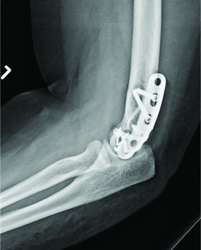
Postoperative lateral roentgenography
The re-operative clinical examinations and radiologic findings of the patients were analyzed retrospectively from the digital patient record system. The initial evaluations of the patients were accomplished by taking elbow X-rays (anteroposterior and lateral) and a computed tomography. On physical examination, the range of flexion, extension, pronation and supination, varus and valgus stress tests, lateral collateral ligament instability, and popping or cracking sensation during the movement of joints were recorded. On the plain radiographs, findings suggestive of fracture healing, loss of reduction, AVN, arthrosis, implant position, and heterotopic ossification were investigated. The outcomes were evaluated using the Mayo Elbow Performance Index (MEPI), visual analog scale (VAS) pain score, and patients’ subjective opinions. Joint arthrosis was staged according to the Broberg and Morrey classification (6).
Surgical technique
All patients were operated the next day after their trauma occurred. The Kocher approach, with a mean longitudinal incision of 7 cm starting from the center of the lateral humeral condyle and extending 5 cm proximally and 2 cm distally, was followed in all cases. The extensor carpi radialis longus and brevis and brachioradialis muscles, as well as the common extensor tendon were anteriorly retracted. In the cases where the lateral collateral ligament (LCL) insertions were intact, the joint capsule was longitudinally incised over the anterior aspect of the lateral ulnar collateral ligament and an ecarteur was placed. The screws in the medial area were placed minimally oblique. If the site of insertion of LCL was fractured, this fragment was everted laterally along with the ligament and the screws were placed anteroposterior in the subluxation position of the elbow. Figure 4 depicts the surgical approach and intraoperative picture of a Dubberley type B fracture. Subsequent to the meticulous anatomical reduction of the fragments, a temporary fixation with K-wires was performed in various directions. Two permanent K-wires, which could particularly fix the small fragments and contribute to the rotational stabilization, were applied from the lateral to medial direction and parallel to the joint line. Two or 3 headless cannulated screws were used to secure the fragments that contained the articular cartilage in an anteroposterior direction. Then, a 3 or 5-hole buttress plate was placed laterally onto the distal humerus. The plate was meticulously located to avoid impingement, which can occur because of the crossing of the bony end of capitellum. The plate holes were selectively filled, placing 2 or 3 screws proximal to the fracture line and 1 or 2 screws on the capitellar fragments. In the cases of partial or complete rupture of the LCL or in the suspected cases, the LCL was repaired by augmentation of the LCL using non-absorbable sutures and passing them through the plate and a tunnel over the insertion of the LCL. The elbow was immobilized with a long arm splint at 90° of flexion after the surgery. The details on the surgical technique are illustrated in Figures 5 and 6.
Figure 4.

Intraoperative picture of surgical approach of a Dubberley type B fracture
Figure 5.

Anteroposterior illustration depicting surgical technique details. Note that Kirschner wires provide rotational control, the plate provides butress effect for the fragment and LCL and long screws provide mediolateral interfragmentary compression
Figure 6.

Illustration of lateral view of the surgical technique, note that the canulated screws provide AP compression of the fragments
Postoperative rehabilitation
All patients received 25 mg indomethacin, 3 times a day for 3 weeks, starting from the day of trauma. After the surgery, hinged elbow braces were used in all the patients. The braces were fixed at a 90° in the first 3 weeks. Subsequently, flexion-extension movements were allowed in the braces. In the first 3 weeks, supination-pronation and extension-flexion exercises were performed in the supine position without braces, depending on the severity of the pain. After the sixth postoperative week, the braces were removed. Weightlifting of more than 1 kg was not allowed, although other daily movements and isometric exercises were permitted. After 6 months, heavy work and sportive activities were allowed.
Statistical analysis
The statistical analysis was performed using Statistical Package for the Social Sciences version 21.0 (IBM SPSS Corp.; Armonk, NY, USA). The data are provided as mean±standard deviation (minimum-maximum) for continuous variables and as frequency for categorical variables.
Results
According to the Dubberley classification, 10 patients were type B. None of the patients had ipsilateral LCL injury or radial head injury. One patient had contralateral transolecranon fracture. All cases were closed fractures because of the fall on the upper extremity without any neurovascular injury.
The mean age of the patients was 43.8±11.1 (34–72) years and the mean follow-up period was 59.6±38.79 (22–127) months.
At the final follow-up, the mean elbow flexion was 137.5°±3° (132°–140°), extension was −17.9°±9.2° (10°–35°), pronation was 72.2°±2.6°(68°–75°), and supination was 78.9°±4.09° (72°–85°). The flexion-extension range (ROM) was determined as 119.6°±9.3° (101°–130°) and mean rotational arc was measured as 151.1°±5.4° (142°–160°). The mean MEPI score was calculated as 95.5±5.98 (85–100). According to the MEPI score, 8 patients were evaluated as excellent and 2 patients as good. The mean VAS pain score was 0.8±1.03 (0–2). The subjective patient evaluation was recorded as excellent in 5 patients, good in 3 patients, and moderate in 2 patients.
One patient developed AVN. This patient underwent implant extraction, capitellum humeral resection, and lateral release 8 months after the initial surgery. The imaging data of the patients before and after the surgery are shown in Figure 7. At the last follow-up, the patient’s elbow flexion was 140°, extension was −14°, pronation was 75°, and supination was 78°. There was no popping sensation nor cracking sound within the elbow range of motion. In addition to accomplishing her daily activities, she reported in her subjective evaluation as also being able to perform challenging activities.
Figure 7. a–h.

Preoperative computerized tomograpgy images of Dubberly type B capitellar and trochlear fracture in distal humerus (a, b). Postoperative x-ray images (c, d). Nonunion and avascular necrosis were seen in computerized tomography images at 8th month after operation (e, f). X-ray image after capitellum humeri resection (g, h)
Grade 3 degenerative elbow arthrosis was diagnosed in a 72-year-old female patient whose subjective evaluation was recorded as moderate at the last check-up. However, she already had degenerative arthrosis before the fracture.
This patient could not perform his daily activities nor accomplish difficult tasks. Grade 2 degenerative arthrosis was observed in another patient. However, this patient stated that he did not have any problems in performing difficult activities.
K-wire migration was observed in a patient who had no complaints other than a popping sensation during the joint movements. Another patient underwent implant extraction at the 9th postoperative month because of hardware irritation under the skin.
Loss of reduction, nonunion, malunion, reflex sympathetic dystrophy or heterotopic ossification were not observed in any of the cases.
Discussion
Various methods have been implemented from the past to present for the surgical treatment of distal humerus coronal plane fractures. These methods have included closed reduction (7), percutaneous pining (8), fragment excision (9), arthroscopic reduction and fixation (10), open reduction and internal fixation (ORIF) (2, 3, 5, 11–22), and elbow arthroplasty (23). Today, ORIF is a widely accepted treatment modality for these complex fractures. Along with the fracture type, the surgeon’s experience plays a definitive role while choosing the implants to be applied in this method. Cancellous screws (11), Herbert screws (14, 16), headless screws (5, 11, 15, 17, 24), metallic pins (18), plates (2, 3, 20, 25, 26, 27), and absorbable pins (28) are among the many options for fixation.
The type B subdivision of the Dubberley classification refers to posterior comminution in the coronal plane. This classification is helpful in decision making and prediction of the outcome (2). The posterior comminution of the capitellum and trochlea leads to deterioration in the blood supply of the fragments (4). The osteosynthesis of these fractures aims to restore the anatomical integrity of the fragments, ensure lateral column stability, prevent interfragmentary rotation, and establish adequate support at the distal humerus. If these objectives cannot be achieved, it will not be possible to initiate early joint motion, and poor functional results will be obtained. This observation explains the challenging nature of Dubberley type B fractures and the need for their meticulous handling.
The literature offers numerous studies that report favorable outcomes for the treatment of coronal plane fractures with the use of headless screws (5, 11, 42, 15, 17). However, this implant is inadequate on its own in the management of Dubberley type B fractures that have multiple fragments at the posterior part of the distal humerus and at the lateral capitellar area where the LCL adheres. A minimum of 5-mm–thick subchondral bone stock is required to ensure stable fixation with a headless screw. If adequate subchondral bone stock is not available, iatrogenic fractures may occur (26). Mighell et al. previously suggested that reconstruction with a posterolateral buttress plate provides a more stable fixation than do screws alone, in comminuted coronal plane fractures (26). Biomechanical studies have shown that headless screws are more stable than cancellous screws when applied in the anterior to posterior (AP) direction (29). It has also been reported that the application of cancellous screws in the posterior to anterior (PA) direction provides a more stable fixation than that in the AP direction (29). Cartilage coverage and subchondral bone may be damaged during the placement of cancellous screws in the AP direction. However, PA applications require extensive soft-tissue dissection to expose the bone and may increase the risk of AVN by disrupting the blood supply (30). Moreover, excessive soft-tissue stripping detaches bone fragments from soft tissue and causes further fragmentation. In our surgical technique, we avoided excessive posterior soft-tissue dissection by applying the headless screws in the AP direction.
The lateral epicondyle is an important anatomical structure that provides a bony attachment for the major stabilizers of the elbow joint. Hence, the correct anatomical reduction and stable fixation of the lateral epicondyle possess paramount importance for the stability of the elbow joint. To restore the lateral pillar, we applied a buttress plate from the lateral aspect of the distal humerus. The screws on plate, which were inserted parallel to the joint line lateromedially, provided a subchondral support and an interlocking mechanism for the distal fragments.
Ensuring sufficient stability with the primary fixation to initiate the early range of motion exercises is the essential in all techniques described in the literature for distal humerus fractures. Therefore, we aimed to provide the highest possible level of stability combining 3 different methods of osteosyntheses. Moreover, the K-wires that were used for the temporary fixation may be cut flush to the cortex and conserved permanently if adequate stabilization cannot be achieved with the plate and screws. In cases of deficient subchondral bone stock, low calibration K-wires may help in the permanent fixation of the small fragments.
In our experience, using a lateral buttress plate, headless screws in the AP direction, interosseous K-wires for rotational stability, and small fragment fixation, we were able to establish a solid fixation, and thus, could begin an early range of motion exercises for the patients with Dubberly type B fractures with posterior comminution.
Several plating techniques with various outcomes have been previously described in the literature for these fractures. Dubberley et al. adapted pelvic reconstruction plates for 6 patients with posterior comminution in their large series of 28 coronal plane fractures (2). Although favorable outcomes were reported in this study with MEPI scores as high as 91±11, these outcomes were not specific to the subgroup with posterior comminution. Sen et al. reported their surgical technique using a lateral compression plate and headless screws applied in the PA direction (25). They suggested that the plate fixation combined with headless screws could provide stability againstshearing forces, ensuring rotational and translational stability to the capitellar fragment. These researchers also reported that this technique allowed them to start early mobilization. In a previously reported case series with 26 patients, 10 patients with Dubberly type B fractures were treated with a dorsal plate to support posterior comminution (3).
These researchers reported fracture healing without using any grafts in every case where a lateral column was buttressed. The authors noted that the mean MEPI score of the posterior comminution patients was 71.3 as compared with the mean MEPI score of 91.3 for the patients without comminution. This study also compared the 1- or 2-part articular fractures to the 3- or 4-part fractures, recording that the comminution was associated with the worse outcomes. Another previous study described the outcomes of 8 Dubberley type B coronal plane fracture patients, treated with a lateral plate and Herbert screws (20). The authors recorded stable fixation and early mobilization in every patient with subjective patient scores as excellent in 2 patients, good in 4, and poor in 2.
In our study, we evaluated the outcomes of 10 patients with Dubberley type B fractures. The mean MEPI score of our patients was 95.5±5.98 (85–100) and the subjective scores were more favorable than those found in previous studies, with scores as excellent in 8 patients and good in 2 patients.
The lateral approach to the elbow joint has many advantages such as the ability to directly visualize the elbow joint, low risk of neurovascular bundle injury, and low risk of AVN because of the preservation of the posterior soft-tissue integrity (30). Moreover, the lateral approach does not require an olecranon osteotomy. The major drawbacks of this approach are the elbow joint instability with iatrogenic LCL injury and the difficulties of fixing medially extended fractures and Dubberley type B fractures (13). Moreover, skin irritation because of a prominent hardware may be encountered. Despite the common recommendation for using the posterior transolecranon approach in Dubberley type B fractures, capitellum and trochlea may be better visualized through the anterolateral approach (31). Additionally, smaller cartilage fragments can be addressed more accurately through anterolateral exposure. Hence, the risk of neurovascular injury is the major limitation of this approach (13, 14).
In our study, we preferred a lateral approach and did not performed any olecranon osteotomy to preserve the posterior soft-tissue integrity. Vascular support from the posterior soft tissues is asserted to be an important source of fragment vascularization in the literature. Damaging these structures is associated with the development of AVN (30). As such, there are several studies reporting AVN in patients who were operated on through the posterior approach (2, 21), along with a single study that does not encounter any AVN (3). In our study, we employed a lateral approach and a lateral buttress plate. Despite preservation of the posterior blood supply, we encountered AVN in 1 patient (Figure 7). Extensive dissection of the posterior soft tissues, fracture comminution, time from trauma to surgery, age, and the intensity of the injury are the factors associated with AVN in the literature. Ashwold et al. included 10 patients with posterior comminution into their series of 26 Duberley type B patients and treated them with posterior plating (3). The authors announced that they did not record any AVN in their entire series (3). However, Dubberley et al. reported 2 AVN, in their series of 28 patients with coronal plane fractures including 6 with posterior comminution who were treated with pelvic reconstruction plates (2). Another study that enrolled 23 patients aged 65 or older, also reported 1 patient complication with AVN (21).
The internal fixation rigid enough to initiate early range of motion exercises is essential in the treatment of distal humerus fractures. The conditions that interfere with early joint motion are associated with the restriction of the final range of motion. Dubberley type B fractures are frequently discussed in the literature. When we compared our results with those of the previous studies that included more simple fractures but not those that could be qualified as the Dubberley type B (17, 22), plate fixation studies that did include Dubberley type B patients along with other types (2, 3, 20), and the studies that enrolled only Dubberley type B fractures, we concluded that we managed to initiate the early range of motion without compromising the fixation. This observation is suggested to be the main reason for our superior outcomes and can be explained by the solid fixation provided by our novel technique. One limitation of the study is the absence of a control group.
In conclusion, our study demonstrates that with the use of a lateral buttress plate and headless cannulated screws, which are introduced in the AP direction with interfragmentary K-wires, the internal fixation of the distal humerus coronal plane fractures is solid and secure. This rigid construction provides early mobilization of the joint and this fact positively affected our final outcomes. We suggest that superior outcomes can be achieved without major complications in these complex fractures using our triple fixation technique.
HIGHLIGHTS.
Coronal plane fractures are rare and complex fractures of the elbow joint.
Favorable outcomes are closely associated with early joint motion.
An internal fixation using lateral buttress plate, headless cannulated screws, and interfragmentary K-wires provides a secure Fixation allowing early postoperative joint motion.
Footnotes
Ethics Committee Approval: Ethics committee approval was received for this study from the Ethical Committee of Biruni University (approval number:2018/20-07).
Informed Consent: Written informed consent was obtained from the patients.
Author Contributions: Concept - H.K., M.T.D.; Design - M.T.D., H.K., S.E.B.; Supervision - H.K.; Resources - H.K.; Materials - M.T.D., O.B.; Data Collection and/or Processing - M.T.D., S.E.B., M.S., Y.P.; Analysis and/or Interpretation - Y.P., M.S.; Literature Search - M.T.D., S.E.B.; Writing Manuscript - M.T.D.; Critical Review - H.K., O.B.
Conflict of Interest: The authors have no conflicts of interest to declare.
Financial Disclosure: The authors declared that this study has received no financial support.
References
- 1.Jupiter JB, Morrey BF. Fractures of The Distal Humerus in Adult. In: Morrey BF, editor. The elbow and its disorders. 2nd edn. WB Saunders; Philadelphia: 1993. pp. 328–366. [Google Scholar]
- 2.Dubberley JH, Faber KJ, Macdermid JC, Patterson SD, King GJ. Outcome after open reduction and internal fixation of capitellar and trochlear fractures. J Bone Joint Surg Am. 2006;88:46–54. doi: 10.2106/00004623-200601000-00007. [DOI] [PubMed] [Google Scholar]
- 3.Ashwood N, Verma M, Hamlet M, Garlapati A, Fogg Q. Transarticular shear fractures of the distal humerus. J Shoulder Elbow Surg. 2010;19:46–52. doi: 10.1016/j.jse.2009.07.061. [DOI] [PubMed] [Google Scholar]
- 4.Ring D, Jupiter JB, Gulotta L. Articular fractures of the distal part of the humerus. J Bone Joint Surg Am. 2003;85:232–8. doi: 10.2106/00004623-200302000-00008. [DOI] [PubMed] [Google Scholar]
- 5.Mighell M, Virani NA, Shannon R, et al. Large coronal Shear fractures of the capitellum and trochlea Treated with headless compression screws. J Shoulder Elbow Surg. 2010;19:38–45. doi: 10.1016/j.jse.2009.05.012. [DOI] [PubMed] [Google Scholar]
- 6.Amini MH, Sykes JB, Olson ST, et al. Reliability testing of two classification systems for osteoarthritis and post-traumatic arthritis of the elbow. J Shoulder Elbow Surg. 2015;24:353–7. doi: 10.1016/j.jse.2014.10.015. [DOI] [PubMed] [Google Scholar]
- 7.Ochner RS, Bloom H, Palumbo RC, Coyle MP. Closed reduction of coronal fractures of the capitellum. J Trauma. 1996;40:199–203. doi: 10.1097/00005373-199602000-00005. [DOI] [PubMed] [Google Scholar]
- 8.Ma YZ, Zheng CB, Zhou TL, Yeh YC. Percutaneous probereduction of frontal fractures of the humeral capitellum. Clin Orthop Relat Res. 1984;183:17–21. doi: 10.1097/00003086-198403000-00004. [DOI] [PubMed] [Google Scholar]
- 9.Fowles JV, Kassab MT. Fracture of the capitulum humeri: Treatment by excision. J Bone Joint Surg Am. 1974;56:794–8. doi: 10.2106/00004623-197456040-00013. [DOI] [PubMed] [Google Scholar]
- 10.Kuriyama K, Kawanishi Y, Yamamoto K. Arthroscopic-assisted reduction and percutaneous fixation for coronal shear fractures of the distal humerus: Report of two cases. J Hand Surg Am. 2010;35:1506–9. doi: 10.1016/j.jhsa.2010.05.021. [DOI] [PubMed] [Google Scholar]
- 11.McKee MD, Jupiter JB, Bamberger HB. Coronal Shear fractures of the distal end of the humerus. J Bone Joint Surg Am. 1996;78:49–54. doi: 10.2106/00004623-199601000-00007. [DOI] [PubMed] [Google Scholar]
- 12.Tarallo L, Mugnai R, Adani R, Zambianchi F, Costanzini CA, Catani F. Shear fractures of the distal humerus: Is the use of intra-articular screws a safe treatment? Musculoskelet Surg. 2015;99:217–23. doi: 10.1007/s12306-015-0386-8. [DOI] [PubMed] [Google Scholar]
- 13.Li Y, Cha YJ, Li T, Gong MQ, Jiang XY. Analysis of anterolateral approach and lateral approach for the treatment of coronal shear fracture of the distal humeral. Beijing Da Xue Xue Bao. 2016;48:1026–31. [PubMed] [Google Scholar]
- 14.Imatani J, Morito Y, Hashizume H, Inoue H. Internal fixation for coronal shear fracture of the distal end of the humerus by the anterolateral approach. J Shoulder Elbow Surg. 2001;10:554–6. doi: 10.1067/mse.2001.118005. [DOI] [PubMed] [Google Scholar]
- 15.Ruchelsman DE, Tejwani NC, Kwon YW, Egol KA. Open reduction and internal fixation of capitellar fractures with headless screws. Surgical technique. J Bone Joint Surg Am. 2009;91(Suppl 2 Pt 1):38–49. doi: 10.2106/JBJS.H.01195. [DOI] [PubMed] [Google Scholar]
- 16.Singh AP, Dhammi IK, Garg V, Singh AP. Outcome of surgical treatment of type IV capitellum fractures in adults. Chin J Traumatol. 2012;15:201–5. [PubMed] [Google Scholar]
- 17.Vaishya R, Vijay V, Jha GK, Agarwal AK. Open reduction and internal fixation of capitellar fracture through anterolateral approach with headless double-threaded compression screws: a series of 16 patients. J Shoulder Elbow Surg. 2016;25:1182–8. doi: 10.1016/j.jse.2016.07.016. [DOI] [PubMed] [Google Scholar]
- 18.Sen RK, Tripahty SK, Goyal T, Aggarwal S. Coronal shear fracture of the humeral trochlea. J Orthop Surg (Hong Kong) 2013;21:82–6. doi: 10.1177/230949901302100121. [DOI] [PubMed] [Google Scholar]
- 19.Bilsel K, Atalar AC, Erdil M, Elmadag M, Sen C, Demirhan M. Coronal plane fractures of the distal humerus involving the capitellum and trochlea treated with open reduction internal fixation. Arch Orthop Trauma Surg. 2013;133:797–804. doi: 10.1007/s00402-013-1718-5. [DOI] [PubMed] [Google Scholar]
- 20.Hu X, Xiang M, Yang G, Chen H, Tang H. Operative treatment of Dubberley type 3B capitulum-trochlea fractures. Zhongguo Xiu Fu Chong Jian Wai Ke Za Zhi. 2014;28:1194–8. [PubMed] [Google Scholar]
- 21.Lopiz Y, Rodríguez-González A, García-Fernández C, Marco F. Open reduction and internal fixation of coronal fractures of the capitellum in patients older than 65 years. J Shoulder Elbow Surg. 2016;25:369–75. doi: 10.1016/j.jse.2015.12.004. [DOI] [PubMed] [Google Scholar]
- 22.Mighell M, Virani NA, Shannon R, Echols EL, Jr, Badman BL, Keating CJ. Large coronal shear fractures of the capitellum and trochlea treated with headless compression screws. J Shoulder Elbow Surg. 2010;19:38–45. doi: 10.1016/j.jse.2009.05.012. [DOI] [PubMed] [Google Scholar]
- 23.Lapner M, King GJ. Elbow arthroplasty for distal humeral fractures. Instr Course Lect. 2014;63:15–26. [PubMed] [Google Scholar]
- 24.Mehdian H, McKee MD. Fractures of capitellum and trochlea. Orthop Clin North Am. 2000;31:115–27. doi: 10.1016/S0030-5898(05)70132-2. [DOI] [PubMed] [Google Scholar]
- 25.Sen MK, Sama N, Helfet DL. Open reduction and internal fixation of coronal fractures of the capitellum. J Hand Surg Am. 2007;32:1462–5. doi: 10.1016/j.jhsa.2007.08.015. [DOI] [PubMed] [Google Scholar]
- 26.Mighell MA, Harkins D, Klein D, Schneider S, Frankle M. Technique for internal fixation of capitellum and lateral trochlea fractures. J Orthop Trauma. 2006;20:699–704. doi: 10.1097/01.bot.0000246411.33047.80. [DOI] [PubMed] [Google Scholar]
- 27.Tang HC, Xiang M, Chen H, Hu XC, Yang S, Yang GY. Cannulated screw combined with buttress plate for the treatment of transarticular shear fractures of the distal humerus. Zhongguo Gu Shang. 2014;27:161–4. [PubMed] [Google Scholar]
- 28.Kraan GA, Krijnen MR, Eerenberg JP. Internal fixation for coronal shear fracture of the capitellum with polylactide resorbable fixation. BMJ Case Rep. 2013. bcr2012006364. [DOI] [PMC free article] [PubMed]
- 29.Elkowitz SJ, Polatsch DB, Egol KA, Kummer FJ, Koval KJ. Capitellum fractures: a biomechanical evaluation of three fixation methods. J Orthop Trauma. 2002;16:503–6. doi: 10.1097/00005131-200208000-00009. [DOI] [PubMed] [Google Scholar]
- 30.Mehidian H, McKee MD. Fractures of capitellum and trochlea. Orthop Clin North Am. 2000;31:115–27. doi: 10.1016/S0030-5898(05)70132-2. [DOI] [PubMed] [Google Scholar]
- 31.Ravishankar MR, Kumar MN, Raut R. Choice of surgical approach for capitellar fractures based on pathoanatomy of fractures: outcomes of surgical management. Eur J Orthop Surg Traumatol. 2017;27:233–42. doi: 10.1007/s00590-016-1877-5. [DOI] [PubMed] [Google Scholar]