Dr Bianca Mazini et al.1 recently published the interesting case report entitled “Isolated superior striate vein thrombosis in adults.” I would like to comment on this article in relation to the deep venous collaterals.
Medullary and subependymal veins
In the early stage of development, venous drainage of the brain is centrifugal towards the surface of the developing encephalon. With the development of the telencephalon, the diencephalon, and the ventricular system together with the choroidal plexuses, a deep venous system with centripetal drainage develops.2 As a result, two venous drainage systems of the brain develop, one draining to the surface and the other deeply, and in both, the drainage occurs by way of medullary veins. It is believed that the term “medullary” vein was first adopted by Duret in 1874.3 Testut gave a detailed description on them in his manual in 1911.4 The superficial cerebral venous system includes the cortical veins and subcortical veins and the medullary veins that drain the superficial white matter up to a depth of about 1 cm, all of which will converge into the pial veins at the surface of the brain. The deep venous system consists of the deep medullary veins draining the deep white matter and the veins draining part of the central gray nuclei. Deep medullary veins are longer than superficial medullary veins and converge at the superior lateral angle of the lateral ventricle towards the subependymal veins. Subependymal veins can be divided into a medial group and a lateral group.5 The medial group, draining the septum pellucidum, the fornix, and the white matter includes (i) the anterior septal veins, which course along the septum pellucidum of the frontal horn; (ii) the posterior septal veins along the body of the lateral ventricle; (iii) the medial atrial vein in the atrium of the lateral ventricle, which also receives tributaries from the medial wall of the occipital horn; and (iv) the hippocampal veins along the choroidal fissure in the temporal horn. The lateral group draining ganglionic nuclei and the white matter includes: (i) the thalamostriate vein, at the level of the foramen of Monro, which is formed by the union of an transverse and longitudinal caudate veins;6–8 (ii) an inconstant direct lateral vein, which may be very conspicuous in cases where the thalamostriate vein is absent; (iii) the lateral atrial vein in the atrium, which also receives tributaries from the lateral wall of the occipital horn; and (iv) the inferior ventricular vein along the roof of the temporal horn. Except for the lateral atrial, hippocampal, and inferior ventricular veins, which are tributaries of the basal vein of Rosenthal, the remaining subependymal veins drain into the internal cerebral vein. The superior striatal veins drain into the thalamostriate vein and the inferior striatal veins drain into the deep middle cerebral vein (MCV) or basal vein of Rosenthal. Embryologically, the inferior striatal veins are primitive veins, but the superior striatal veins are secondary formed.
Trans-cerebral and trans-striatal veins
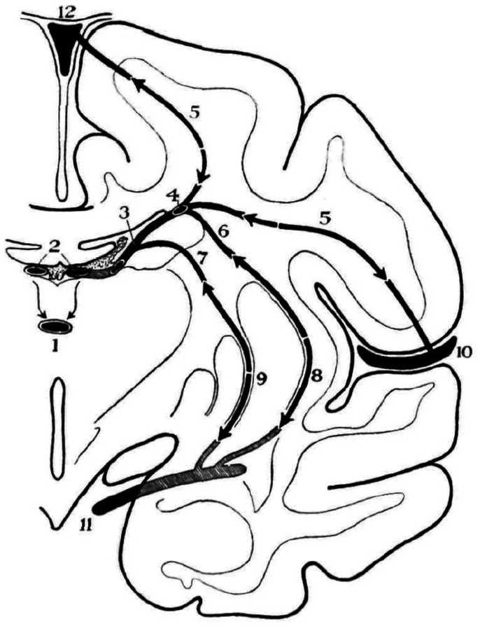
Venular anastomoses exist between the superficial and deep medullary veins in the white matter.6,8 In addition, there are direct anastomotic veins, that is, trans-cerebral veins, which connect superficial pial veins to subependymal veins directly.2,4,6,8,9 It is not clearly defined whether medullary veins include striatal veins as striatal veins also drain the white matter. For instance, the superior striate veins drain parts of the internal capsule. Nevertheless, it is clearer to define medullary veins as being confined to parenchymal veins in the white matter. Trans-cerebral veins allow for connections between the superficial pial veins, i.e., superficial MCVs, and the subependymal veins. Secondary possibly post-natal anastomosis between the superior and inferior striatal veins may exist providing an anastomotic route between pial veins (deep MCV or basal vein of Rosenthal) and subependymal veins10,11 (Figure 1).
Figure 1.
Collateral pathways between the superficial and deep venous system of a Rhesus monkey. Trans-cerebral (5) and trans-striatal routes (8 and 9) are illustrated. Both medullary and striatal venous collaterals connect the superficial (pial) venous system (10 and 11) and deep (subependymal) venous system (3 and 4) directly and indirectly, respectively. 1: Great vein of Galen; 2: small veins of Galen (internal cerebral veins); 3: transverse caudate vein; 4: longitudinal caudate vein; 5: trans-cortical vein; 6 and 7: superior striatal veins; 8 and 9: inferior striatal veins; 10: superficial middle cerebral vein; 11: deep middle cerebral vein or basal vein of Rosenthal; 12: superior sagittal sinus. Source: reproduced with permission from Oxford University Press.6.
Alternative deep venous drainage to the vein of Galen system
In cases of normal deep venous outflow restriction or arterialized inflow into the deep venous system, alternative venous routes may develop to drain the deep venous system towards the superficial cerebral venous system, the dural venous sinuses and finally the extracranial venous outflow pathways.12,13 Possible causes of deep venous system outflow restriction include vein of Galen-straight sinus venous thrombosis, tumor (mostly benign slow growing tumor like falco-tentorial meningioma), and surgical complication. Retrograde blood flow into the deep venous system through the vein of Galen can be caused by dural arteriovenous fistula or brain arteriovenous malformation, which is commonly accompanied by chronic venous sinus occlusion of transverse and/or sigmoid sinus(es).
In the above-mentioned situations, venous blood in the deep venous system may be drained through the following venous routes14: (a) direct cortical route: superficial cortical veins directly connected to the vein of Galen, which include internal occipital, posterior pericallosal, and superior vermian veins; (b) basal vein route connected (i) anteriorly to the deep MCV, then to the dural sinuses of the middle cranial fossa, that is the cavernous sinus, the laterocavernous sinus or the paracavernous sinus; (ii) infratentorially towards the pial brainstem venous system and superior petrosal sinus; (c) terminal venous arcade from superior to the inferior terminal veins, then to basal vein of Rosenthal; (d) thalamic route from superior to inferior/posterior thalamic veins, then to basal vein of Rosenthal; (e) trans-striatal route from superior to inferior striatal veins, then to deep MCV or basal vein of Rosenthal; (f) hippocampal route from posterior to anterior longitudinal hippocampal veins, then to basal vein of Rosenthal; and (g) trans-cortical venous route as described above.3 The terminal venous arcade runs along the stria terminalis between the thalamus and caudate nucleus. It is composed of superior, middle, and inferior terminal veins connecting thalamostriate vein to inferior ventricular vein. Discrimination between terminal arcade route and thalamic route is often difficult. Theoretically, (h) choroid plexus route might connect the deep venous system to basal vein of Rosenthal via superior and inferior choroidal veins. However, I have never seen such a collateral route in any patients. Alternative routes (a), (d), and (e) are reported as direct, dorso-caudal (diencephalic) and ventro-rostral (telencephalic) draining pathways, respectively, in vein of Galen absence or unavailability by Lasjaunias et al.15 Thalamic, trans-striatal, and trans-cortical routes can be regarded as diencephalic, trans-subpallial, and trans-pallial venous routes, respectively (Figure 2).
Figure 2.
Collateral pathways in case of occlusion of the vein of Galen and/or straight sinus or retrograde blood flow to the deep venous system due to dural arteriovenous fistulas or brain arteriovenous malformation. Collateral pathways from deep to superficial venous system are as follows: (a) direct cortical venous route, (b) basal vein route (to deep middle cerebral vein, cavernous sinus, and so forth), (c) terminal venous arcade, (d) thalamic (diencephalic) route, (e) trans-striatal (subpallial) route, (f) hippocampal venous route (through posterior and anterior longitudinal hippocampal veins), and (g) trans-cortical (pallial) venous route. BV: basal vein of Rosenthal; ICV: internal cerebral vein; SS: straight sinus; VOG: great vein of Galen.
Alternative routes in high-flow arteriovenous shunts
Terminal venous arcade and thalamic route are often observed in the cases of completely cured vein of Galen aneurysmal malformations after embolization together with trans-cortical route in the absence of falcine and/or straight sinus drainage.16 Retrograde blood flow from the torcular Herophili to the deep venous system through the straight sinus is often observed in the patients with dural arteriovenous fistulas. In this situation, trans-striatal route and trans-cortical route play as main drainage routes to the extracranial venous system (Figure 3).
Figure 3.
A nine-year-old boy with multiple dural arteriovenous fistulas at superior sagittal sinus and bilateral transverse-sigmoid sinuses. Bilateral transverse-sigmoid sinuses were occluded. After three sessions of intervention, entire superior sagittal sinus, bilateral transverse-sigmoid sinuses, and straight sinus were occluded with coils, and dural arteriovenous fistulas are almost occluded. Normal venous drainages of left cerebral hemisphere were through the prominent left superficial middle cerebral veins and left trans-striatal route (arrow) to the cavernous sinus. (a) Left internal carotid angiogram (lateral view, venous phase). (b) Three-dimensional rotational angiography of left internal carotid injection (lateral view, venous phase). (c) Three-dimensional rotational angiography of left internal carotid injection (lateral view, venous phase, superficial veins are removed manually). Arrow indicates trans-striatal route collecting enlarged deep medullary venous flow (arrowheads).
Vascular territories between superior striatal veins and deep medullary veins might not be sharply demarcated. Venous ischemic lesion reported by Dr Bianca Mazini et al.1 may include not only venous thrombosis of superior striatal veins but that of deep medullary veins as well. Good prognosis of the two reported patients could be explained by good collateral to inferior striatal veins and/or superficial medullary veins draining to deep and superficial MCVs, respectively.
Declaration of conflicting interests
The author declared no potential conflicts of interest with respect to the research, authorship, and/or publication of this article.
Funding
The author received no financial support for the research, authorship, and/or publication of this article.
ORCID iD
Masaki Komiyama https://orcid.org/0000-0003-0998-6315
References
- 1.Mazini B, Bonvin C, Gailloud P, et al. Isolated superior striate vein thrombosis in adults. Interv Neuroradiol. Epub ahead of print 22 January 2020. DOI: 10.1177/1591019919900825. [DOI] [PMC free article] [PubMed]
- 2.Jimenez JL, Lasjaunias P, Terbrugge K, et al. The trans-cerebral veins: normal and non-pathologic angiographic aspects. Surg Radiol Anat 1989; 11: 63–72. [DOI] [PubMed] [Google Scholar]
- 3.Duret H. Recherches anastomiques sur la circulation de l’encéphale. Arch Physiol Norm Pathol 1874; 6: 316–353. [Google Scholar]
- 4.Testut L. Traité d’anatomie humaine 19116e édition Vol II Paris: Doin. [Google Scholar]
- 5.Wolf BS, Huang YP. The subependymal veins of the lateral ventricles. Am J Roentgenol 1964; 91: 406–426. [PubMed] [Google Scholar]
- 6.Schlesinger B. The venous drainage of the brain, with special reference to the galenic system. Brain 1939; 62: 274–291. [Google Scholar]
- 7.Rosa M, Borzone M. The venous system of the corpus striatum. Neuroradiology 1973; 6: 219–230. [DOI] [PubMed] [Google Scholar]
- 8.Okudera T, Huang YP, Fukusumi A, et al. Micro-angiographical studies of the medullary venous system of the cerebral hemisphere. Neuropathology 1999; 19: 93–111. [DOI] [PubMed] [Google Scholar]
- 9.Kaplan HA. The transcerebral venous system. AMA Arch Neurol 1959; 1: 148–152. [DOI] [PubMed] [Google Scholar]
- 10.Padget D. The development of the cranial venous system in man from the viewpoint of comparative anatomy. Contr Embryol Carneg Inst 1957; 611: 79–140. [Google Scholar]
- 11.Hédon E. Étude anatomique sur la circulation veineuse de l’encéphale, Bordeaux: Bellier, 1888. [Google Scholar]
- 12.Dandy WE, Blackfan KD. An experimental, clinical and pathological study. Part I. Experimental studies. Am J Dis Child 1914; 8: 406–482. [Google Scholar]
- 13.Bedford THB. The venous system of the velum interpositum of the rhesus monkey and the effect of experimental occlusion of the great vein of Galen. Brain 1934; 57: 255–265. [Google Scholar]
- 14.Komiyama M. Functional venous anatomy of the brain for neurosurgeons. Jpn J Neurosurg (Tokyo) 2017; 26: 488–495. [Google Scholar]
- 15.Lasjaunias P, Garcia-Monaco R, Rodesch G, et al. Deep venous drainage in great cerebral vein (vein of Galen) absence and malformations. Neuroradiology 1991; 33: 234–238. [DOI] [PubMed] [Google Scholar]
- 16.Lasjaunias P, Berenstein A and ter Brugge KG. Intracranial venous system. In: Clinical vascular anatomy and variations. Berlin: Springer, 2001, pp. 631–713.


