

SECTION 1 – QUIZ
Case description
A 54-year-old premenopausal woman, G3 P3, was referred to the gynecology outpatient clinic due to abnormal uterine bleeding and sonographic suspicion of adenomyosis and leiomyomas. In the past 2 years, she noticed shortening of the menstrual cycles and menorrhagia. She was obese (body mass index: 39 kg/cm2) and had chronic hypertension, depression, and fibromyalgia. She was medicated with duloxetine 30 mg/day, trazodone 100 mg, and olmesartan + hidroclorotiazida 40/12.5 mg.
On physical examination, the patient had a globus cervix with no visible lesions. Bimanual palpation was impossible due to the patient's biotype.
Pelvic magnetic resonance imaging revealed an uterus sized 155 mm × 104 mm × 90 mm with three nodules in the uterine wall suggestive of leiomyoma; the biggest sized 72 mm at the right portion of the fundus, another left anterior sized 45 mm, and one right sized 40 mm, all of them FIGO type 5; thickening of the junction zone due to probable adenomyosis; and an agglomerate of cystic formations at the uterine cervix with a total largest diameter of 50 mm [Figures 1 and 2].
Figure 1.

Pelvic magnetic resonance imaging – Sagittal view – Two nodules in the uterine wall suggestive of leiomyomas, thickening of the junction zone due to probable adenomyosis, and an agglomerate of cystic formations at the uterine cervix
Figure 2.

Pelvic magnetic resonance imaging – Sagittal view – Uterus deformed by leiomyomas and agglomerate of cystic formations at the uterine cervix
Meanwhile, screening for human papillomavirus (HPV) cervical detection came out positive for HPV 33 and 52, and reflective cervical cytology revealed low-grade squamous intraepithelial lesion (LSIL). Colposcopy was adequate, with a type 3 transformation zone, and no abnormal findings were observed. The endocervical curettage was negative to dysplasia or malignancy. A loop electrosurgical excision procedure followed, and histology revealed LSIL involved in mucous.
The patient was submitted to total hysterectomy by laparotomy due to growing symptomatic leiomyomas. After removal of the specimen, the cervix was inspected – it was enlarged with multiple cystic lesions filled with mucous [Figures 3-5].
Figure 3.
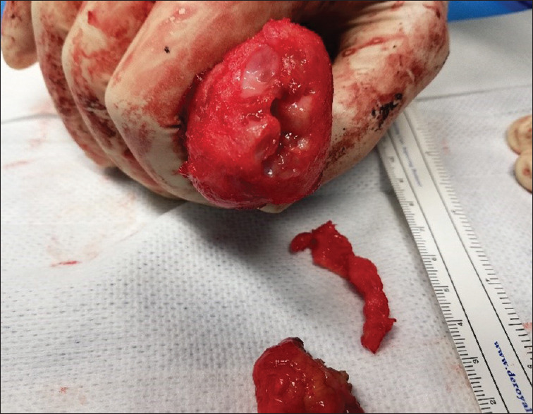
Surgical specimen – Enlarged cervix with multiple cystic lesions filled with mucous
Figure 5.

Surgical specimen – Uterus deformed by multiple leiomyomas
Figure 4.

Surgical specimen – Uterus deformed by multiple leiomyomas and an enlarged cervix with multiple cystic lesions filled with mucous
WHAT IS THE DIAGNOSIS?
Declaration of patient consent
The authors certify that they have obtained all appropriate patient consent forms. In the form the patient(s) has/have given his/her/their consent for his/her/their images and other clinical information to be reported in the journal. The patients understand that their names and initials will not be published and due efforts will be made to conceal their identity, but anonymity cannot be guaranteed.
Financial support and sponsorship
Nil.
Conflicts of interest
There are no conflicts of interest.