PLOS ONE logoLink to PLOS ONE
. 2020 Aug 25;15(8):e0238103. doi: 10.1371/journal.pone.0238103

Survival and clinicopathological characteristics of different histological grades of oral cavity squamous cell carcinoma: A single-center retrospective study

Nan-Chin Lin 1,2, Jui-Ting Hsu 1, Kuo-Yang Tsai 2,3,*
Editor: Dipak Sapkota4
PMCID: PMC7447052  PMID: 32841288

Abstract

The TNM staging system for oral squamous cell carcinoma (OSCC) provides clinicians a dependable foundation for patient prognosis and management decisions, but in clinical practice, treatment outcomes of patients with OSCC are sometimes unsatisfactory. This retrospective study investigated the association between survival and clinicopathological characteristics and histological grades of 2535 patients with OSCC. Additionally, the present study aimed to compare the predictive abilities of histological grades with other common prognostic factors. The enrolled patients were divided into three groups by two experienced pathologists into well-differentiated, moderately differentiated, and poorly differentiated groups, according to the WHO classification. Finally, we designed an observational, retrospective study based on the histological grading of tumors to compare their clinicopathological characteristics and conducted survival analysis among the three groups. Advanced tumor stage was diagnosed in 23.9%, 44.0%, and 55.1% of patients with grades 1–3 OSCC, respectively. By T status, T3 or T4 tumors were found in approximately 22%, 34%, and 40% of patients with grades 1–3 OSCC, respectively. By N status, lymph node metastases were found in 6.1%, 29.3%, and 45.9% of patients with grades 1–3 OSCC, respectively. Thus, significant survival differences were observed based on different OSCC histological grades. Meanwhile, in the multivariate (adjusted) analysis, N1 and N2 stages, extranodal spread, and poor differentiation were associated with a higher recurrence risk than the other common prognostic factors. In conclusion, 5% of patients in our study presented with poorly differentiated OSCC at diagnosis. Furthermore, grade 3 OSCC has worse prognosis and is more aggressive than grades 1 and 2 OSCC. In the future, we should focus on modifying individual therapy for poorly differentiated OSCC to achieve improved outcomes.

Introduction

The TNM staging system of oral squamous cell carcinoma (OSCC) has historically provided clinicians a dependable foundation for patient prognosis and management decisions. Generally, the relative risk of neck lymph node metastasis of T1 and T2 tumors is 10% and 30%, respectively, whereas that of T3 and T4 tumors is distinctly higher [1, 2]. This is significant because the most important prognostic factor in OSCC is the presence of cervical lymph node metastasis, which results in the reduction of the overall survival of these patients by 50% [3, 4].

However, in clinical practice, the treatment outcome of OSCC is sometimes not satisfactory. Several studies have shown that even an early-stage tumor may cause a fatal outcome [57]. New parameters such as depth of invasion was included in the 8th edition of the American Joint Committee on Cancer’s (AJCC) Cancer Staging Manual to improve their predictive value and different stage stratification and to explain patients previously considered to have early-stage tumor but with a poor survival.

The pathological parameters including pathological stage, histopathological grading as per the World Health Organization (WHO) [8], presence of vascular and perineural invasion, extracapsular spread, and positive surgical margins have been used as tumor prognostic factors in OSCC. In this situation, histological grade can be an important prognostic factor for treatment outcome. Evaluation of histological characteristics of a tumor plays an important role in the diagnosis of ablated tumor specimens, and efforts have been undertaken to predict clinical outcomes from histological findings.

In the present retrospective study, we sought to address the association between clinicopathological characteristics and survival of OSCC with different histological grades. We also aimed to compare the predictive abilities of histological grade to other common prognostic factors. To this end, we performed survival analysis based on different histological grading of tumors.

Patients and methods

Patients

This retrospective cohort study was approved by the Institutional Review Board (IRB) and Ethical Committee of Changhua Christian Hospital on April 22, 2020 (IRB number: 200312). All clinical data were obtained through chart review and the cancer registry center. In total, we identified 3248 patients who were diagnosed with OSCC and received treatment and follow-up at Changhua Christian Hospital between January 1, 2008 and December 31, 2018. The follow-up duration was from indexing to June 30, 2019. Exclusion criteria included patients who did not receive treatment per the AJCC cancer treatment guidelines (n = 41), whose anatomic site of the lip did not involve the mucosa (n = 121), who were lost to follow-up or had incomplete data (n = 140), who were initially diagnosed with recurrence or distant metastasis (n = 70), and who had not receive treatment at Changhua Christian Hospital (n = 341). Finally, 2535 patients were included and subsequently analyzed.

Histopathological evaluation

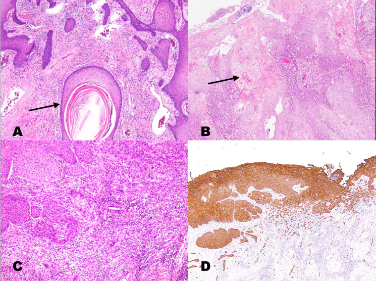
Final pathological reports of resected tumor specimens from all 2535 patients were further investigated. Hematoxylin and eosin-stained slides were reviewed and independently graded by two experienced pathologists under a microscope; if they could not reach a consensus, the intervention of a third pathologist was engaged, and a final agreement was obtained for histopathological features according to the 2017 The College of American Pathologists oral cavity cancer guidelines [9]. Immunohistochemical confirmation for cytokeratin was performed to detect poorly differentiated OSCC because features of squamous differentiation were minimal or absent. The enrolled patients were divided into three groups according to the degree of keratinization, nuclear pleomorphism, and mitosis rate based on the current WHO classification criteria of tumors of the head and neck: well differentiated (grade 1, n = 435), moderately differentiated (grade 2, n = 1964), and poorly differentiated (grade 3, n = 136) [8]. The WHO grading system, revised on the basis of Broders’ classification [10] in 2005, involved microscope based searches for differences between tumor tissue and normal epithelium because of the lack of cellular differentiation. This classification was used routinely in biopsy and surgical specimen analyses [8]. The characteristics of the three grades are shown in Fig 1.

Fig 1.

Fig 1

a. A keratinizing (well-differentiated) squamous cell carcinoma showing large keratin pearls (arrows) and well-defined tumor cell outlines with slender cytoplasmic connections (intercellular bridges). Nuclear pleomorphism was mild, and the mitotic rate was low. b. Moderately differentiated OSCC (grade 2) presenting nests of basaloid tumor cells with peripheral palisading at the stromal interface. Intercellular bridges may have been present, but keratinization was focal (arrow). c. Poorly differentiated OSCC (grade 3) presenting confluent nests of tumor cells lacking keratinization or intercellular bridges. There is a slight background lymphocytic infiltrate, likely representing a host response. d. The identification of this grade 3 tumor as squamous carcinoma may require confirmatory immunostaining for cytokeratin, p63, or p40 (the same patient in Fig 1C with immunostaining for cytokeratin).

Clinicopathological parameters

The characteristics of the three groups that were analyzed included age at OSCC diagnosis, survival time, sex, pathological AJCC anatomic site, AJCC TNM stage (7th and 8th edition), recurrence, close margin (safe margin ≦ 1 mm), and extranodal spread as well as behaviors, including smoking, chewing betel nuts, and alcohol consumption. The anatomic site were subclassified into the alveolar ridge, anterior two-thirds of the tongue, buccal mucosa, hard palate, floor of the mouth, retromolar trigone, and lip mainly involving the mucosa. Death information was retrieved from the cancer registry center of Changhua Christian Hospital as well as the data renewed annually by the Health Bureau of Changhua city. Finally, we designed an observational, retrospective study based on the histological grading of tumors to compare their clinicopathological characteristics as well as survival analysis among the three groups.

Statistical analysis

Continuous variables are presented as means ± standard deviation, and categorical variables are presented as percentages. One-way analysis of variance was used to compare the continuous variables among the three groups. Chi-square test was used to compare the differences in the categorical variables among the three groups. The effects of clinicopathological factors on recurrence among patients with OSCC were examined using univariate and multivariate Cox proportional hazard models. Hazard ratios (HRs) and confidence intervals (CIs) were subsequently calculated. Estimates of the overall survival rates (OS) were calculated using Kaplan–Meier analyses. Comparisons of the group survival functions were performed using log rank tests based on OS. A p-value of <0.05 was considered statistically significant. All statistical analyses were performed using statistical package SPSS for Windows (version 16, SPSS, Chicago, IL, USA).

Results

Our retrospective study enrolled 2535 patients who were divided into three groups: well differentiated (grade 1; group 1), moderately differentiated (grade 2; group 2), and poorly differentiated (grade 3; group 3). The results of our histology grading showed 435, 1964, and 136 patients with grade 1 (17%), grade 2 (78%), and grade 3 (5%) OSCC, respectively.

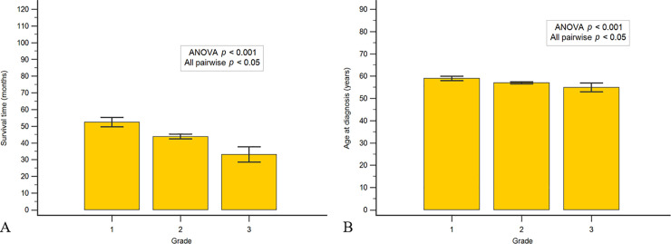
Fig 2A shows that the mean age at diagnosis was 59.0, 57.0, and 54.9 years for grade 1, grade 2, and grade 3 OSCC, respectively. Fig 2B shows that the mean survival time over 120 months (January 1, 2008–June 30, 2019) was 52.4, 43.9, and 33.1 months for grade 1, grade 2, and grade 3 OSCC, respectively. There was statistically significant difference in the mean age at diagnosis of cases and survival time (p < 0.001).

Fig 2.

Fig 2

a. Mean age at diagnosis of 59.0, 57.0, and 54.9 years with grade 1 (n = 435), grade 2 (n = 1964), and grade 3 (n = 136) OSCC, respectively. b. Mean survival time over 120 months (from OSCC diagnosis to death or to June 30, 2019) of 52.4, 43.9, and 33.1 months with grade 1, grade 2, and grade 3 OSCC, respectively.

The clinicopathological characteristics are listed in Tables 1 and 2. Table 1 summarizes sex, stratified data of age at diagnosis, anatomic site of the tumor and the associations among the grade of tumor cell differentiation and habits of lifestyle, including smoking, betel nut chewing, and alcohol consumption. There was no significant sex difference among the three groups (p = 0.076). Comparing among three groups, the proportion of females was slightly increased with an increasing trend in grade of tumor cell differentiation (p trend = 0.024). Age at diagnosis was divided into five groups, and there were significant differences among the three groups (p < 0.001). In grades 1 and 2 OSCC, age at diagnosis was mainly in group 3 (51–60 years), followed by group 4 (61–70 years), and in grade 3 OSCC, age at diagnosis was mainly in group 3 (51–60 years), followed by group 2 (41–50 years). There were significant differences in the anatomic site of the tumor among the three groups (p < 0.001). Most tumors of grades 1 and 2 were located on the buccal mucosa (44.4% and 33.6%, respectively), and grade 3 tumors were mostly located on the anterior two-thirds of the tongue (38.2%). Regarding lifestyle habits, Table 1 shows no significant association between the grade of tumor cell differentiation and habits of smoking, betel nut chewing, and alcohol consumption. Nevertheless, with increasing tumor grade, we noted an increase in the proportion of drinkers (p = 0.027).

Table 1. Summarized, stratified data of age at diagnosis, anatomic tumor site, and sex and associations among the grade of tumor cell differentiation and habits of smoking, betel nut chewing, and alcohol consumption.

Grade
Grade 1 (n = 435) Grade 2 (n = 1964) Grade 3 (n = 136) Total (n = 2535) p-value
N % N % N % N %
Age at diagnosis ≤40 15 3.4 137 7.0 11 8.1 163 6.4 <0.001
41–50 75 17.2 434 22.1 37 27.2 546 21.5
51–60 156 35.9 701 35.7 48 35.3 905 35.7
61–70 127 29.2 448 22.8 20 14.7 595 23.5
≧71 62 14.3 244 12.4 20 14.7 326 12.9
Site of tumors Alveolar ridge 71 16.3 328 16.7 16 11.8 415 16.4 <0.001
Anterior tongue 82 18.9 587 29.9 52 38.2 721 28.4
Buccal mucosa 193 44.4 659 33.6 39 28.7 891 35.1
Hard palate 16 3.7 53 2.7 4 2.9 73 2.9
Floor of mouth 3 0.7 67 3.4 7 5.1 77 3.0
Retromolar trigone 15 3.4 110 5.6 7 5.1 132 5.2
Mucosa of the Lips 55 12.6 160 8.1 11 8.1 226 8.9
Gender Female 12 2.8 98 5.0 9 6.6 119 4.7 0.076
Male 423 97.2 1866 95.0 127 93.4 2416 95.3
Smoking No 97 26.6 418 25.8 28 23.9 543 25.8 0.839
Yes 267 73.4 1204 74.2 89 76.1 1560 74.2
Unknown 432
Betel nut No 171 47.0 687 42.4 48 41.0 906 43.1 0.246
Yes 193 53.0 935 57.6 69 59.0 1197 56.9
Unknown 432
Alcohol No 183 56.0 714 49.7 47 46.1 944 50.6 0.079
Yes 144 44.0 723 50.3 55 53.9 922 49.4
Unknown 669

p-value by chi-square test. Regarding sex, p trend = 0.024In the alcohol group, p trend = 0.027.

Grade 1: well differentiated; Grade 2: moderately differentiated; Grade 3: poorly differentiated.

In the smoking and betel nut groups, 432 cases were excluded due to incomplete data.

In the alcohol group, 669 cases were excluded due to incomplete data.

Table 2. Associations among the grade of tumor cell differentiation and tumor pathological stage, including overall stage, T stage, N stage, recurrence status, and extranodal spread.

Grade
Grade 1 (n = 435) Grade 2 (n = 1964) Grade 3 (n = 136) Total (n = 2535) p-value
N % N % N % N %
Stage 1 249 59.1 712 36.8 27 20.5 988 39.7 <0.001
2 70 16.6 359 18.6 31 23.5 460 18.5
3 30 7.1 164 8.5 21 15.9 215 8.6
4 72 17.1 699 36.1 53 40.2 824 33.1
Missing data 48
T stage 1 262 60.2 812 41.5 36 26.5 1110 43.9 <0.001
2 77 17.7 477 24.4 46 33.8 600 23.7
3 25 5.7 109 5.6 17 12.5 151 6.0
4 71 16.3 560 28.6 37 27.2 668 26.4
No residual tumor 6
N stage 0 261 93.9 1096 70.7 59 54.1 1416 73.1 <0.001
1 10 3.6 154 9.9 18 16.5 182 9.4
2 7 2.5 273 17.6 29 26.6 309 16.0
3 0 0.0 27 1.7 3 2.8 30 1.5
Incomplete data 598
N stage Negative 261 93.9 1096 70.7 59 54.1 1416 73.1 <0.001
Positive 17 6.1 454 29.3 50 45.9 521 26.9
Incomplete data 598
Extra-nodal spread No 271 97.5 1318 85.0 80 73.4 1669 86.2 <0.001
Yes 7 2.5 232 15.0 29 26.6 268 13.8
Incomplete data 598
Recurrence No 375 86.2 1563 79.4 94 69.1 2028 80.0 <0.001
Yes 60 13.8 405 20.6 42 30.9 507 20.0

p-value by chi-square test. In the overall stage, N positive or negative group and extra-nodal spread group, p trend < 0.001.

Grade 1: well differentiated; Grade 2: moderately differentiated; Grade 3: poorly differentiated.

In the T stage group, 6 cases received curative chemoradiotherapy first, and the final pathological report showed no residual tumor.

In the overall stage, 48 cases were excluded due to poor data quality.

In the N stage and extranodal spread group, 550 cases were excluded due to operation without neck dissection and 48 cases were excluded due to poor data quality.

Table 2 shows a significant association between the grade of tumor cell differentiation and pathological stage (overall stage, T stage, and N stage), extranodal spread, and recurrence (p < 0.001). More than half (56.1%) of the patients with grade 3 OSCC but only 24.2% and 44.6% patients with grades 1 and 2 OSCC, respectively, were diagnosed with advanced tumor stage. By T status, T3 or T4 tumors were found in approximately 22%, 34%, and 40% of patients with grades 1–3 OSCC, respectively. Out of the patients with grade 3 OSCC, 45.9% were diagnosed with lymph node metastasis (N status positive), whereas only 6.1% with grade 1 and 29.3% with grade 2 were N status positive. Simultaneously, pathological N2 and N3 status were found in 29.4% of patients with grade 3 OSCC.

Table 2 also shows that there was a significant difference between recurrence status and extranodal spread among the three study groups (p < 0.001). Recurrence was recorded in 13.8%, 20.6%, and 30.9% of patients with grades 1–3 OSCC, respectively. Extranodal spread was recorded in 26.9%, 15.5%, and only 2.6% of patients with grade 3, grade 2, and grade 1, respectively.

Comparison of predictive power between pathologic grade and other common prognostic factors are shown in Table 3. In the multivariate (adjusted) analysis, during N1 and N2 stage, extranodal spread and poor differentiation were observed to be associated with a higher recurrence risk than the other common prognostic factors listed in Table 3 (aHR = 1.5, 2.4, 1.7, and 2.0, respectively).

Table 3. The effect of clinicopathological factors on recurrence and non-adjusted and adjusted hazard ratio among OSCC patients.

Cox proportional-hazards regression analysis of recurrence
Recurrence Univariate analysis (crude) Multiple analysis (adjusted)
Total N % Hazard ratio 95% CI P-value Hazard ratio 95% CI P-value
Stage 1 988 160 16.1 1.000
2 460 88 19.1 1.235 0.952–1.603 0.111
3 215 35 16.2 1.085 0.752–1.564 0.663
4 824 220 26.7 1.954 1.593–2.396 <0.001
T stage 1 1110 188 16.9 1.000 0.158–2.566 0.526
2 600 126 21.0 1.322 0.208–3.405 0.810
3 151 28 18.5 1.301 0.198–3.481 0.798
4 668 170 25.4 1.749 0.276–4.489 0.880
N stage 0 1416 209 14.8 1.000
1 182 46 25.3 1.851 1.345–2.547 <0.001 1.523 1.019 - 2.276 0.040
2 309 111 35.9 3.174 2.520–3.999 <0.001 2.426 1.629 - 3.613 <0.001
3 30 10 33.3 5.259 2.777–9.962 <0.001 3.140 0.939 - 10.500 0.063
Extranodal spread No 1669 266 15.9 1.000 1.000
Yes 268 102 38.0 3.156 2.509–3.970 <0.001 1.666 1.129 - 2.458 0.010
Close margin No 2455 491 20.0 1.000
Yes 80 23 28.8 1.527 1.005–2.320 0.047
Grade Well 435 60 13.8 1.000 1.000
Moderately 1964 405 20.6 1.672 1.275–2.194 <0.001 1.167 0.793 - 1.715 0.433
Poor 136 42 30.9 3.029 2.041–4.495 <0.001 1.973 1.167 - 3.336 0.011

Following period was from time at OSCC diagnosed to June 30, 2019.

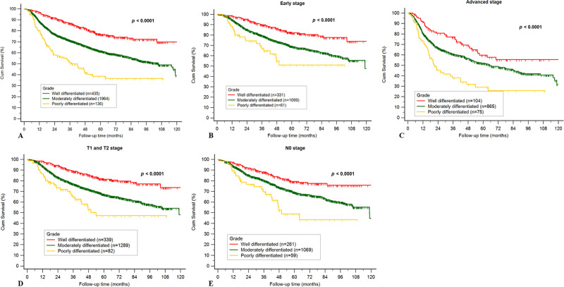
Kaplan–Meier curves for the three different histological grade tumors based on overall stage, early stages (stages I and II), advanced stages (stages III and IV), early T stages (T1 and T2), and without neck metastasis (pN0) are presented in Fig 3. These results show significant differences in overall stage, early stage, advanced stage, early T stage, and N0 stage (p < 0.001). Based on the Kaplan–Meier analysis, a better survival condition was observed in patients with well-differentiated OSCC than in those with moderately differentiated or poorly differentiated OSCC.

Fig 3.

Fig 3

a. Kaplan–Meier curve for different grades of tumor cell differentiation in the overall stage. b. Kaplan–Meier curve for different grades of tumor cell differentiation at early stage. c. Kaplan–Meier curve for different grades of tumor cell differentiation at advanced stage. d. Kaplan–Meier curve for different grades of tumor cell differentiation with early T stage (T1 and T2). e. Kaplan–Meier curve for different grades of tumor cell differentiation at N0 stage.

Discussion

Despite improved treatment modalities, clinicopathological prognostic factors remain insufficient for predicting recurrence and survival of OSCC; hence, the survival of patients has remained unchanged over the last few decades [11]. OSCC is known to present with verifiable histological behavior patterns. In our study, when compared with grades 1 and 2 OSCC, grade 3 OSCC was found in younger patients and was mostly located on the anterior two-thirds of the tongue. Patients with OSCC diagnosed as grade 3 tumors showed poor survival condition and higher risk of recurrence than other groups. Besides, histological grade of OSCC was observed to be a stronger predictive factor for predicting recurrence when compared with the other common prognostic factors.

In the present study, we found that the presence of high-grade histology was strongly associated with advanced T stage, neck lymph node metastasis, and extranodal spread, which subsequently contributed to advanced stage and prevalent recurrence. Similar conclusions have been deduced in previous research [1218]. Kademani et al. presented 10-year survival among three different histological grade OSCCs in 233 patients, and their results were similar to those of our study [12]. However, our study compares survival among not only three grades of tumors but also with respect to different clinicopathological parameters. Larsen et al. showed that T1 and T2 tumors had better local control than that of T3 and T4 tumors; however, T4 tumors also had better local control than that of T3 tumors [13]. These results may have been related to sample quantity (total sample size, n = 142). Contrarily, we studied a larger sample size and emphasized the distinct survival differences among the three groups of early stage, early T stage, and N0. Niu et al. showed that T stage, N stage, and histopathological grade of tumor were significantly associated with recurrence among 168 patients in a North Chinese population [16]. In the present study, histopathological grade of tumor, as opposed to T stage, was a more powerful prognostic factor to predict recurrence. The same research team, Xu et al., reported similar survival conditions among three different histological grade OSCCs in 2036 patients, and also reported that histopathological grade was associated with many clinicopathological features [16]. Simultaneously, they revealed that histopathological grade was an independent prognostic factor for patients with early-stage OSCC but not for patients with advanced-stage OSCC [17]. However, in our study, histopathological grade was associated with not only the survival condition of patients with early-stage OSCC but also of those with advanced stage OSCC.

Importantly, we observed that grade 3 OSCC may be important in early-stage cases for indicating potentially poor treatment outcomes because survival of patients with early-stage tumor and grade 3 tumor was distinctly poorer than that of patients with grades 1 and 2 tumor. The commonly accepted TNM staging for cancer is currently used by clinicians to guide their management decisions, but treatment outcomes are sometimes unsatisfactory for patients with early-stage tumor. In clinical practice, histopathological grade of OSCC is not regarded as a prognostic factor in National Comprehensive Cancer Network (NCCN) guideline; however, histological grade may be used as a supplementary material to clinical practice and provide additional information for decision making.

Several studies have indicated that patients with grade 3 OSCC are more likely to present with neck lymph node metastasis and be associated with decreased survival as compared to the patients with other tumor grades [1219]. However, the sample sizes of these studies were not as large as that in our study. Moreover, to the best of our knowledge, the current study is the first to describe the association among age, anatomic tumor site, alcohol consumption, and tumor grade in Taiwan. In clinical practice, relatively young patients with early TNM OSCC stage, especially in the tongue, should be more intensively followed up if they have higher differentiation degree of tumors because this may lead to occult metastasis, extranodal spread, and poor survival.

Regarding prognostic factors, we found that N stage, extranodal spread, and grade 3 tumors were strongly associated with recurrence. No significant correlation was found between recurrence and overall TNM and T stage, and there was a surprisingly close margin in our study. This finding may be explained by the advances in radiotherapy. In clinical practice, when facing close margins in resected tumors, clinicians can further consider wider re-excision or radiotherapy to decrease recurrence. Perhaps, this could explain that the role of close margin was not as important as other prognostic factors.

There are several limitations of our study. First, in this study, data were collected from a single medical center in Taiwan. Oral cavity cancer is highly associated with betel nuts, popular in Taiwan, which leads to buccal mucosa cancer, indicating a predilection at our site that may differ from other geographical regions. According to data from the Health Promotion Administration, Ministry of Health and Welfare in Taiwan, oral cavity cancer mostly occurs in the buccal mucosa followed by the tongue [20]. Second, the gross and histologic features of squamous cell carcinoma involving the head and neck are similar to those of squamous cell carcinoma in other organs. However, the histological grading of invasive squamous cell carcinoma remains controversial and there is no general agreement on the best system or scoring scheme [21, 22]. Finally, in the present study, although pathological slides were graded by two experienced pathologists, at best, grading attempts were subjective, and not absolutely reproducible. However, the modifiers “well,” “moderately,” and “poorly” differentiated carcinoma are used by many pathologists. “Moderately differentiated” is a commonly utilized fallback term in the diagnostic field; thus, owing to its frequency, it becomes clinically and pathologically useless. Practically, perhaps simpler scoring protocols will increase the reproducibility [23].

In conclusion, 5% of the patients in our study presented with poorly differentiated OSCC at diagnosis. As previously stated, grade 3 tumors have worse prognosis and are more aggressive than grades 1 and 2 tumors; alternatively, grade 3 tumors are more sensitive to radiotherapy than other grade tumors [2427]. The ability to predict which primary lesions are capable of early metastasis or poor prognosis would enable more individualized and aggressive therapy to be delivered to patients at higher risk of locoregional disease recurrence and death. In the future, we should focus on how to modify individual therapy for poorly differentiated OSCC to achieve improved outcomes.

Supporting information

S1 File

(PDF)

Acknowledgments

The authors would like to thank Enago (www.enago.com) for the English language review.

Data Availability

All relevant data are within the paper and its Supporting Information files.

Funding Statement

The authors received no specific funding for this work.

References

  • 1.Shah JP, Cendon RA, Farr HW, Strong EW. Carcinoma of the oral cavity. Factors affecting treatment failure at the primary site and neck. Am J Surg. 1976;132: 504–507. 10.1016/0002-9610(76)90328-7 [DOI] [PubMed] [Google Scholar]
  • 2.Leemans CR, Tiwari R, Nauta JJ, van der Waal I, Snow GB. Recurrence at the primary site in head and neck cancer and the significance of neck lymph node metastasis as a prognostic factor. Cancer. 1994;73: 187–190. [DOI] [PubMed] [Google Scholar]
  • 3.Shah JP. Cervical lymph node metastasis, its diagnostic, therapeutic and prognostic implications. Oncol Williston Park. 1990;4: 61–69; discussion 72, 76. [PubMed] [Google Scholar]
  • 4.Grandi C, Alloisio M, Moglia D, Podrecca S, Sala L, Salvatori P, et al. Prognostic significance of lymphatic spread in head and neck carcinomas: therapeutic implications. Head Neck Surg. 1985;8: 67–73. 10.1002/hed.2890080202 [DOI] [PubMed] [Google Scholar]
  • 5.Partridge M, Li SR, Pateromichelakis S, Francis R, Phillips E, Huang XH, et al. Detection of minimal residual cancer to investigate why oral tumors recur despite seemingly adequate treatment. Clin Cancer Res. 2000;6: 2718–2725. [PubMed] [Google Scholar]
  • 6.Anneroth G, Batsakis J, Luna M. Review of the literature and a recommended system of malignancy grading in oral squamous cell carcinomas. Scand J Dent Res. 1987;95(3):229–249. 10.1111/j.1600-0722.1987.tb01836.x [DOI] [PubMed] [Google Scholar]
  • 7.Kudo Y, Kitajima S, Ogawa I, Hiraoka M, Sargolzaei S, Keikhaee MR, et al. Invasion and metastasis of oral cancer cells require methylation of E-cadherin and/or degradation of membranous beta-catenin. Clin Cancer Res. 2004;10: 5455–5463. 10.1158/1078-0432.CCR-04-0372 [DOI] [PubMed] [Google Scholar]
  • 8.El-Naggar A. K. (Hg.). WHO classification of head and neck tumours: International Agency for Research on Cancer. (2017). [Google Scholar]
  • 9.Seethala RR, Weinreb I, Bullock JM. College of American Pathologists Protocol for the examination of specimens from patients with cancers of the lip and oral cavity Version LipOralCavity 4.0. 0.1; 2017. [cited June 17, 2020] Available from: https://cap.objects.frb.io/protocols/cp-headandneck-liporalcavity- 17protocol-4001.pdf [Google Scholar]
  • 10.Broders AC. The microscopic grading of cancer. Surg Clin North Am. 1941;21: 947–962. [Google Scholar]
  • 11.Neville BW, Day TA. Oral cancer and precancerous lesions. CA Cancer J Clin. 2002;52: 195–215. 10.3322/canjclin.52.4.195 [DOI] [PubMed] [Google Scholar]
  • 12.Kademani D, Bell RB, Bagheri S, Holmgren E, Dierks E, Potter B, et al. Prognostic factors in intraoral squamous cell carcinoma: the influence of histologic grade. J Oral Maxillofac Surg. 2005;63: 1599–1605. 10.1016/j.joms.2005.07.011 [DOI] [PubMed] [Google Scholar]
  • 13.Larsen SR, Johansen J, Sørensen JA, Krogdahl A. The prognostic significance of histological features in oral squamous cell carcinoma. J Oral Pathol Med. 2009;38: 657–662. 10.1111/j.1600-0714.2009.00797.x [DOI] [PubMed] [Google Scholar]
  • 14.Jögi A, Vaapil M, Johansson M, Påhlman S. Cancer cell differentiation heterogeneity and aggressive behavior in solid tumors. Ups J Med Sci. 2012;117: 217–224. 10.3109/03009734.2012.659294 [DOI] [PMC free article] [PubMed] [Google Scholar]
  • 15.Woolgar JA. Histopathological prognosticators in oral and oropharyngeal squamous cell carcinoma. Oral Oncol. 2006;42: 229–239. 10.1016/j.oraloncology.2005.05.008 [DOI] [PubMed] [Google Scholar]
  • 16.Niu LX, Feng Z, Li JN, Li CZ, Peng X, Guo CB. Prognostic factors of squamous cell carcinoma of the buccal mucosa: a retrospective study of 168 cases in North China. J Oral Maxillofac Surg. 2014;72: 2344–2350. 10.1016/j.joms.2014.05.021 [DOI] [PubMed] [Google Scholar]
  • 17.Xu QS, Wang C, Li B, Li JZ, Mao MH, Qin LZ, et al. Prognostic value of pathologic grade for patients with oral squamous cell carcinoma. Oral Dis. 2018;24:335–346. 10.1111/odi.12727 [DOI] [PubMed] [Google Scholar]
  • 18.Rodrigues RM, Bernardo VG, Da Silva SD, Camisasca DR, Faria PA, Dias FL, et al. How pathological criteria can impact prognosis of tongue and floor of the mouth squamous cell carcinoma. J Appl Oral Sci. 2019;28: e20190198 10.1590/1678-7757-2019-0198 [DOI] [PMC free article] [PubMed] [Google Scholar]
  • 19.Bryne M, Jenssen N, Boysen M. Histological grading in the deep invasive front of T1 and T2 glottic squamous cell carcinomas has high prognostic value. Virchows Arch. 1995;427: 277–281. 10.1007/BF00203395 [DOI] [PubMed] [Google Scholar]
  • 20.Cancer registry annual report. Taiwan; 2016 [cited Dec 2019]. Available from: https://www.hpa.gov.tw/Pages/Detail.aspx?nodeid=269&pid=10227date.
  • 21.Bryne M, Koppang HS, Lilleng R, Kjaerheim A. Malignancy grading of the deep invasive margins of oral squamous cell carcinomas has high prognostic value. J Pathol. 1992;166: 375–381. 10.1002/path.1711660409 [DOI] [PubMed] [Google Scholar]
  • 22.Akhter M, Hossain S, Rahman QB, Molla MR. A study on histological grading of oral squamous cell carcinoma and its co-relationship with regional metastasis. J Oral Maxillofac Pathol. 2011;15: 168–176. 10.4103/0973-029X.84485 [DOI] [PMC free article] [PubMed] [Google Scholar]
  • 23.Steigen SE, Søland TM, Nginamau ES, Laurvik H, Costea DE, Johannessen AC, et al. Grading of oral squamous cell carcinomas–Intra and interrater agreeability: simpler is better? [published online ahead of print, 2020 Jan 3]. J Oral Pathol Med. 2020;10.1111/jop.12990. 10.1111/jop.12990 [DOI] [PubMed] [Google Scholar]
  • 24.Liao CT, Chang JT, Wang HM, Ng SH, Hsueh C, Lee LY, et al. Analysis of risk factors of predictive local tumor control in oral cavity cancer. Ann Surg Oncol. 2008;15: 915–922. 10.1245/s10434-007-9761-5 [DOI] [PubMed] [Google Scholar]
  • 25.Liao CT, Wang HM, Ng SH, Yen TC, Lee LY, Hsueh C, et al. Good tumor control and survivals of squamous cell carcinoma of buccal mucosa treated with radical surgery with or without neck dissection in Taiwan. Oral Oncol. 2006;42: 800–809. 10.1016/j.oraloncology.2005.11.020 [DOI] [PubMed] [Google Scholar]
  • 26.Liao CT, Huang SF, Chen IH, Kang CJ, Lin CY, Fan KH, et al. Outcome analysis of patients with pN2 oral cavity cancer. Ann Surg Oncol. 2010;17: 1118–1126. 10.1245/s10434-009-0894-6 [DOI] [PubMed] [Google Scholar]
  • 27.Liao CT, Chang JT, Wang HM, Ng SH, Hsueh C, Lee LY, et al. Salvage therapy in relapsed squamous cell carcinoma of the oral cavity: how and when? Cancer. 2008;112: 94–103. 10.1002/cncr.23142 [DOI] [PubMed] [Google Scholar]

Decision Letter 0

Dipak Sapkota

16 Jun 2020

PONE-D-20-10673

Survival and Clinicopathological Characteristics of Different Histological Grades of Oral Cavity Squamous Cell Carcinoma: A Single-Center Retrospective Study

PLOS ONE

Dear Dr. Tsai,

Thank you for submitting your manuscript to PLOS ONE. After careful consideration, the reviewers and i feel that it has merit but does not fully meet PLOS ONE’s publication criteria as it currently stands. Therefore, we invite you to submit a revised version of the manuscript that addresses the points raised during the review process.

Please submit your revised manuscript by Jul 31 2020 11:59PM. If you will need more time than this to complete your revisions, please reply to this message or contact the journal office at plosone@plos.org. When you're ready to submit your revision, log on to https://www.editorialmanager.com/pone/ and select the 'Submissions Needing Revision' folder to locate your manuscript file.

Please include the following items when submitting your revised manuscript:

  • A rebuttal letter that responds to each point raised by the academic editor and reviewer(s). You should upload this letter as a separate file labeled 'Response to Reviewers'.

  • A marked-up copy of your manuscript that highlights changes made to the original version. You should upload this as a separate file labeled 'Revised Manuscript with Track Changes'.

  • An unmarked version of your revised paper without tracked changes. You should upload this as a separate file labeled 'Manuscript'.

If you would like to make changes to your financial disclosure, please include your updated statement in your cover letter. Guidelines for resubmitting your figure files are available below the reviewer comments at the end of this letter.

If applicable, we recommend that you deposit your laboratory protocols in protocols.io to enhance the reproducibility of your results. Protocols.io assigns your protocol its own identifier (DOI) so that it can be cited independently in the future. For instructions see: http://journals.plos.org/plosone/s/submission-guidelines#loc-laboratory-protocols

We look forward to receiving your revised manuscript.

Kind regards,

Dipak Sapkota, PhD

Academic Editor

PLOS ONE

Journal Requirements:

When submitting your revision, we need you to address these additional requirements.


1. Please ensure that your manuscript meets PLOS ONE's style requirements, including those for file naming. The PLOS ONE style templates can be found at

https://journals.plos.org/plosone/s/file?id=wjVg/PLOSOne_formatting_sample_main_body.pdf and

https://journals.plos.org/plosone/s/file?id=ba62/PLOSOne_formatting_sample_title_authors_affiliations.pdf

2. In the ethics statement in the manuscript and in the online submission form, please provide additional information about the patient records used in your retrospective study, including: a) whether all data were fully anonymized before you accessed them; b) the date range (month and year) during which patients' medical records were accessed; and c) the source of the medical records analyzed in this work (e.g. hospital, institution or medical center name). If patients provided informed written consent to have data from their medical records used in research, please include this information.

3. Thank you for stating the following financial disclosure:

 [The funders had no role in study design, data collection and analysis, decision to publish, or preparation of the manuscript.].

At this time, please address the following queries:

  1. Please clarify the sources of funding (financial or material support) for your study. List the grants or organizations that supported your study, including funding received from your institution.

  2. State what role the funders took in the study. If the funders had no role in your study, please state: “The funders had no role in study design, data collection and analysis, decision to publish, or preparation of the manuscript.”

  3. If any authors received a salary from any of your funders, please state which authors and which funders.

  4. If you did not receive any funding for this study, please state: “The authors received no specific funding for this work.”

4. We note that you have indicated that data from this study are available upon request. PLOS only allows data to be available upon request if there are legal or ethical restrictions on sharing data publicly. For information on unacceptable data access restrictions, please see http://journals.plos.org/plosone/s/data-availability#loc-unacceptable-data-access-restrictions.

In your revised cover letter, please address the following prompts:

a) If there are ethical or legal restrictions on sharing a de-identified data set, please explain them in detail (e.g., data contain potentially identifying or sensitive patient information) and who has imposed them (e.g., an ethics committee). Please also provide contact information for a data access committee, ethics committee, or other institutional body to which data requests may be sent.

b) If there are no restrictions, please upload the minimal anonymized data set necessary to replicate your study findings as either Supporting Information files or to a stable, public repository and provide us with the relevant URLs, DOIs, or accession numbers. Please see http://www.bmj.com/content/340/bmj.c181.long for guidelines on how to de-identify and prepare clinical data for publication. For a list of acceptable repositories, please see http://journals.plos.org/plosone/s/data-availability#loc-recommended-repositories.

We will update your Data Availability statement on your behalf to reflect the information you provide.

Please include your amended statements within your cover letter; we will change the online submission form on your behalf.

[Note: HTML markup is below. Please do not edit.]

Reviewers' comments:

Reviewer's Responses to Questions

Comments to the Author

1. Is the manuscript technically sound, and do the data support the conclusions?

The manuscript must describe a technically sound piece of scientific research with data that supports the conclusions. Experiments must have been conducted rigorously, with appropriate controls, replication, and sample sizes. The conclusions must be drawn appropriately based on the data presented.

Reviewer #1: Yes

Reviewer #2: Partly

Reviewer #3: Yes

**********

2. Has the statistical analysis been performed appropriately and rigorously?

Reviewer #1: Yes

Reviewer #2: Yes

Reviewer #3: Yes

**********

3. Have the authors made all data underlying the findings in their manuscript fully available?

The PLOS Data policy requires authors to make all data underlying the findings described in their manuscript fully available without restriction, with rare exception (please refer to the Data Availability Statement in the manuscript PDF file). The data should be provided as part of the manuscript or its supporting information, or deposited to a public repository. For example, in addition to summary statistics, the data points behind means, medians and variance measures should be available. If there are restrictions on publicly sharing data—e.g. participant privacy or use of data from a third party—those must be specified.

Reviewer #1: Yes

Reviewer #2: Yes

Reviewer #3: No

**********

4. Is the manuscript presented in an intelligible fashion and written in standard English?

PLOS ONE does not copyedit accepted manuscripts, so the language in submitted articles must be clear, correct, and unambiguous. Any typographical or grammatical errors should be corrected at revision, so please note any specific errors here.

Reviewer #1: Yes

Reviewer #2: No

Reviewer #3: No

**********

5. Review Comments to the Author

Please use the space provided to explain your answers to the questions above. You may also include additional comments for the author, including concerns about dual publication, research ethics, or publication ethics. (Please upload your review as an attachment if it exceeds 20,000 characters)

Reviewer #1: The authors present a complete and well organised manuscript that highlights the importance of providing histological grade of oral squamous carcinomas. Although the significance of the findings might be deeply related to the demographical and social characteristics of the cohort (betel chewing for example), the main suggestions about the value of the histological grading in OSCC in this report are still universally valuable and in line with practice with other type of malignancies affecting other organs/systems and that are stratified by histological grade.

The authors exclude from the cohort cases of carcinomas of the lip not affecting labial mucosa, but Table 5 reports tumors of the lips, which is confusing. Indicating this group as "labial mucosa" sounds more correct and takes away this confusing.

The authors correctly point that the histological grade might be to a certain degree subjective, and here the grading is performed by only one individual, which should be pointed also in the discussion as one of the challenges with this manuscript.

Finally, the authors use OSCC but also SCC and should keep one form; in page 2 and page 15 "should not be disregarded" might be better expressed as for "should be followed more closely than OSCC in early stages but higher differentiation degree"; in page 12 it should be "poor differentiation" not "poor differentiate"; in page 15 I guess authors mean "diagnostic field" and not "diagnostic filed".

Reviewer #2: Dear authors,

Thank you for the opportunity to review this manuscript in a very interesting topic. I have several concerns about your manuscript pointed bellow. I strongly recommend an English language review. You should considerer to reestructure the manuscript, this atual version is presented in a very confuse way.

Abstract

- The abstract section is very confuse and do not reflect clearly the content of the study.

Introduction

- The introduction section is a little poor, the authors should consider to include one or two more paragraphs to state the problem to the readers.

Patients and Methods

- The authors have to add the number and the date of the ethical board protocol aprouvement. (page 3)

- In my point of view include lip squamous cell carcinoma is a bias in your study. It is well known that, in this location, the neoplasia have a complete different biological behaviour. (page 4)

- The authors have to explain better the histopathological classification, especially how it has been performed, this is the central part of the study. Only one pathologist performed the analysis? How many slides per day? Directly in a microscope? With an image manager program? (page 4)

- Has the immunohistochemistry been performed in a regular basis? How many cases? In wich conditions?(page 5)

- As mentioned, the inclusion of the lip is a bias in your study. In my point of view, the authors should exclude this region. (page 5)

Results

- In general the results were presented in a very confuse way, the authors should rewrite this part of the manuscript. The tables are very difficult to understant and some of them are unnecessary.

Discussion

- In my point of view the discussion section is a bit confuse and include a restrict number of references, considering this is a classic topic (only 18 references in total).

- The authors should rewrite the discussion section according to the reestructured results section in a possible future revised version.

Reviewer #3: Although the finding are significant, the manuscript is not written well. Therefore I advise you to get the help from a language expert. There is a significant amount of literature missing in the reference list.

**********

[NOTE: If reviewer comments were submitted as an attachment file, they will be attached to this email and accessible via the submission site. Please log into your account, locate the manuscript record, and check for the action link "View Attachments". If this link does not appear, there are no attachment files.]

While revising your submission, please upload your figure files to the Preflight Analysis and Conversion Engine (PACE) digital diagnostic tool, https://pacev2.apexcovantage.com/. PACE helps ensure that figures meet PLOS requirements. To use PACE, you must first register as a user. Registration is free. Then, login and navigate to the UPLOAD tab, where you will find detailed instructions on how to use the tool. If you encounter any issues or have any questions when using PACE, please email PLOS at figures@plos.org. Please note that Supporting Information files do not need this step.

PLoS One. 2020 Aug 25;15(8):e0238103. doi: 10.1371/journal.pone.0238103.r002

Author response to Decision Letter 0


25 Jun 2020

General response:

We sincerely thank the editor and all reviewers for their valuable feedback that we have used to improve the quality of our manuscript. The reviewer comments are laid out below in italicized font and specific concerns have been numbered. Our response is given in normal font and changes/additions to the manuscript are given in ”Track changes”.

We have revised the manuscript extensively based on the reviewer’s comments. If there are any other modifications we could make, we would like very much to do so and we greatly appreciate your help. We hope that our manuscript will be considered for publication in your journal. Thank you very much.

Sincerely,

Kuo-Yang Tsai

Department of Oral and Maxillofacial Surgery, Changhua Christian Hospital

No. 235, Xuguang Rd, Changhua City, Changhua County 500, Taiwan

Phone: +886 933127916

Email: 72837@cch.org.tw

Attachment

Submitted filename: Response to Reviewers.docx

Decision Letter 1

Dipak Sapkota

21 Jul 2020

PONE-D-20-10673R1

Survival and Clinicopathological Characteristics of Different Histological Grades of Oral Cavity Squamous Cell Carcinoma: A Single-Center Retrospective Study

PLOS ONE

Dear Dr. Tsai,

Thank you for submitting your manuscript to PLOS ONE. After careful consideration, we still feel that the reviewers' comments are not satisfactorily addressed in the current version. Therefore, we invite you to submit a revised version of the manuscript that addresses the points raised during the review process.

Please submit your revised manuscript by Sep 04 2020 11:59PM. If you will need more time than this to complete your revisions, please reply to this message or contact the journal office at plosone@plos.org. When you're ready to submit your revision, log on to https://www.editorialmanager.com/pone/ and select the 'Submissions Needing Revision' folder to locate your manuscript file.

Please include the following items when submitting your revised manuscript:

  • A rebuttal letter that responds to each point raised by the academic editor and reviewer(s). You should upload this letter as a separate file labeled 'Response to Reviewers'.

  • A marked-up copy of your manuscript that highlights changes made to the original version. You should upload this as a separate file labeled 'Revised Manuscript with Track Changes'.

  • An unmarked version of your revised paper without tracked changes. You should upload this as a separate file labeled 'Manuscript'.

If you would like to make changes to your financial disclosure, please include your updated statement in your cover letter. Guidelines for resubmitting your figure files are available below the reviewer comments at the end of this letter.

If applicable, we recommend that you deposit your laboratory protocols in protocols.io to enhance the reproducibility of your results. Protocols.io assigns your protocol its own identifier (DOI) so that it can be cited independently in the future. For instructions see: http://journals.plos.org/plosone/s/submission-guidelines#loc-laboratory-protocols

We look forward to receiving your revised manuscript.

Kind regards,

Dipak Sapkota, PhD

Academic Editor

PLOS ONE

[Note: HTML markup is below. Please do not edit.]

Reviewers' comments:

Reviewer's Responses to Questions

Comments to the Author

1. If the authors have adequately addressed your comments raised in a previous round of review and you feel that this manuscript is now acceptable for publication, you may indicate that here to bypass the “Comments to the Author” section, enter your conflict of interest statement in the “Confidential to Editor” section, and submit your "Accept" recommendation.

Reviewer #1: (No Response)

Reviewer #2: (No Response)

**********

2. Is the manuscript technically sound, and do the data support the conclusions?

The manuscript must describe a technically sound piece of scientific research with data that supports the conclusions. Experiments must have been conducted rigorously, with appropriate controls, replication, and sample sizes. The conclusions must be drawn appropriately based on the data presented.

Reviewer #1: Partly

Reviewer #2: Partly

**********

3. Has the statistical analysis been performed appropriately and rigorously?

Reviewer #1: Yes

Reviewer #2: Yes

**********

4. Have the authors made all data underlying the findings in their manuscript fully available?

The PLOS Data policy requires authors to make all data underlying the findings described in their manuscript fully available without restriction, with rare exception (please refer to the Data Availability Statement in the manuscript PDF file). The data should be provided as part of the manuscript or its supporting information, or deposited to a public repository. For example, in addition to summary statistics, the data points behind means, medians and variance measures should be available. If there are restrictions on publicly sharing data—e.g. participant privacy or use of data from a third party—those must be specified.

Reviewer #1: Yes

Reviewer #2: Yes

**********

5. Is the manuscript presented in an intelligible fashion and written in standard English?

PLOS ONE does not copyedit accepted manuscripts, so the language in submitted articles must be clear, correct, and unambiguous. Any typographical or grammatical errors should be corrected at revision, so please note any specific errors here.

Reviewer #1: Yes

Reviewer #2: No

**********

6. Review Comments to the Author

Please use the space provided to explain your answers to the questions above. You may also include additional comments for the author, including concerns about dual publication, research ethics, or publication ethics. (Please upload your review as an attachment if it exceeds 20,000 characters)

Reviewer #1: I commend the authors for having worked with the text and accepting our suggestions, and the presentation of the data and the discussion are more complete and precise. The manuscript has improved on the language side as well, but there are still mistakes in the text (page 8 "...grade 2, years, and grade 3 OSCC..."; page 9 "...most..." instead of "...mostly..."; page 16 "re-wide excision" never heard before, I guess the authors mean "wider re-excision").

In page 17 the authors write that "...grading attempts are inexact, subjective, not reproducible." It is also my experience that most squamous carcinomas fall in the definition of moderate grade of differentiation, but the authors are quite sharp in their definition. And just using keratinising vs non keratinising could give the same problem and be potentially subjective. This has been dealt with by other authors who suggested a to-tier grading to improve the concordance of the grading [I can quickly suggest for example Krishnan, Karpagalvi...Patil, J Oral Maxillofac Pathology (2016) and Steigen, Søland...Olsen, J Oral Pathol Med (2020)].

Also, in the text the authors now write that two pathologists classified the material and not just one, but the numbers of the groups were the same as far as I can see, and the authors write nothing on how the two pathologists came to an agreement. Did the pathologists agree on all cases? In this case why do the authors write the grading is not reproducible?

This point (only one pathologist) and a point previously addressed (grade 3 cases should not be "disregarded") have not been addressed in the abstract, that should be corrected as well.

The text is written in good English all in all but the imprecisions deter from the scientific value of the study, real piety since I find the study really useful also in clinical settings, reminding pathologists to provide clinicians with a histological variable that can be evaluated on routinely provided HE slides.

Reviewer #2: The main questions addressed in the first round of revision were not properly answered. Including the lip OSCC is one of the major concern in the manuscript.

**********

7. PLOS authors have the option to publish the peer review history of their article (what does this mean?). If published, this will include your full peer review and any attached files.

If you choose “no”, your identity will remain anonymous but your review may still be made public.

Do you want your identity to be public for this peer review? For information about this choice, including consent withdrawal, please see our Privacy Policy.

Reviewer #1: No

Reviewer #2: No

[NOTE: If reviewer comments were submitted as an attachment file, they will be attached to this email and accessible via the submission site. Please log into your account, locate the manuscript record, and check for the action link "View Attachments". If this link does not appear, there are no attachment files.]

While revising your submission, please upload your figure files to the Preflight Analysis and Conversion Engine (PACE) digital diagnostic tool, https://pacev2.apexcovantage.com/. PACE helps ensure that figures meet PLOS requirements. To use PACE, you must first register as a user. Registration is free. Then, login and navigate to the UPLOAD tab, where you will find detailed instructions on how to use the tool. If you encounter any issues or have any questions when using PACE, please email PLOS at figures@plos.org. Please note that Supporting Information files do not need this step.

PLoS One. 2020 Aug 25;15(8):e0238103. doi: 10.1371/journal.pone.0238103.r004

Author response to Decision Letter 1


24 Jul 2020

General response:

We sincerely thank the editor and all reviewers for their valuable feedback that we have used to improve the quality of our manuscript. The reviewer comments are laid out below in italicized font and specific concerns have been numbered. Our response is given in normal font and changes/additions to the manuscript are given in ”Track changes”.

Point-to-point response:

Reviewer #1: I commend the authors for having worked with the text and accepting our suggestions, and the presentation of the data and the discussion are more complete and precise. The manuscript has improved on the language side as well, but there are still mistakes in the text (page 8 "...grade 2, years, and grade 3 OSCC..."; page 9 "...most..." instead of "...mostly..."; page 16 "re-wide excision" never heard before, I guess the authors mean "wider re-excision").

Response: Thank you very much for your positive comments and suggestions. I have revised the manuscript accordingly.

In page 17 the authors write that "...grading attempts are inexact, subjective, not reproducible." It is also my experience that most squamous carcinomas fall in the definition of moderate grade of differentiation, but the authors are quite sharp in their definition. And just using keratinising vs non keratinising could give the same problem and be potentially subjective. This has been dealt with by other authors who suggested a to-tier grading to improve the concordance of the grading [I can quickly suggest for example Krishnan, Karpagalvi...Patil, J Oral Maxillofac Pathology (2016) and Steigen, Søland...Olsen, J Oral Pathol Med (2020)].

Response: Thank you very much for your suggestion. I found the conclusion in Steigen et al. was similar to my point of view, they suggested that simpler/uncomplicated scoring protocols will increase the reproducibility. In their study, Intrarater comparison of WHO grading system, after in binary categorization, mean agreement could reach 92.5%. I have added the article into my reference and revised the manuscript in smooth tone. Thanks again for your valuable help.

Also, in the text the authors now write that two pathologists classified the material and not just one, but the numbers of the groups were the same as far as I can see, and the authors write nothing on how the two pathologists came to an agreement. Did the pathologists agree on all cases? In this case why do the authors write the grading is not reproducible?

Response: In the present study, we investigated the grade of OSCC in retrospective fashion (based on final pathological report). The pathological report was accomplished by two pathologists within 10 days after surgery had been done. And the intervention of third pathologist was engaged if the first two pathologists could not reach a consensus. In our study we only presented the final agreement of the grade of tumors documented on the pathological report. Grading was achieved by human through microscope and it is inevitable that human made different judgment in those borderline cases. Steigen et al. also reported that simpler/uncomplicated scoring protocols will increase the reproducibility (two options better than three options, and three options better than fine or four options). In our study, we did not verify the reproducibility of OSCC grading, however, it is very hard to achieve 100% the same result when all our cases graded in the other center or other country.

This point (only one pathologist) and a point previously addressed (grade 3 cases should not be "disregarded") have not been addressed in the abstract, that should be corrected as well. The text is written in good English all in all but the imprecisions deter from the scientific value of the study, real piety since I find the study really useful also in clinical settings, reminding pathologists to provide clinicians with a histological variable that can be evaluated on routinely provided HE slides.

Response:

Thank you very much for your suggestion. I have revised the Abstract accordingly. In the present study, I would like to suggest clinicians do not overlook the importance of the grade of OSCC, in 2018 AJCC Head and Neck guideline, tumor invade depth was added in TNM staging. In somedays, histological grade of OSCC or a new grading system maybe added into TNM stage.

Reviewer #2: The main questions addressed in the first round of revision were not properly answered. Including the lip OSCC is one of the major concern in the manuscript.

Response: Thanks for your comments. In AJCC 2018 head and neck cancer guideline, oral cavity had included the mucosal lip. In Southeast Asia, the main factors contributing to mucosal lip SCC are betel nuts chewing, smoking and alcohol consumption and these factors are the same in the oral cavity SCC. Meanwhile, I have reviewed many researches for OSCC, and found these article also included mucosal lip SCC into their study (I take two study for example: Brinkman D, Callanan D, O'Shea R. Impact of 3 mm margin on risk of recurrence and survival in oral cancer. Oral Oncol. 2020;110:104883. doi:10.1016/j.oraloncology.2020.104883 and Mochizuki Y, Harada H, Ikuta M, et al. Clinical characteristics of multiple primary carcinomas of the oral cavity. Oral Oncol. 2015;51(2):182-189. doi:10.1016/j.oraloncology.2014.11.013 ). If you insist excluding the entity of the mucosal lip in the present study, we would like very much to do so and we greatly appreciate your help.

In the first round of revision, we have revised the manuscript extensively based on your suggestions. In the present study, we investigated the grade of OSCC in retrospective fashion (based on final pathological report). The pathological report was accomplished by two pathologists within 10 days after surgery had been done. And the intervention of third pathologist was engaged if the first two pathologists could not reach a consensus. Immunohistochemical confirmation for cytokeratin was performed to detect poorly differentiated OSCC because features of squamous differentiation were minimal or absent. In our study we only presented the final agreement of the grade of tumors documented on the pathological report. In the present study, I would like to suggest clinicians do not overlook the importance of the grade of OSCC, in 2018 AJCC Head and Neck guideline, tumor invade depth was added in TNM staging. In somedays, histological grade of OSCC or a new grading system maybe added into TNM stage. If there are any other modifications we could make, we would like very much to do so.

We have revised the manuscript extensively based on the reviewer’s comments. If there are any other modifications we could make, we would like very much to do so and we greatly appreciate your help. We hope that our manuscript will be considered for publication in your journal. Thank you very much.

Sincerely,

Kuo-Yang Tsai

Department of Oral and Maxillofacial Surgery, Changhua Christian Hospital

No. 235, Xuguang Rd, Changhua City, Changhua County 500, Taiwan

Phone: +886 933127916

Email: 72837@cch.org.tw

Attachment

Submitted filename: Response to Reviewers.docx

Decision Letter 2

Dipak Sapkota

30 Jul 2020

PONE-D-20-10673R2

Survival and clinicopathological characteristics of different histological grades of oral cavity squamous cell carcinoma: a single-center retrospective study

PLOS ONE

Dear Dr. Tsai,

Thank you for revising the manuscript according to the reviewers suggestions. The manuscript has improved significantly, but i feel that the manuscript will immensely benefit if you could improve the language (especially the consistent use of 'tense' in the method, result and the discussion sections) of your manuscript. Therefore, i invite you to submit a revised version of the manuscript. 

Please submit your revised manuscript by Sep 13 2020 11:59PM. If you will need more time than this to complete your revisions, please reply to this message or contact the journal office at plosone@plos.org. When you're ready to submit your revision, log on to https://www.editorialmanager.com/pone/ and select the 'Submissions Needing Revision' folder to locate your manuscript file.

Please include the following items when submitting your revised manuscript:

  • A rebuttal letter that responds to each point raised by the academic editor and reviewer(s). You should upload this letter as a separate file labeled 'Response to Reviewers'.

  • A marked-up copy of your manuscript that highlights changes made to the original version. You should upload this as a separate file labeled 'Revised Manuscript with Track Changes'.

  • An unmarked version of your revised paper without tracked changes. You should upload this as a separate file labeled 'Manuscript'.

If you would like to make changes to your financial disclosure, please include your updated statement in your cover letter. Guidelines for resubmitting your figure files are available below the reviewer comments at the end of this letter.

If applicable, we recommend that you deposit your laboratory protocols in protocols.io to enhance the reproducibility of your results. Protocols.io assigns your protocol its own identifier (DOI) so that it can be cited independently in the future. For instructions see: http://journals.plos.org/plosone/s/submission-guidelines#loc-laboratory-protocols

We look forward to receiving your revised manuscript.

Kind regards,

Dipak Sapkota, PhD

Academic Editor

PLOS ONE

Editor Comments:

Thank you for revising the manuscript according to the reviewers suggestions. The manuscript has improved significantly, but the manuscript still appears inconsistent with respect to the use of 'tense' in the method, result and the discussion sections. Please use a consistent tense to describe the current results as well as results from other researchers.

[Note: HTML markup is below. Please do not edit.]

While revising your submission, please upload your figure files to the Preflight Analysis and Conversion Engine (PACE) digital diagnostic tool, https://pacev2.apexcovantage.com/. PACE helps ensure that figures meet PLOS requirements. To use PACE, you must first register as a user. Registration is free. Then, login and navigate to the UPLOAD tab, where you will find detailed instructions on how to use the tool. If you encounter any issues or have any questions when using PACE, please email PLOS at figures@plos.org. Please note that Supporting Information files do not need this step.

PLoS One. 2020 Aug 25;15(8):e0238103. doi: 10.1371/journal.pone.0238103.r006

Author response to Decision Letter 2


4 Aug 2020

General response:

We sincerely thank the editor and all reviewers for their valuable feedback that we have used to improve the quality of our manuscript. The editor comments are laid out below in italicized font and specific concerns have been numbered. Our response is given in normal font and changes/additions to the manuscript are given in ”Track changes”.

Point-to-point response:

Editor: Thank you for revising the manuscript according to the reviewers suggestions. The manuscript has improved significantly, but the manuscript still appears inconsistent with respect to the use of 'tense' in the method, result and the discussion sections. Please use a consistent tense to describe the current results as well as results from other researchers.

Response: Thank you very much for your positive comments and suggestions. I have revised the manuscript accordingly. The changes to the manuscript are given in “Track changes”.

We have revised the manuscript extensively based on the yours comments. If there are any other modifications we could make, we would like very much to do so and we greatly appreciate your help. We hope that our manuscript will be considered for publication in your journal. Thank you very much.

Sincerely,

Kuo-Yang Tsai

Department of Oral and Maxillofacial Surgery, Changhua Christian Hospital

No. 235, Xuguang Rd, Changhua City, Changhua County 500, Taiwan

Phone: +886 933127916

Email: 72837@cch.org.tw

Decision Letter 3

Dipak Sapkota

11 Aug 2020

Survival and clinicopathological characteristics of different histological grades of oral cavity squamous cell carcinoma: a single-center retrospective study

PONE-D-20-10673R3

Dear Dr. Tsai,

We’re pleased to inform you that your manuscript has been judged scientifically suitable for publication and will be formally accepted for publication once it meets all outstanding technical requirements.

Within one week, you’ll receive an e-mail detailing the required amendments. When these have been addressed, you’ll receive a formal acceptance letter and your manuscript will be scheduled for publication.

An invoice for payment will follow shortly after the formal acceptance. To ensure an efficient process, please log into Editorial Manager at http://www.editorialmanager.com/pone/, click the 'Update My Information' link at the top of the page, and double check that your user information is up-to-date. If you have any billing related questions, please contact our Author Billing department directly at authorbilling@plos.org.

If your institution or institutions have a press office, please notify them about your upcoming paper to help maximize its impact. If they’ll be preparing press materials, please inform our press team as soon as possible -- no later than 48 hours after receiving the formal acceptance. Your manuscript will remain under strict press embargo until 2 pm Eastern Time on the date of publication. For more information, please contact onepress@plos.org.

Kind regards,

Dipak Sapkota, PhD

Academic Editor

PLOS ONE

Additional Editor Comments (optional):

Reviewers' comments:

Acceptance letter

Dipak Sapkota

17 Aug 2020

PONE-D-20-10673R3

Survival and clinicopathological characteristics of different histological grades of oral cavity squamous cell carcinoma: a single-center retrospective study

Dear Dr. Tsai:

I'm pleased to inform you that your manuscript has been deemed suitable for publication in PLOS ONE. Congratulations! Your manuscript is now with our production department.

If your institution or institutions have a press office, please let them know about your upcoming paper now to help maximize its impact. If they'll be preparing press materials, please inform our press team within the next 48 hours. Your manuscript will remain under strict press embargo until 2 pm Eastern Time on the date of publication. For more information please contact onepress@plos.org.

If we can help with anything else, please email us at plosone@plos.org.

Thank you for submitting your work to PLOS ONE and supporting open access.

Kind regards,

PLOS ONE Editorial Office Staff

on behalf of

Dr. Dipak Sapkota

Academic Editor

PLOS ONE

Associated Data

    This section collects any data citations, data availability statements, or supplementary materials included in this article.

    Supplementary Materials

    S1 File

    (PDF)

    Attachment

    Submitted filename: Response to Reviewers.docx

    Attachment

    Submitted filename: Response to Reviewers.docx

    Data Availability Statement

    All relevant data are within the paper and its Supporting Information files.


    Articles from PLoS ONE are provided here courtesy of PLOS

    RESOURCES