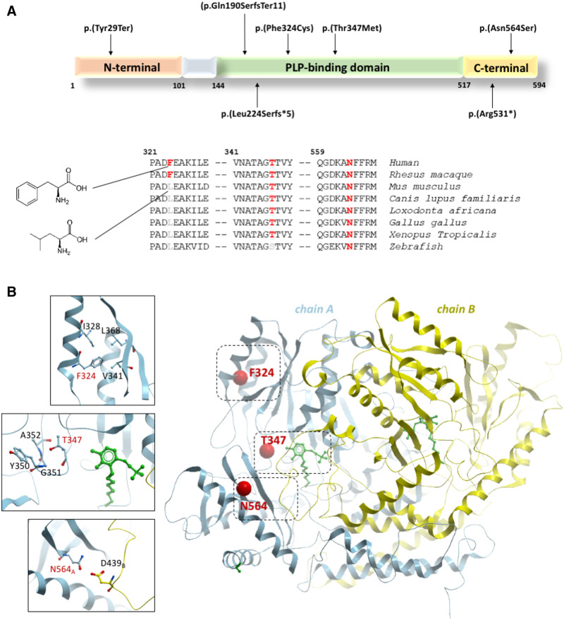
Figure 2.
Schematic and cartoon representation of GAD1. (A) Schematic representation of the GAD1 isoform GAD67 (NP_000808.2) with the pathogenic variants identified in this study. Of the six variants, four fall within the PLP-binding domain, a conserved region that is essential for the binding of the crucial cofactor pyridoxal 5′-phosphate (PLP). The remaining variants affect the C-terminal domain, which contains the catalytic site of the enzyme. Conservation status among different species is shown for the missense variants. (B) Cartoon representation of human GAD1 dimer (PDB: 2okj) with the two subunits in blue and yellow. Sites of the three missense mutations in this study are shown as red spheres, and close-up views of their nearby atomic environment are shown as insets. The PLP cofactor is shown in green.

