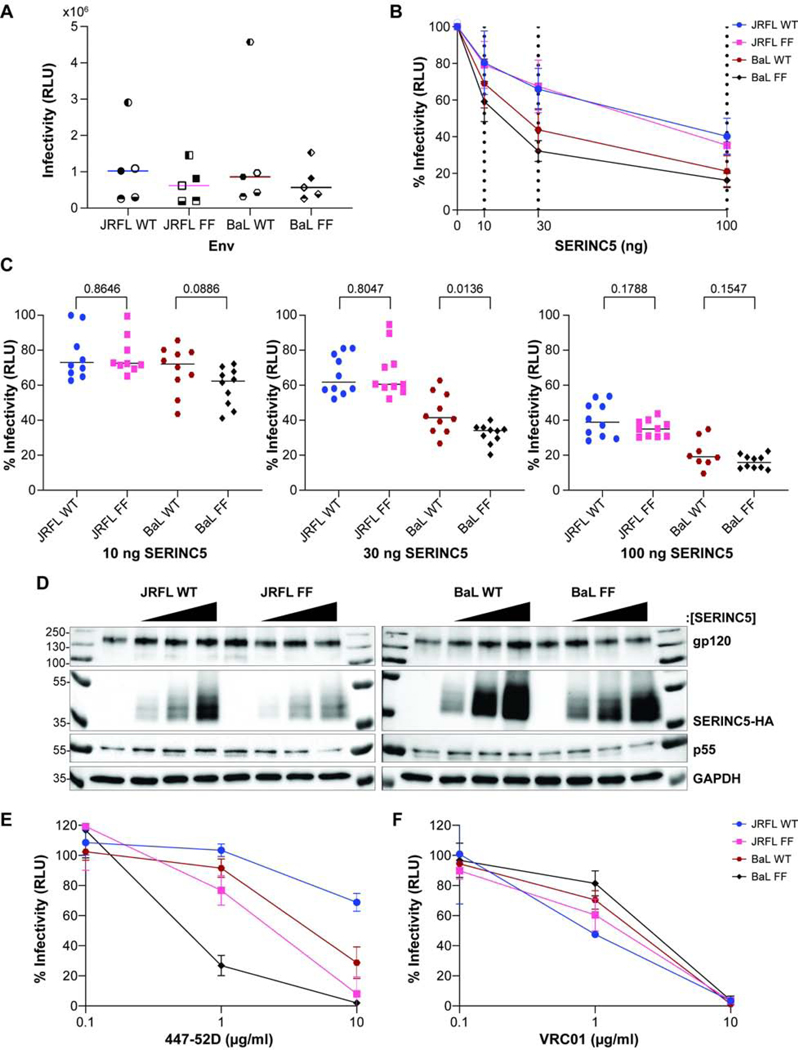
Figure 2. Effect of V2 tyrosine substitutions on the sensitivity 447–52D antibody and to SERINC5.

A) The infectivity of pseudovirions containing WT or mutant “FF” (YY173, 177 FF) Envs of JRFL or BaL. The mean values from five independent experiments are shown for each Env; and each assay was run in triplicate. Experiment 1 – filled symbol; 2 – symbol blank; 3 – the top half of symbol filled; 4 – the bottom half of symbol filled; 5 – left half of the symbol filled. B) Inhibition of infectivity by expression of SERINC5. The relative infectivity of pseudovirions was quantified each of the indicated Envs and compared to no added SERINC5 (0 ng SERINC5 = 100%). The dashed lines indicate the amount of SERINC5 added (0, 10, 30, 100 ng). Data are shown from five independent experiments; each data point represents 10 independent values for each Env (the infectivity of each pseudovirion preparation measured at 1:3 and 1:9 dilutions, each in triplicate). C) Relative infectivity of pseudovirions containing WT and mutated Envs at increasing concentrations of SERINC5. Pairwise comparisons were made between each Env and its corresponding tyrosine-to-phenylalanine mutant. P-values were determined by Welch’s t-test. D) Expression of the key proteins was measured in the pseudovirion-producer cells. Molecular masses in kDa are indicated on the left; Proteins are indicated on the right. Each Env was grouped by increasing SERINC5 protein concentration: 0, 10, 30, 100 ng from left to right. E) Neutralization of infectivity by the antibody 447–52D. Data are presented as in Figure 1E. Data shown are from two independent experiments; each neutralization assay was run in duplicate. F) Neutralization of infectivity by the antibody VRC01. The data shown represent one experiment performed in duplicate.
