
Figure 1:
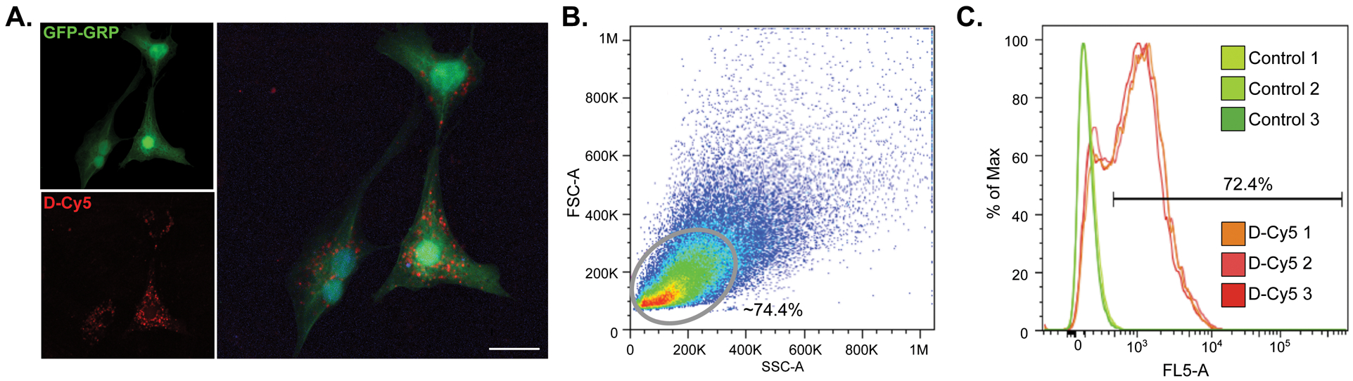
GFP-GRPs show uptake of Cy5-conjugated dendrimer (D-Cy5) after a 12 hr exposure. A) Confocal imaging of D-Cy5 exposed GRPs; scale bar equals 20 μm. B) Flow cytometry was used to confirm dendrimer uptake. Gating was conducted on GFP-expressing GRPs which reveals an average of 72.4% of D-Cy5 exposed cells across three replicates show accumulation of the conjugate (C).