Description
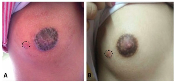
A 10-year-old girl presented with the complaints of right-sided nipple skin crusting, itching and serous discharge for 4 months (figure 1A). The nipple discharge occurred intermittently throughout the day and required padding under the brassiere. There was no history of blood or pus in the discharge, no history of palpable breast lump, nor any history of local trauma, local drug application or oral drug intake. The contralateral breast was normal. A provisional diagnosis of breast abscess was made in a local hospital, and the patient underwent incision and drainage (I&D) on two occasions for the same. However, due to inadequate response to treatment, she was referred to our hospital. On examination, her height and weight were average for age. In sexual maturity rating, she had stage III maturity of breasts and pubic hair. Local examination of the right breast revealed excoriated, cracked and thickened skin overlying the entire areola oozing serous discharge. There was a scar mark of around 5 mm lateral to the areola attributable to previous I&D. The rest of the right breast and left breast were normal. Ultrasonography of the right breast showed normal breast parenchyma and glandular pattern with no mass lesion, duct dilation or fluid collection. A differential diagnosis of nipple eczema versus Paget’s disease was considered. However, Paget’s disease is generally seen in postmenopausal women, and it presents with eczematous lesions on the breast with bloody discharge and destruction of the nipple areolar complex. Also, ultrasonography in Paget’s disease often shows parenchymal heterogeneity with hypoechoic areas, discrete masses, skin thickening and dilated ducts.1 Since our case showed no such findings except an eczematous lesion with serous discharge, a clinical diagnosis of nipple eczema was made. Also, the lesion showed significant improvement with the application of topical steroids and emollients within 8 weeks, and the nipple had normalised by 9–10 weeks, thus confirming our diagnosis (figure 1B).
Figure 1.

Right breast photograph showing nipple discharge, erosions, scaling and crusting (A); normal nipple after treatment (B); dotted circles show sites of incision and drainage.
Nipple eczema refers to localised dermatitis of the nipple and areola, and is usually seen in lactating mothers and is often bilateral.2 It presents with erythematous papules, crusting, oozing and erosions, and rarely with nipple discharge, if it becomes infected.2 Diagnosis is made clinically, and topical steroids and emollients form the mainstay of therapy.3 However, if the discharge is purulent, swabs must be sent for culture. Moreover, if the lesions do not resolve even after 3 months of treatment, alternate diagnoses such as allergic contact dermatitis, psoriasis and Paget’s disease must be considered.4 Our case highlights atypical presentation of nipple eczema at puberty with persistent unilateral discharge, of which paediatricians need to be familiar.
Patient’s perspective.
We were really anxious since even after two breast surgeries, our daughter continued to have persistent discharge. It’s a big relief to know that it was a mild medical condition which would require just a couple of topicals.
Learning points.
Unilateral nipple discharge is an atypical presentation of nipple eczema in pubertal girls which paediatricians should be familiar with.
Patient’s history and clinical examination play a crucial role in the diagnosis of breast eczema.
Footnotes
Contributors: AK: patient management, literature review and manuscript preparation; RK: clinician in charge, critical review of the manuscript and final approval of the version to be published; SG: patient management and literature review.
Funding: The authors have not declared a specific grant for this research from any funding agency in the public, commercial or not-for-profit sectors.
Competing interests: None declared.
Patient consent for publication: Parental/guardian consent obtained.
Provenance and peer review: Not commissioned; externally peer reviewed.
References
- 1.Karakas C. Paget's disease of the breast. J Carcinog 2011;10:31. 10.4103/1477-3163.90676 [DOI] [PMC free article] [PubMed] [Google Scholar]
- 2.Song HS, Jung S-E, Kim YC, et al. Nipple eczema, an indicative manifestation of atopic dermatitis? A clinical, histological, and immunohistochemical study. Am J Dermatopathol 2015;37:284–8. 10.1097/DAD.0000000000000195 [DOI] [PubMed] [Google Scholar]
- 3.Barankin B, Gross MS. Nipple and areolar eczema in the breastfeeding woman. J Cutan Med Surg 2004;8:126–30. 10.1177/120347540400800209 [DOI] [PubMed] [Google Scholar]
- 4.Kim SK, Won YH, Kim S-J. Nipple eczema: a diagnostic challenge of allergic contact dermatitis. Ann Dermatol 2014;26:413–4. 10.5021/ad.2014.26.3.413 [DOI] [PMC free article] [PubMed] [Google Scholar]


