Abstract
Oral rehabilitation of partially edentulous arches requires careful treatment planning before any prosthodontic intervention. The connection of the metal framework of fixed (fixed dental prosthesis (FPD)) and removable partial denture using adhesive attachments is a good alternative prosthetic option when solely fixed prosthesis (FPD or implant) cannot be used due to anatomical limitation. Attachments are the tiny interlocking devices that act as a hybrid link to join removable prosthesis to the abutment and direct the masticatory forces along the long axis of the abutment. This joint acts as a non-rigid stress breaker, which helps in distributing the occlusal load. Precision and semiprecision attachment have always been bordered by an aura of mystery due to technique sensitive procedure and lack of knowledge. The following case describes a combined contemporary and conventional approach and treatment sequence with the use of attachments for the rehabilitation of partially edentulous arches.
Keywords: dentistry and oral medicine, rehabilitation medicine, mouth
Background
Prosthetic rehabilitation of partially edentulous arch aims at restoration of function, preservation of periodontium and aesthetics.1 Various treatment modalities are available for partially edentulous patients, such as clasp retained removable partial denture (RPD), RPDs with adhesive attachments, a fixed dental prosthesis (FPD) and an implant-retained prosthesis.
Conventional fixed partial denture is not recommended in a long-edentulous span with compromised bone support from a residual ridge. The dental implant as a treatment of choice is also limited due to need of ridge augmentation. Therefore, combining fixed prosthesis with removable denture using precision attachments always remains an alternative treatment modality to conventional clasp-retained removable prosthesis. To counteract the damaging effect of lateral forces, attachments along with clasp may also be used to retain and stabilise the prosthesis.2
As per glossary of prosthodontics term (GPT) an attachment can be defined as ‘a retainer consisting of a metal receptacle (matrix) and a closely fitting part (patrix), the matrix is usually contained within the normal expanded contour of the crown on abutment teeth and patrix is attached the removable partial denture framework’.3 Further, an attachment can be classified according to the method of fabrication as precision or semiprecision and mode of retention as intracoronal and extracoronal.4 In an intracoronal attachment the matrix is fitted within the contour of crown whereas, in extracoronal attachment, the matrix is outside the contoured crown.
Few advantages of intracoronal attachments are appearance, unaffected retention by crown contour and minimum stresses on abutment teeth but intracoronal attachment requires extensive abutment preparation and therefore becomes the reason for early and easy wear off.
Extracoronal attachments have few advantages over intracoronal as they can be universally used (no restriction in size), have greater freedom in design and can be fashioned to give greater retention.2 4
Unlike removable restoration, fixed-removable prosthesis (RPD with attachment) offers considerable advantages of preventing lateral movement and selective movement of prosthesis on occlusal loading. Therefore, minimise the transfer of stress over abutment tooth and provide a biomechanical advantage in long-span partial edentulism.4 The attachment retained RPD significantly improves the retention, functional efficiency corresponding to a fixed prosthesis and aesthetics and hygiene maintenance of a removable prosthesis. However, the biomechanical factor must be taken into account during treatment planning for therapeutic results.5
From the patient's perspective, a removable prosthesis with attachment offers more retention, masticatory efficiency and aesthetics due to decreased mobility of prosthesis than the one fabricated without attachment.5
The use of precision attachments among dental professionals has been very limited since its introduction because of the limited curriculum in under graduation and technique sensitive laboratory needed for precise placement of attachment. A reliable laboratories support for incorporating the appropriate attachment and insight of proper treatment planning in a particular clinical situation is must.
Case report
A 41-year-old woman reported to the Prosthodontic Postgraduate Section, Faculty of Dental Sciences, Banaras Hindu University with the chief complaint of inability to chew food from right back tooth region associated with a dull pain on biting hard food. On clinical examination, the patient revealed the dental history of the fixed partial denture in all four quadrants for last 9 months. In the maxillary arch, there was dislodged metal-ceramic bridge prosthesis bilaterally and in the mandibular arch, it was clearly evident that the porcelain facing was fractured with exposed metal coping on 35 and 38 abutment teeth. Both maxillary and mandibular arch had bilateral saddle areas surrounded by prepared teeth anteriorly and posteriorly.
On examination of the maxillary arch, on the right side a total of 5 teeth were present that is, 18, 14, 13, 12, 11 and among these, the teeth used as abutment were 14 and 18 for the metal-ceramic prosthesis, similarly on the left side a total of 5 teeth present were 21, 22, 23, 27, 28 while tooth number 23 and 27 were serving as an abutment for the previous prosthesis.
In the mandibular arch, on the left side a total of 6 teeth were present, that is, 38, 35, 34, 33, 32, 31, while 2 teeth 36 and 37 were missing. Similarly, on the right side, a total of 6 teeth were present, that is, 41, 42, 43, 44, 45, 48 while teeth missing were 46 and 47. Therefore, as per Applegate's rule, both maxillary and mandibular arches had Kennedy Class III modification 1 edentulous spaces.
Investigations
To make a definitive treatment plan, we should always analyse the three most important components of diagnostic procedure especially in prosthetic dentistry which are6
Thorough clinical examination.
Radiographical examination.
Diagnostic mounting.
To achieve all the three objectives, a comprehensive clinical examination was done for evaluating the condition of abutment teeth after removing the old prosthesis. Radiographic evaluation and its correlation with the clinical condition were done with orthopantomagram X-ray as an aid in diagnostic tool (figure 1). Diagnostic impressions of the maxillary and mandibular arches were made using irreversible hydrocolloid (Zelgen, Dentsply, Germany). The impressions were poured with type 3 gypsum product, that is dental stone (Kalstone, Kalabhai, Karson, India). The facebow transfer was done and the bite registration record was made using bite registration paste (O-bite, DMG). Diagnostic mounting was done using maxillary and mandibular cast in the centric bite record (figure 2A, B).
Figure 1.
Preoperative orthopantomagram X-ray.
Figure 2.
(A, B) Diagnostic models; (C) diagnostic mounting with wax-up.
Treatment
Before starting the treatment, all the modalities of treatment options were explained to the patient but the patient agreed to treatment protocols of the present case report.
In case of the maxillary arch, it was decided to fabricate cast partial denture (CPD) along with individual porcelain fused metal (PFM) jacket crown for 13, 14, and 18 (right side) and 23 and 27 (left side).
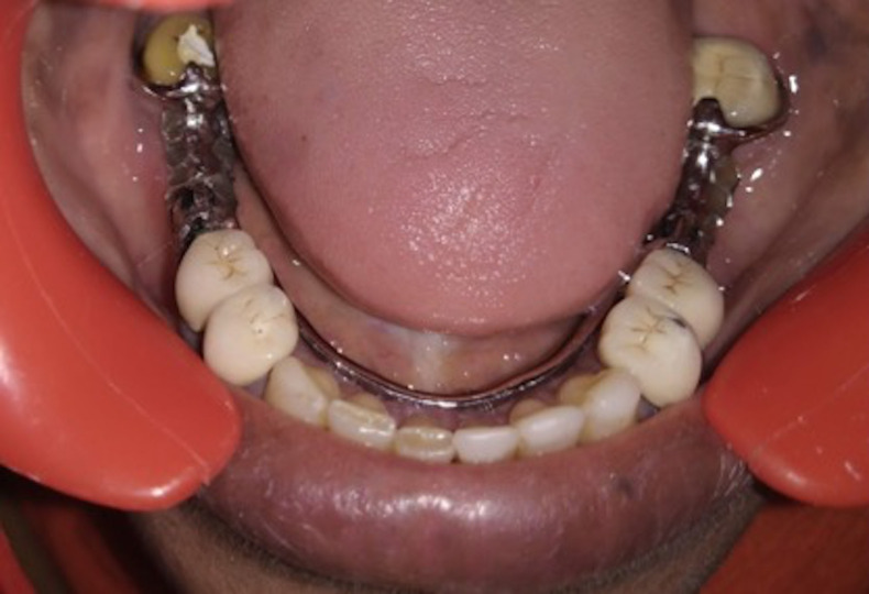
In case of the mandibular arch, contemporary technique with fixed-removable dental prosthesis was planned. It was decided to attach extracoronal attachment on the proximal surface of the second premolar bilaterally. First premolars (34, 44) were also used along with second premolar (35, 45) as a abutment and splinted together with metal porcelain jacket crown to aid in the retention of the fixed component of the above-mentioned combination.
For another important component of the prosthesis that is, removable component, a CPD fabrication was planned after evaluating available existing space.
Entire procedure along with appointment schedule was elaborated to the patients, and bilingual informed consent was obtained.
To estimate the existing space for extracoronal resilient attachment, a diagnostic wax pattern was fabricated and a putty index was made from addition to silicone putty material (Aquasil, Dentsply, Germany) (figure 2C). The abutment teeth 34, 35, 38, 44, 45 and 48 were prepared to receive PFM restoration. A two-stage putty-wash impression was made and poured in type 4 gypsum stone (Kalstone, Kalabhai, Karson). The provisional polymethyl methacrylate prosthesis was fabricated with the help of putty index and cementation was done using temporary luting agent (Temp-Bond; Kerr Corporation, Romulus) (figure 3). Wax patterns were fabricated for all the prepared teeth and matrix portion of the extraoral attachment was attached on the distal wall of 35 and 45 wax pattern using a dental surveyor. The selection criteria for choosing a particular size was based on available space aiming at providing 2 mm of clearance from the gingival margin to preserve the periodontium of abutment teeth. Wax copings with Precision attachments were cast using the lost wax technique of casting. Special attention was given during finishing and sandblasting procedure of casted prosthesis to avoid wear of extracoronal attachment
Figure 3.

Provisional restorations.
Metal Try-in of the splinted crown with patrix component was done to check for margin and occlusal clearance (figure 4). Pick-up impression was made using putty consistency of additional silicon and poured to fabricate CPD framework (figure 5). Attachment positioning plastic burn out caps (matrix component) were positioned on the cast and a new refractory was made to cast a lingual bar CPD. A retentive cap was inserted in CPD after try-in in the patient mouth.
Figure 4.
Splinted metal-ceramic crown on 34, 35, 44,45 with attachment component.
Figure 5.
Pick-up impression with putty for the fabrication of removable prosthesis.
In the maxillary arch, finish line margins of all the prepared abutments were refined, Surveying of the wax pattern was accomplished and thereafter PFM crown. Cast partial framework was also designed and fabricated (figure 6).
Figure 6.
Cast partial prosthesis trial.
A second facebow transfer was done after final cementation of crowns in maxillary arch and jaw relation was recorded with bite registration paste followed by articulation and teeth arrangement. Waxed denture trial was done followed by acrylisation with heat-polymerised acrylic resin (figure 7). Lab remounting, along with clinical remounting procedures, were performed in both centric and eccentric positions. To evaluate any kind of high points and premature contacts, marks were obtained using 40 micron articulating paper (BAUSH articulating paper).
Figure 7.
Final mandibular prosthesis.
After occlusal correction, Cementation of PFM crowns in the mandibular arch was done using luting (type 1) glass ionomer cement (GC Gold Label 1, Japan) and CPD was also inserted simultaneously. The patient was asked to close in centric occlusion. After completion of cementation, occlusal contacts were again marked to check for any high point.
Outcome and follow-up
At recall visits at 1 week, 1 month and 3 months for follow-up, the prosthesis was found to satisfactory in terms of function and aesthetics (figure 8).
Figure 8.
Final maxillary and mandibular prosthesis.
Discussion
There are the various treatment modalities present for the rehabilitation of partially edentulous state, such as clasp-retained RPD, FDP and an implant-retained prosthesis. The dental implant was not taken as treatment of choice due to anatomical and financial barrier. Tooth supported FDP was not a viable option because of compromised biomechanics of the prosthesis in the long-edentulous span. Removable prosthesis with the attachment was planned as treatment of choice due to the benefit of aesthetic, retention and improved functional efficiency over conventional clasp retained RPD. Moreover, this particular case presented as unconventional Kennedy’s class III as tooth posterior to the edentulous span does not display suitable contour for retention on surveying. These unconventional class III act as Kennedy’s class II on application of unseating force and class III with seating force.7 On posterior abutment non-retentive clasp is given to enhance stability and attachment is given on anterior abutment to improve retention. However, a conventional treatment modality of CPD was planned for the maxillary arch.
A fixed-removable prosthesis is an efficient and cost-effective treatment option for long span partially edentulous ridge. There are multiple advantages of such prosthesis, namely, retention and stabilising qualities of a fixed prosthesis along with freedom in teeth arrangement, hygiene maintenance and aesthetics of removable prosthesis. Besides these advantages, the attachment allows the prosthesis to be inserted and removed several times without losing retention. On the same time, it also splints the teeth and provides favourable biomechanics.8 9
However, the biomechanical factor must be taken into considerations when using attachment-RPD design. Repeated removal and placement of prosthesis result in wear of the retention clip, requiring periodic replacement of the clip. Other possible disadvantages include extensive tooth preparation, splinting of teeth together, minimum abutment height of 5–6 mm required for attachment functionality and laboratory techniques need an expert dexterity in fabricating this prosthesis. Daily oral hygiene maintenance and care of the prosthesis are required on the part of the patient. The long-term success of the prosthesis requires knowledge of important laboratory techniques, clinical skills, and proper execution of all the clinical and laboratory procedures. In terms of clinical success, the fixed-removable bridge meets all the demands of function and aesthetic appearance with the added benefit of facilitating the careful postoperative evaluation of oral soft tissue.
Dr Herman ES Chayes introduced Chayes' attachment in 1912, which forms the basis of modern friction grip attachment. These two joints are so arranged that they articulate to a precise but separable joint, where the matrix envelops the patrix.10
Both intracoronal and extracoronal attachments are biomechanically favourable for the abutment. All the biomechanics of extracoronal attachments lie outside the crown structure and are attached to the proximal surface of the crown. They are mainly indicated in those areas where the loss of tooth tissue renders it incapable of intracoronal attachments.11
Selection of attachment is based on many factors11 12 -
Crown root ratio desired.
Type of coping.
Vertical space available.
Available teeth support.
Available bone support.
Location of abutments.
Cohn states that ‘The precision attachments prevent lateral stresses to periodontium of abutment teeth when inserting or removing the denture. It distributes stress vertically to the tooth during function and stabilises the abutment teeth during lateral stresses’.13 Precision attachments provide better vertical support and stimulation to the underlying tissue through intermittent vertical massage.14
Conclusion
Although FPD is better tolerated by the patients in comparison to RPD, the latter is still prevalent in partially dentate people. The acceptance of RPDs has increased when used along with precision attachments. FPD/RPD with attachments are a great therapeutic treatment option in this case with limiting anatomical consideration of bone factor and unretentive abutment. Adherence to precision techniques, a proper diagnosis will result in successful treatment and preservation of the patient's existing dentition.
Learning points.
Before planning for extracoronal attachment, crown height of teeth adjacent to the edentulous span should be sufficient to allow room for placement of the attachment.
Careful positioning of attachment will increase the longevity of the prosthesis by avoiding unnecessary wear of attachment on lateral loading.
Abutment use for attachment placement should have sound periodontal health and if needed should be splinted to provide better support.
Footnotes
Contributors: The case was done by RS and NR. Supervised by AB Co-supervised, edited and communicated by PD.
Funding: The authors have not declared a specific grant for this research from any funding agency in the public, commercial or not-for-profit sectors.
Competing interests: None declared.
Patient consent for publication: Obtained.
Provenance and peer review: Not commissioned; externally peer reviewed.
References
- 1.Carr AB, McGivney GP, Brown DT. McCracken’s Removable Partial Prosthodontics. 11th edn St. Louis, Missouri: Elsevier Mosby, 2005. [Google Scholar]
- 2.Preiskel HW, Preiskel A. Precision attachments for the 21st century. Dent Update 2009;36:221–7. 10.12968/denu.2009.36.4.221 [DOI] [PubMed] [Google Scholar]
- 3.The Academy of Denture Prosthetics Glossary of prosthodontics term. J Prosthet Dent 2005;94:37. [Google Scholar]
- 4.Burns DR, Ward JE. A review of attachments for removable partial denture design: Part 1. Classification and selection. Int J Prosthodont 1990;3:98–102. [PubMed] [Google Scholar]
- 5.Hedzelek W, Rzatowski S, Czarnecka B. Evaluation of the retentive characteristics of semi-precision extracoronal attachments: evaluation of semi-precision attachments. Journal of Oral Rehabilitation 2011;38:462–8. [DOI] [PubMed] [Google Scholar]
- 6.Peter E. Dawson, Funtional occlusion from TMJ to SMILE. St. Louis, Missouri: Elsevier Mosby, 2007: P.238. [Google Scholar]
- 7.Phoenix RD, Cagna DR, DeFreest CF, et al. Stewart's clinical removable partial prosthodontics. 4th edn Chicago: Quintessence, 2008. [Google Scholar]
- 8.Dittmann B, Rammelsberg P. Survival of abutment teeth used for telescopic abutment retainers in removable partial dentures. Int J Prosthodont 2008;21:319–21. [PubMed] [Google Scholar]
- 9.el Charkawi HG, Wakad MT. Effect of splinting on load distribution of extracoronal attachment with distal extension prosthesis in vitro. J Prosthet Dent 1996;76:315–20. 10.1016/S0022-3913(96)90178-X [DOI] [PubMed] [Google Scholar]
- 10.da Cruz Perez LE, Alfenas BFM. Maxillary rehabilitation using fixed and removable partial dentures with attachments: a clinical report: using FPDs and RPDs with attachments. Journal of Prosthodontics 2014;23:58–63. [DOI] [PubMed] [Google Scholar]
- 11.Zitzmann NU, Rohner U, Weiger R, et al. When to choose which retention element to use for removable dental prostheses. Int J Prosthodont 2009;22:161. [PubMed] [Google Scholar]
- 12.Vaidya S, Kapoor C, Bakshi Y, et al. Achieving an esthetic SMILE with fixed and removal prosthesis using extracoronal castable precision attachments. J Indian Prosthodont Soc 2015;15:284. 10.4103/0972-4052.155048 [DOI] [PMC free article] [PubMed] [Google Scholar]
- 13.Gupta N, Bhasin A, Gupta P, et al. Combined prosthesis with extracoronal castable precision attachments. Case Rep Dent 2013;2013:1–4. 10.1155/2013/282617 [DOI] [PMC free article] [PubMed] [Google Scholar]
- 14.Hedzelek W, Rzatowski S, Czarnecka B. Evaluation of the retentive characteristics of semi-precision extracoronal attachments: evaluation of SEMI-PRECISION attachments. Journal of Oral Rehabilitation 2011;38:462–8. [DOI] [PubMed] [Google Scholar]


