Abstract
The supramalleolar osteotomy is a joint-preserving surgical procedure. It is a very good treatment option for the asymmetric varus ankle and medial compartment osteoarthritis. The primary objective of the procedure is to shift medial concentration of stress toward the lateral intact articular cartilage to redistribute the joint loads during ambulation. Several studies have shown that deformities of the ankle result in uneven load distribution in the ankle joint, which eventually leads to articular cartilage degeneration. Since the lateral articular cartilage is intact, joint-sacrificing procedures such as total ankle replacement or ankle arthrodesis are not the most appropriate treatment choices for medial compartment arthritis. Results of supramalleolar osteotomies are very promising in terms of functional outcome and pain relief. In younger patients with medial compartment varus ankle osteoarthritis or even with a normal tibial anterior surface angle, supramalleolar osteotomies can be performed to realign the ankle to promote regeneration of the asymmetrically damaged cartilage. In this review article, we will discuss the indications, complications, surgical techniques, and outcomes of the supramalleolar osteotomy reported in the current literature.
Keywords: Supramalleolar osteotomy, Medial compartment arthritis, Ankle joint
Osteoarthritis (OA) of the ankle represents 1% of all OA cases in the world.1) Unlike OA of the hip and knee, OA of the ankle is usually a consequence of trauma.2,3) The ankle joint carries around 4 times the body weight in the stance phase. It has a small contact area of approximately 522 mm,3) which is 33% of the hip or knee.4) Biomechanically, the ankle joint is highly congruent. The cartilage of the ankle joint is thinner than that of the hip and knee, but it can withstand greater shear and tensile forces, which enables the ankle joint's contact pressure to be at a low level.5) This is why symptomatic ankle joint OA is 9 times less common than knee OA.5) Uneven stress accumulation on the joint surface in the lower extremities can cause asymmetric cartilage wear and joint deformities, which may increase the contact pressure even further, creating a vicious circle that will result in OA.6) If the joint surface area is reduced or the congruency is lost, the pressure increases exponentially, resulting in OA. Posterior malleolar fractures with 33% involvement of its articulating surface and 1-mm talar tilt reduce the tibiotalar contact by 13% and 42%, respectively.2,3) However, transection of the deltoid ligament does not affect tibiotalar contact.7)
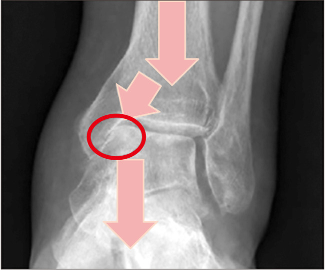
Different operative methods for different stages of ankle OA have been introduced. Such methods can be divided into 2 main groups: joint-preserving and joint-sacrificing procedures. Joint-preserving procedures include ankle arthroscopic debridement, distraction arthroplasty, osteochondral resurfacing procedures, and corrective osteotomies. Ankle arthrodesis and ankle replacement have own advantages but they also have some complications and long-term problems.8,9,10) Ankle OA patients are in general younger than hip and knee OA patients, because of the higher incidence of the posttraumatic ankle OA. Patients with posttraumatic ankle OA manifest symptoms approximately 12 to 15 years earlier than do patients with knee or hip OA.11) Thus, long-lasting pain relieving surgery is very important for the treatment of ankle OA. Among the surgical options, joint conservation procedures will be the best treatment option for ankle OA. The supramalleolar osteotomy (SMO) is a joint-preserving technique indicated for ankle OA with asymmetric axial joint overloading, more commonly in the medial compartment in Asians with varus ankles. Ankle varus malalignment brings about focal static, dynamic overload in the medial compartment, especially in the medial gutter (Fig. 1).12)
Fig. 1. Focal static and dynamic overload in the medial compartment, especially the medial gutter.

The objective of SMO is to realign the varus or valgus deformed ankles by distal tibial and fibular osteotomies to shift the asymmetrically overloaded joint contact pressure in the arthritic area toward the cartilage-preserved joint area. The SMO is mainly indicated for the incongruent osteoarthritic ankle (either varus or valgus) with intact medial or lateral tibiotalar joint cartilage. The SMO can be also used to correct the alignment of the severely deformed end-stage osteoarthritic ankle before total ankle arthroplasty.12,13,14) Hindfoot instability that cannot be managed with ligament reconstruction is an absolute contraindication of the SMO. Other absolute contraindications include severe vascular and neurologic insufficiency in the affected extremity, inflammatory joint disease, neuroarthropathy, and acute or chronic infection of the ankle.15) Relative contraindications include elderly patients (70 years of age), advanced tibiotalar OA, and tobacco smoking, which predisposes patients to nonunion.16)
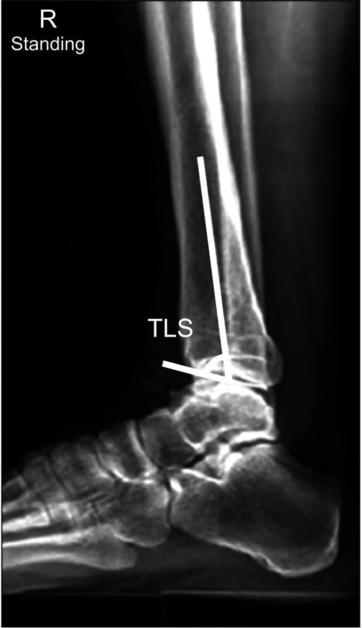
Weight-bearing radiographs including AP, lateral (Fig. 2) and mortise views of the ankle and whole leg radiographs are obtained to assess concomitant deformities of the lower extremity and to guide surgical planning (Fig. 3).
Fig. 2. Lateral view demonstrating measurement of the tibial lateral surface (TLS) angle.
Fig. 3. Radiographic assessment of the foot and ankle. Concomitant deformities of the lower extremity were assessed on weight-bearing radiographs including the anteroposterior view of the ankle (A), ankle mortise (B), lateral view of the ankle (C), and whole leg radiograph (D).
SURGICAL TECHNIQUES
Medial Opening-Wedge Osteotomy
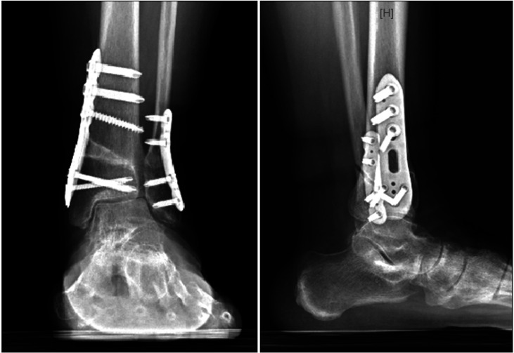
All forms of corrective osteotomies can be performed under either general or regional anesthesia. The patient is in the supine position with the foot placed at the edge of the table. Due to intraoperative fluoroscopy, a radiolucent operating table should be used. The ipsilateral back is raised until the foot is in a purely upward position. A pad for elevation and fluoroscopy is placed under the lower leg during surgery. A tourniquet is applied to the ipsilateral thigh (usually 300 mmHg). Intraoperative arthroscopic examination of the ankle and synovectomy are performed initially (Fig. 4). Medial opening-wedge osteotomy is then performed at the distal tibia through a medial longitudinal incision. The medial skin incision is used with minimal stripping of the periosteum just enough to complete the osteotomy. Using an oscillating saw, a tibial oblique cut is made while preserving the lateral cortex and periosteum, which serve as a fulcrum for the opening wedges and thus improve stability (Fig. 5). Gentle distraction is done at the osteotomy site using a laminar spreader under the guidance of fluoroscopy until desired correction of the deformity is achieved. One or two tricortical bone block graft of appropriate size is placed in the space created by the low tibial osteotomy. The aim of corrected tibial anterior surface (TAS) angle is 94°–95° aiming for 3°–5° of overcorrection. A low-profile medial locking plate is applied to the medial osteotomy (Fig. 6). A small lateral incision is used, and a fibular oblique osteotomy at the same level with the tibial osteotomy is then subsequently performed. A plate with appropriate bending is then applied to the fibular osteotomy site after valgus angulation.
Fig. 4. Arthroscopic examination of the denuded tibiotalar articular cartilage of the ankle and synovectomy were performed before the supramalleolar osteotomy procedure.
Fig. 5. Fluoroscopic images showing the level of oblique tibial osteotomy using a Kirschner-wire and the osteotomy performed using the oscillating saw.
Fig. 6. Eight-month postoperative weight-bearing ankle radiographs showing osseous healing at the distal tibia medial opening-wedge osteotomy site and the valgus angulated distal fibular osteotomy site with an increased tibial anterior surface angle and the medial gutter space.
Lateral Closing-Wedge Osteotomy
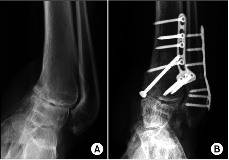
In patients with compromised medial soft tissue, a lateral closing-wedge osteotomy can be used instead of a medial opening wedge osteotomy (Fig. 7). Advantages of closingwedge osteotomy include improved stability, no need for allograft, and the possibility to perform a synchronous fibular osteotomy through the same incision. Disadvantage of this procedure is that it might require supplemental medial plate fixation as the medial cortex of the distal tibia is weak and soft-tissue contractures might be present. An additional biplanar cut might be needed for a coexisting sagittal defortmity.15)
Fig. 7. A 37-year-old woman had posttraumatic 28° varus ankle osteoarthritis and a tibial anterior surface angle of 59° after an ankle fracture that had occurred 30 years ago. She complained of medial ankle pain (visual analog scale 5). She was treated with a lateral closing-wedge osteotomy of the tibia and fibula. Preoperative (A) and postoperative (B) radiographs.
A medial opening-wedge osteotomy may not be feasible in patients with a preoperative varus malalignment of more than 10° as the fibula may restrict the degree of overcorrection as well as the medial soft-tissue compromise due to excessive widening of the medial osteotomy gap of the distal tibia.17) In case of a large degree of varus correction, an anterior crescentic osteotomy can be performed to correct by rotation. The lateral closing-wedge osteotomy involves a lateral wedge closing osteotomy or a fibula osteotomy. A cut is made on the distal fibula's anterior rim. A Z-shaped fibular osteotomy is done using a narrow oscillating saw where the fibula is shortened by cutting the bone base.18) There is considerably less flexibility in the straight transverse fibular osteotomy, which can result in fibular malposition.17,19) Kirschner wires are drilled into the tibia in the preoperatively defined direction after the fibula is removed. The periosteum is incised and elevated after testing of the Kirschner wire position by fluoroscopy.
Postoperative Management
Patients are immobilized with walker boots for 12 weeks. Early ankle range of motion exercises are allowed at postoperative 2 weeks with toe-touch weight-bearing for 6 postoperative weeks. Partial weight-bearing with 50% body weight is allowed at postoperative 6 to12 weeks. When early radiographic bony healing is noted, full weight-bearing and rehabilitation exercises are allowed, such as muscle strengthening, gait training, passive and active range of motion, and coordination and proprioception muscular activities.15) However, if there is a delayed bony union at postoperative 12 weeks, partial weight-bearing is extended for 4 to 8 more weeks with further immobilization in a walking cast or boots. Computed tomography (CT) is performed to confirm the radiographic bony healing of the opening-wedge bone graft usually at postoperative 3 months or at 6 or 12 months in case of delayed union.15)
Complications
Intraoperative complications include injuries of tendons and neurovascular structures. Surgeons must have a full grasp of surgical approaches used. If superficial and deep infections occur, intravenous antibiotics and/or surgical debridement may be used for treatment. Medial skin compromise or skin necrosis is not uncommon after medial opening-wedge osteotomy of the distal tibia fixed with a medial locking plate in patients especially with a thin soft-tissue layer in the medial part of the distal tibia with a varus deformity. Medial skin necrosis can be catastrophic and often lead to salvage surgery of free flap reconstruction.
Poor fixation and ill-advised weight-bearing in the early postoperative phase or wedge bone graft with poor quality allogenous bone can result in malunion, delayed union, or nonunion. Loss of correction or undercorrection can result from implant failure, delayed union/nonunion, or failure to address concomitant pathology, such as ligamentous incompetence, muscular dysfunction, and inframalleolar deformities.12,13) Metal removal is recommended in patients with painful hardware, once clinical and radiographic healing has been confirmed by CT. Another complication is the progression from Takakura Stage 3b to 4, which requires ankle arthrodesis or total ankle replacement. In a study by Knupp et al.,20) the outcomes of SMO were unsuccessful in 10 out of 94 ankles, which required conversion to total ankle replacement (9 ankles) or ankle arthrodesis (1 ankle). Worse outcomes were noted in patients with type I valgus deformity (talar tilt 4°, congruent joint) where the length of the fibula was not changed, patients with type III varus deformity, and patients with ankle joint instability.
RESULTS
The impact of SMO such as survival rates and clinical outcomes among different patient profiles are well documented in the literature.12,20,21,22,23) One study by Takakura et al.22) described the mid-term results of the procedure involving medial opening-wedge osteotomy of the tibia for varus ankle OA. In the cohort study, results were excellent in 6 ankles, good in 9, and fair in 3, and patients reported to have substantial functional improvement and postoperative relief.22) Follow-up results of these patients showed that the procedure improved the condition by providing better pain control management and functional improvement.
In the study of Cheng et al.,21) good to excellent improvement was observed in 18 patients at 48 months postoperatively. Another major finding observed by Pagenstert et al.23) was that 91% of their patients were able to avoid developing ankle arthrodesis or ankle arthroplasty. A further postoperative follow-up of 5 years revealed that patients reported significant pain relief and functional improvement including improved range of motion scores although it was also noted in the study that 10 ankles had to undergo ankle revision surgery for correction.23)
These favorable outcomes were corroborated by the results of the study by Knupp at al.20) They assessed 94 ankles and found significant improvement in American Orthopaedic Foot and Ankle Society (AOFAS) ankle-hindfoot score and a significant reduction in pain at 3 years after surgery. In this study, it was further discussed that there was no change in the radiologic signs of arthritis in patients with incongruent ankles with talar tilt, whereas patients with congruent joints showed a reduction of radiological signs of arthritis (p < 0.05) regardless of demographic characteristics such as age and sex.20)
It was also observed from the literature that clinical improvement does not necessarily depend on anatomic radiographic correction despite the use of talar tilt correction. For patients with greater deformities, such as a greater talar tilt, the results of supramalleolar correction were less favorable.26) Kim et al.30) retrospectively analyzed 31 patients who underwent SMO coupled with a marrow stimulation procedure (BMSP) and reported that considerable pain relief was achieved at 2 years of follow-up. Lee et al.26) performed SMO combined with the fibular osteotomy in 16 patients for treatment of moderate medial ankle OA. The 2- to 6-year follow-up results revealed that the mean AOFAS score, mean Takakura OA stage, and mean values of all radiographic parameters improved significantly after the surgery. In a similar study, patients with minimal talar tilt and neutral or varus heel alignment had a better postoperative outcome.28)
Stamatis et al.28) conducted a study of SMO for treating distal tibial malalignment of at least 10° in patients who had ankle pain. In the study, 13 patients were treated and followed up for 34 weeks. AOFAS scores significantly improved from 54 to 87 points, as well as Takakura ankle scores from 57 to 82 points. Valgus malalignment was also corrected from a preoperative TAS angle of 107° to a postoperative TAS angle of 92.6°. Varus malalignment also improved from a TAS angle of 72° preoperatively to 86.6° postoperatively. Neumann et al.34) also performed supramalleolar lateral closing-wedge osteotomy in 27 patients with varus OA of the ankle. The findings of the study also revealed that the mean ankle varus deformity of 27° preoperatively improved to a mean of 6° varus postoperatively. Also, it was noted that during the course of the study, 3 patients underwent total ankle replacement and 3 patients underwent ankle arthrodesis (Table 1).
Table 1. Literature Review Regarding Functional Outcome in Patients Who Underwent Supramalleolar Osteotomies.
| Study | LOE | Patient | Follow-up (yr) | Surgical technique | Pain relief | Functional outcome | ROM (°) |
|---|---|---|---|---|---|---|---|
| Cheng et al. (2001)21) | IV | 18 (18 Ankles) | 4.0 (2.1–6.8) | Medial opening-wedge OT with oblique OT of the fibula (18) | 24.4 → 47.5* | 25.2 → 41.0† | NA |
| Harstall et al. (2007)24) | IV | 9 (9 Ankles) | 4.7 (1.3–7.3) | Lateral closing-wedge OT (9) | 16 ± 8.8 → 30 ± 7.1‡ | 48 ± 16.0 → 74 ± 11.7§ | NA |
| Hintermann et al. (2011)16) | IV | 48 (48 Ankles) | 7.1 (2–15) | Medial closing-wedge OT (45), lateral opening-wedge OT (3) | 41 Patients, pain-free; 6 patients, VAS 2.1 | 48 → 86§ | 41.2 → 40.1 |
| Knupp et al. (2011)20) | II | 92 (94 Ankles) | 3.6 (1.0–10.5) | Medial closing-wedge OT (61), lateral closing-wedge OT or medial opening-wedge OT (33) | 4.6 ± 1.9 → 2.8 ± 2.3∥ | 55.6 ± 17.2 → 72.8 ± 18.9§ | NA |
| Knupp et al. (2012)25) | IV | 14 (14 Ankles) | 4.2 (2.0–8.2) | Medial closing-wedge OT (14) | 4.1 ± 1.7 → 2.2 ± 1.5∥ | 51.6 ± 12.3 → 77.8 ± 11.8§ | 25 ± 12 → 29 ± 9 |
| Lee et al. (2011)26) | IV | 16 (16 Ankles) | 2.3 (1.0–6.5) | Medial opening-wedge OT with oblique OT of the fibula (16) | NA | 62.3 ± 8.9 → 82.1 ± 11.4§ | NA |
| Pagenstert et al. (2008)27) | II | 35 (35 Ankles) | 5.0 (3.0–10.5) | NA | 7.0 ± 1.6 → 2.7 ± 1.6∥ | 38.5 ± 17.2 → 85.4 ± 12.4§ | 32.8 ± 14.0 → 37.7 ± 9.4 |
| Stamatis et al. (2003)28) | IV | 12 (13 Ankles) | 2.8 (1.0–4.9) | Medial closing-wedge OT (7), medial opening-wedge OT (6) | 14.6 ± 10.5 → 32.3 ± 5.9‡ | 53.8 ± 19.3 → 87.0 ± 10.1§ | NA |
| Takakura et al. (1995)22) | IV | 18 (18 Ankles) | 6.9 (2.7–12.1) | Medial opening-wedge OT (18) | 16.4 ± 4.6 → 34.6 ± 5.3¶ | 39.3 ± 4.1 → 48.4 ± 3.9** | NA |
| Takakura et al. (1998)29) | IV | 9 (9 Ankles) | 7.3 (2.3–13.2) | Medial opening-wedge OT (9) | 20.0 ± 7.1 → 34.4 ± 5.3¶ | 48.9 ± 15.3 → 52.8 ± 12.0** | 62.9 ± 9.6 → 54.5 ± 9.8 |
| Kim et al. (2014)30) | IV | 31 (31 Ankles) | 2.3 (2.0–2.9) | Medial opening-wedge OT | 62.9–83.1‡ | ||
| Nuesch et al. (2015)31) | III | 8 (8 Ankles) | 8.4 (7.6–9.3) | Medial closing-wedge OT | 7.1–3.4‡ | 83.5§ | NA |
| Colin et al. (2014)32) | IV | 83 (83 Ankles) | 3.5 (1–14) | Varus: lateral closing-wedge OT (41); medial opening-wedge OT (21) | 2.1‡ | 58–73§ | NA |
| Valgus: medial closing-wedge OT (12); lateral opening-wedge OT (9) | 66–80§ | ||||||
| Jung et al. (2017)33) | IV | 22 (22 Ankles) | 2.5 (1–7) | Medial closing-wedge OT | 6.5–5.1∥ | 60.7–87.1§ | 17/40 → 17/43 |
Values are presented as median (range) or mean ± standard deviation.
LOE: level of evidence, ROM: range of motion, OT: osteotomy, NA: not available, VAS: visual analog scale.
*Using 50-point pain scale. †Using 50-point functional scale: functional outcome (40) and ROM (10). ‡Using American Orthopedic Foot and Ankle Society (AOFAS) pain subscale. §Using AOFAS hindfoot score. ∥Using VAS. ¶Using 40-point pain scale. **Using 60-point functional scale: walking (20), activities of daily living (20), and ROM (20).
Modified from Barg et al. Int Orthop. 2013;37(9):1683-95. with permission of Springer Nature.12)
In a 2007 study by Harstall et al.,24) among the 9 patients treated with lateral closing-wedge osteotomy for varus ankle OA, it was noted that the average time to bony union was 10 weeks, the mean AOFAS score increased from 48 to 74 points, and the mean TAS angle improved from 6.9° varus preoperatively to 0.6° valgus postoperatively. Further, they reported 2 of the 9 patients had radiographic progression of ankle arthrosis at the final follow-up. Jung et al.33) performed second-look arthroscopy in 14 patients at 1 year after SMO without BMSP. There was regeneration of the articular cartilage in the medial compartment of the ankle joint in 12 of 14 patients (85%), and none of the patients were noted to have cartilage degeneration (Fig. 8).
Fig. 8. A 68-year-old female patient with medial ankle osteoarthritis (OA) underwent supramalleolar osteotomy (SMO). (A) Preoperative anteroposterior view of the ankle showing medial gutter OA with Takakura stage IIIA. (B) Postoperative 1-year radiograph showing realignment with improvement to stage II. (C) Postoperative 2-year radiograph. (D) Full exposure of the medial talar subchondral bone before the SMO (grade 4). (E) Regeneration of the talar dome articular cartilage at 12 months after SMO (grade 2).
DISCUSSION
The SMO has been widely performed in patients with early and mid-stage ankle arthritis since nearly 50% of all patients with ankle OA have deformities or malalignment. The goal of this technique is to enhance biomechanics and restore natural ankle alignment. Improvement in the supramalleolar alignment has been shown to result in functional improvement in patients with varus or valgus arthritic ankles while decreasing ankle pain and radiological signs of arthritis. The SMO should be considered as a viable treatment of choice for patients with early- to midstage asymmetric ankle OA with a valgus or varus deformity. Surgeons should have a full knowledge of this kind of procedure to consider and evaluate all indications and contraindications, which will help them identify patients who will benefit from this procedure. A careful and individual medical and radiographic evaluation is required for each patient.
Recent studies have suggested that ankle arthritis is often not the result of deformation in a single plane; it may involve complex deformation of the ankle joint as well as the neighboring joints. Additional calcaneal osteotomy or ligament stabilization procedures and biplanar osteotomies may also be required for asymmetric ankle OA, especially with severe coronal deformities or ligament instability. Realignment to redistribute the eccentric axial overloading can restore the normal joint biomechanics with, most importantly, pain relief of the ankle and functional improvement, thereby slowing the joint degenerative process. Successful reconstruction of the limb without secondary deformities depends on strict adherence to the recommendations for deformity correction. Based on published clinical research, the SMO can be considered an effective joint-sparing procedure with few complications.
In conclusion, the SMO is an effective joint-preserving procedure for patients with eccentric OA of the ankle. It does not only correct the deformity and improve the functional outcome but also preserve range of motion and redistribute the forces away from the affected ankle compartment, thus preventing the progression of degenerative arthritis of the ankle.
Footnotes
CONFLICT OF INTEREST: No potential conflict of interest relevant to this article was reported.
References
- 1.Peyron JG. The epidemiology of osteoarthritis. In: Moskowitz RW, Howell DS, Goldberg VM, Mankin HJ, editors. Osteoarthritis: diagnosis and treatment. Philadelphia: WB Saunders; 1984. pp. 9–27. [Google Scholar]
- 2.Ramsey PL, Hamilton W. Changes in tibiotalar area of contact caused by lateral talar shift. J Bone Joint Surg Am. 1976;58(3):356–357. [PubMed] [Google Scholar]
- 3.Lloyd J, Elsayed S, Hariharan K, Tanaka H. Revisiting the concept of talar shift in ankle fractures. Foot Ankle Int. 2006;27(10):793–796. doi: 10.1177/107110070602701006. [DOI] [PubMed] [Google Scholar]
- 4.Kimizuka M, Kurosawa H, Fukubayashi T. Load-bearing pattern of the ankle joint: contact area and pressure distribution. Arch Orthop Trauma Surg. 1980;96(1):45–49. doi: 10.1007/BF01246141. [DOI] [PubMed] [Google Scholar]
- 5.Thomas RH, Daniels TR. Ankle arthritis. J Bone Joint Surg Am. 2003;85(5):923–936. doi: 10.2106/00004623-200305000-00026. [DOI] [PubMed] [Google Scholar]
- 6.Barg A, Knupp M, Henninger HB, Zwicky L, Hintermann B. Total ankle replacement using HINTEGRA, an unconstrained, three-component system: surgical technique and pitfalls. Foot Ankle Clin. 2012;17(4):607–635. doi: 10.1016/j.fcl.2012.08.006. [DOI] [PubMed] [Google Scholar]
- 7.Hartford JM, Gorczyca JT, McNamara JL, Mayor MB. Tibiotalar contact area: contribution of posterior malleolus and deltoid ligament. Clin Orthop Relat Res. 1995;(320):182–187. [PubMed] [Google Scholar]
- 8.Coester LM, Saltzman CL, Leupold J, Pontarelli W. Long-term results following ankle arthrodesis for post-traumatic arthritis. J Bone Joint Surg Am. 2001;83(2):219–228. doi: 10.2106/00004623-200102000-00009. [DOI] [PubMed] [Google Scholar]
- 9.Glazebrook MA, Ganapathy V, Bridge MA, Stone JW, Allard JP. Evidence-based indications for ankle arthroscopy. Arthroscopy. 2009;25(12):1478–1490. doi: 10.1016/j.arthro.2009.05.001. [DOI] [PubMed] [Google Scholar]
- 10.Haddad SL, Coetzee JC, Estok R, Fahrbach K, Banel D, Nalysnyk L. Intermediate and long-term outcomes of total ankle arthroplasty and ankle arthrodesis: a systematic review of the literature. J Bone Joint Surg Am. 2007;89(9):1899–1905. doi: 10.2106/JBJS.F.01149. [DOI] [PubMed] [Google Scholar]
- 11.Horisberger M, Valderrabano V, Hintermann B. Posttraumatic ankle osteoarthritis after ankle-related fractures. J Orthop Trauma. 2009;23(1):60–67. doi: 10.1097/BOT.0b013e31818915d9. [DOI] [PubMed] [Google Scholar]
- 12.Barg A, Pagenstert GI, Horisberger M, et al. Supramalleolar osteotomies for degenerative joint disease of the ankle joint: indication, technique and results. Int Orthop. 2013;37(9):1683–1695. doi: 10.1007/s00264-013-2030-2. [DOI] [PMC free article] [PubMed] [Google Scholar]
- 13.Barg A, Saltzman CL. Single-stage supramalleolar osteotomy for coronal plane deformity. Curr Rev Musculoskelet Med. 2014;7(4):277–291. doi: 10.1007/s12178-014-9231-1. [DOI] [PMC free article] [PubMed] [Google Scholar]
- 14.Pagenstert GI, Hintermann B, Barg A, Leumann A, Valderrabano V. Realignment surgery as alternative treatment of varus and valgus ankle osteoarthritis. Clin Orthop Relat Res. 2007;462:156–168. doi: 10.1097/BLO.0b013e318124a462. [DOI] [PubMed] [Google Scholar]
- 15.Hintermann B, Knupp M, Barg A. Supramalleolar osteotomies for the treatment of ankle arthritis. J Am Acad Orthop Surg. 2016;24(7):424–432. doi: 10.5435/JAAOS-D-12-00124. [DOI] [PubMed] [Google Scholar]
- 16.Hintermann B, Barg A, Knupp M. Corrective supramalleolar osteotomy for malunited pronation-external rotation fractures of the ankle. J Bone Joint Surg Br. 2011;93(10):1367–1372. doi: 10.1302/0301-620X.93B10.26944. [DOI] [PubMed] [Google Scholar]
- 17.Knupp M, Pagenstert G, Valderrabano V, Hintermann B. Osteotomies in varus malalignment of the ankle. Oper Orthop Traumatol. 2008;20(3):262–273. doi: 10.1007/s00064-008-1308-9. [DOI] [PubMed] [Google Scholar]
- 18.Barg A, Pagenstert G, Leumann A, Valderrabano V. Malleolar osteotomy: osteotomy as approach. Orthopade. 2013;42(5):309–321. doi: 10.1007/s00132-012-2007-7. [DOI] [PubMed] [Google Scholar]
- 19.Knupp M, Stufkens SA, Pagenstert G, Hintermann B, Valderrabano V. Supramalleolar osteotomy for tibiotalar varus malalignment. Tech Foot Ankle Surg. 2009;8(1):17–23. [Google Scholar]
- 20.Knupp M, Stufkens SA, Bolliger L, Barg A, Hintermann B. Classification and treatment of supramalleolar deformities. Foot Ankle Int. 2011;32(11):1023–1031. doi: 10.3113/FAI.2011.1023. [DOI] [PubMed] [Google Scholar]
- 21.Cheng YM, Huang PJ, Hong SH, et al. Low tibial osteotomy for moderate ankle arthritis. Arch Orthop Trauma Surg. 2001;121(6):355–358. doi: 10.1007/s004020000243. [DOI] [PubMed] [Google Scholar]
- 22.Takakura Y, Tanaka Y, Kumai T, Tamai S. Low tibial osteotomy for osteoarthritis of the ankle: results of a new operation in 18 patients. J Bone Joint Surg Br. 1995;77(1):50–54. [PubMed] [Google Scholar]
- 23.Pagenstert G, Knupp M, Valderrabano V, Hintermann B. Realignment surgery for valgus ankle osteoarthritis. Oper Orthop Traumatol. 2009;21(1):77–87. doi: 10.1007/s00064-009-1607-9. [DOI] [PubMed] [Google Scholar]
- 24.Harstall R, Lehmann O, Krause F, Weber M. Supramalleolar lateral closing wedge osteotomy for the treatment of varus ankle arthrosis. Foot Ankle Int. 2007;28(5):542–548. doi: 10.3113/FAI.2007.0542. [DOI] [PubMed] [Google Scholar]
- 25.Knupp M, Barg A, Bolliger L, Hintermann B. Reconstructive surgery for overcorrected clubfoot in adults. J Bone Joint Surg Am. 2012;94(15):e1101–e1107. doi: 10.2106/JBJS.K.00538. [DOI] [PubMed] [Google Scholar]
- 26.Lee WC, Moon JS, Lee K, Byun WJ, Lee SH. Indications for supramalleolar osteotomy in patients with ankle osteoarthritis and varus deformity. J Bone Joint Surg Am. 2011;93(13):1243–1248. doi: 10.2106/JBJS.J.00249. [DOI] [PubMed] [Google Scholar]
- 27.Pagenstert G, Leumann A, Hintermann B, Valderrabano V. Sports and recreation activity of varus and valgus ankle osteoarthritis before and after realignment surgery. Foot Ankle Int. 2008;29(10):985–993. doi: 10.3113/FAI.2008.0985. [DOI] [PubMed] [Google Scholar]
- 28.Stamatis ED, Cooper PS, Myerson MS. Supramalleolar osteotomy for the treatment of distal tibial angular deformities and arthritis of the ankle joint. Foot Ankle Int. 2003;24(10):754–764. doi: 10.1177/107110070302401004. [DOI] [PubMed] [Google Scholar]
- 29.Takakura Y, Takaoka T, Tanaka Y, Yajima H, Tamai S. Results of opening-wedge osteotomy for the treatment of a post-traumatic varus deformity of the ankle. J Bone Joint Surg Am. 1998;80(2):213–218. doi: 10.2106/00004623-199802000-00008. [DOI] [PubMed] [Google Scholar]
- 30.Kim YS, Park EH, Koh YG, Lee JW. Supramalleolar osteotomy with bone marrow stimulation for varus ankle osteoarthritis: clinical results and second-look arthroscopic evaluation. Am J Sports Med. 2014;42(7):1558–1566. doi: 10.1177/0363546514530669. [DOI] [PubMed] [Google Scholar]
- 31.Nuesch C, Huber C, Paul J, et al. Mid- to long-term clinical outcome and gait biomechanics after realignment surgery in asymmetric ankle osteoarthritis. Foot Ankle Int. 2015;36(8):908–918. doi: 10.1177/1071100715577371. [DOI] [PubMed] [Google Scholar]
- 32.Colin F, Gaudot F, Odri G, Judet T. Supramalleolar osteotomy: techniques, indications and outcomes in a series of 83 cases. Orthop Traumatol Surg Res. 2014;100(4):413–418. doi: 10.1016/j.otsr.2013.12.027. [DOI] [PubMed] [Google Scholar]
- 33.Jung HG, Lee DO, Lee SH, Eom JS. Second-look arthroscopic evaluation and clinical outcome after supramalleolar osteotomy for medial compartment ankle osteoarthritis. Foot Ankle Int. 2017;38(12):1311–1317. doi: 10.1177/1071100717728573. [DOI] [PubMed] [Google Scholar]
- 34.Neumann HW, Lieske S, Schenk K. Supramalleolar, subtractive valgus osteotomy of the tibia in the management of ankle joint degeneration with varus deformity. Oper Orthop Traumatol. 2007;19(5-6):511–526. doi: 10.1007/s00064-007-1025-7. [DOI] [PubMed] [Google Scholar]


