Abstract
Robotic-assisted laparoscopic partial nephrectomies (RAPN) have come up to standard treatment for small renal tumors, with a growing indication to accomplish this procedure. Although a horseshoe kidney is one of the most common congenital renal fusion anomalies, surgical planning for tumors is considered difficult because of its poor mobility and abnormal vascular supply. We showed our experience of RAPN in combination with conventional laparoscopic kidney mobilization and dissection for a patient with renal cell carcinoma in a horseshoe kidney. The patient was an otherwise healthy 66-year-old man with 26 mm right renal mass on the lower pole of the horseshoe kidney. Robotic assistance allows for proper tissue dissection, easy to aware unconfirmed vasculatures, and meticulous fine suturing and would overcome the potential challenges involved in the minimally invasive management of such complex anomalies as shown in the patient.
Keywords: Robotic partial nephrectomy, Renal cell carcinoma, Hybrid technique, Horseshoe kidney
Introduction
Horseshoe kidney is a relatively common renal fusion anomaly [1]. Because of meager mobility of the kidney and its multiple arterial blood supplies, minimally invasive surgery for renal tumors in this anomaly can be challenging [2]. We describe a case of a right-side lower-pole renal cell carcinoma in a horseshoe kidney managed by a hybrid technique, which was dissected kidney and secured arteries by conventional laparoscopy and resected tumor and sutured parenchyma by robot platform. Robot-assisted laparoscopic surgery would allow for fine and careful dissection by the three-dimensional view and joint function; however, a limited range of motion of the robotic arm is one of concern. Conventional laparoscopic could enable us to change the camera and working port each other easily and flexibly and can compensate for this weakness of the robot. Fusion-related limited mobility during the procedure, as well as variable blood supply, requires careful planning.
We showed our experience of robotic-assisted laparoscopic partial nephrectomy (RAPN) in combination with conventional laparoscopy for a patient with renal cell carcinoma in a horseshoe kidney.
Case report
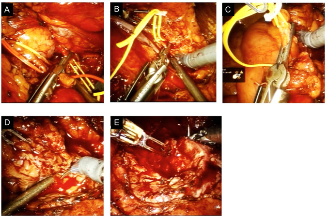
A 66-year-old male was referred to our hospital with right incidental renal mass. A CT scan revealed 26 mm renal mass (cT1aN0M0) in the posterior of the right lower pole of the horseshoe kidney (Fig. 1a) and two right renal arteries (Fig. 1b). Port placement was lower than the standard RAPN because the tumor on the lower pole was located at the aortic bifurcation level (Fig. 2). By conventional laparoscopic procedure without robotic assistance, the two right renal arteries were exposed and secured by vascular tapes (Fig. 3a, b). The right ureter and an unexpected artery were identified and secured by vascular tapes (Fig. 3c). After the right lobe of the horseshoe kidney was mobilized and dissected, the robot was docked and the tumor was exposed. After the excision margin was marked by coagulation, the three arteries were clamped individually (Fig. 3a–c) and the tumor was successfully removed with a 0.5 cm rim of normal parenchyma (Fig. 3d). The inner medullary hemostatic sutures were performed by a running 3-0 V-Loc (Fig. 3e), followed by early unclamping and additional hemostatic sutures.
Fig. 1.
Tumor and arteries finding in computed tomography. A CT scan revealed 26 mm renal mass (cT1aN0M0) in the posterior of the right lower pole of the horseshoe kidney (a) and two right renal arteries (b)
Fig. 2.

Port placement of a hybrid laparoscopic (conventional and robot-assisted) partial nephrectomy
Fig. 3.
The view of surgical procedure in the present patient. The cranial artery from the abdominal aorta was clamped (a). The caudal artery from the abdominal aorta was clamped (b). The incidental artery from the right common iliac artery was clamped (c). The tumor was resected precisely (d). The renal parenchyma was sutured by 3-0 V-Loc running fashion (e)
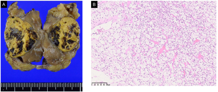
Total operative time was 290 min and warm ischemia time was 12 min. The postoperative course was uneventful. Pathological diagnosis was T1a, grade 2, clear cell renal carcinoma with a negative surgical margin (Fig. 4a, b).
Fig. 4.
Gross and microscopic features of renal cell carcinoma. The tumors were well-circumscribed, yellow semi-translucent in appearance and surrounded by a pseudo capsule; a formalin-fixed appearance. b The neoplasm displayed an admixture of epithelial cells with clear cytoplasm, trabecular pattern, central nucleus, and delicate branching vasculatures
Discussion
We paid attention to the following two major points to cope with this rare situation. Firstly, the horseshoe kidney should be dissected extensively to handle its poor mobility [1]. Second, to find and to secure an unexpected vasculature which had not been pointed out by preoperative evaluations [3].
To achieve the above objectives, we planned conventional and robot-assisted laparoscopic procedures as a hybrid technique [4]. At the beginning of surgery, ports were placed lower than standard RAPN and started dissected by the conventional laparoscopic approach. The right-lobe parenchyma was dissected appropriately from the cranial top to the caudal bottom. The conventional laparoscopic technique can allow for enough dissection because it could afford both easy exchanging instruments and access to all ports. Besides, a high variety of vasculature in horseshoe kidneys is another obstacle to achieve the hemostatic field at the time of tumor resection [5]. Because of fused dysgenesis of a kidney, horseshoe kidney is supplied by multiple unique arteries which originated not only from abdominal aorta but also mesenteric artery and iliac artery. Three-dimensional (3D) CT is useful for identifying variety vasculatures. However, undetectable by 3DCT but non-negligible artery could emerge just at the surgery in our case. A laparoscopic approach is also able to access the majority of variations by taking advantage of its flexibility in terms of instruments and ports [4].
With respect to RAPN, it has been accepted as a standard procedure in the management of small renal mass because it makes intracorporeal tumor resection and suture easier than the conventional laparoscopic approach. Reported advantages compared to conventional laparoscopic include decreased estimated blood loss, decreased incidence of perioperative adverse events, and decreased warm ischemic time [6]. Robotic surgery is especially helpful for finer movements required in delicate procedures such as resection of the tumor or suturing of renal parenchyma and helps address many of the shortcomings associated with laparoscopic partial nephrectomy, including two-dimensional visualization, poor surgeon ergonomics, and decreased dexterity. Disadvantages to a RAPN include difficulties with a limited range of motion of the robotic arm. In our experience, we were convinced that the techniques of laparoscopic and robotic surgery complement each other.
Although only two cases of RAPN for renal cell carcinoma with the horseshoe kidney were reported [7, 8], this did not indicate its difficulty but just rarity. We could make use of each superiority of both conventional and robot-assisted laparoscopy and showed the usefulness and feasibility of the hybrid technique in managing such complex urogenital anomaly with renal cell carcinoma.
Renal cell carcinoma in a horseshoe kidney is a rare entity and minimally invasive surgery for that can be challenging because of its poor mobility and various vasculatures. RAPN in combination with conventional laparoscopic technique, which endows sufficient dissection and identification of unexpected vasculatures, is feasible and may be a better option in the treatment of a small tumor in the horseshoe kidney.
Funding
This work was supported by Grants-in-Aid for Scientific Research, Japan (Grant no.: 17K11121).
Compliance with ethical standards
Conflict of interest
No competing financial interests exist for Kazuyuki Numakura, Yumina Muto, Ryohei Yamamoto, Atsushi Koizumi, Taketoshi Nara, Sohei Kanda, Mitsuru Saito, Shintaro Narita, Takamitsu Inoue, or Tomonori Habuchi.
Ethical approval and informed consent
In Akita University hospital, we announce that clinical studies are performed in this hospital and that their results are published. We explained him and got the written informed consent in accordance with the Declaration of Helsinki.
Footnotes
Publisher's Note
Springer Nature remains neutral with regard to jurisdictional claims in published maps and institutional affiliations.
Kazuyuki Numakura and Yumina Muto contributed equally to this work.
References
- 1.Ohtake S, Kawahara T, Noguchi G, et al. Renal cell carcinoma in a horseshoe kidney treated with laparoscopic partial nephrectomy. Case Rep Oncol Med. 2018;2018:7135180 . doi: 10.1155/2018/7135180. [DOI] [PMC free article] [PubMed] [Google Scholar]
- 2.Raman A, Kuusk T, Hyde ER, Berger LU, Bex A, Mumtaz F. Robotic-assisted laparoscopic partial nephrectomy in a horseshoe kidney. A case report and review of the literature. Urology. 2018;114:e3–e5 . doi: 10.1016/j.urology.2017.12.003. [DOI] [PubMed] [Google Scholar]
- 3.Nikoleishvili D, Koberidze G. Retroperitoneoscopic partial nephrectomy for a horseshoe kidney tumor. Urol Case Rep. 2017;13:31–33. doi: 10.1016/j.eucr.2017.03.013. [DOI] [PMC free article] [PubMed] [Google Scholar]
- 4.Kim H, Kim JR, Han Y, Kwon W, Kim SW, Jang JY (2017) Early experience of laparoscopic and robotic hybrid pancreaticoduodenectomy. Int J Med Robot 13(3). [Epub 2017/03/10] [DOI] [PubMed]
- 5.Benidir T, Coelho de Castilho TJ, Cherubini GR, de Almeida LM. Laparoscopic partial nephrectomy for renal cell carcinoma in a horseshoe kidney. Can Urol Assoc J. 2014;8(11–12):E918–E920. doi: 10.5489/cuaj.2289. [DOI] [PMC free article] [PubMed] [Google Scholar]
- 6.Novara G, La Falce S, Kungulli A, Gandaglia G, Ficarra V, Mottrie A. Robot-assisted partial nephrectomy. Int J Surg. 2016;36(Pt C):554–559. doi: 10.1016/j.ijsu.2016.05.073. [DOI] [PubMed] [Google Scholar]
- 7.Rogers CG, Linehan WM, Pinto PA. Robotic nephrectomy for kidney cancer in a horseshoe kidney with renal vein tumor thrombus: novel technique for thrombectomy. J Endourol. 2008;22(8):1561–1563. doi: 10.1089/end.2008.0043. [DOI] [PMC free article] [PubMed] [Google Scholar]
- 8.Kumar S, Singh S, Jain S, Bora GS, Singh SK. Robot-assisted heminephrectomy for chromophobe renal cell carcinoma in L-shaped fused crossed ectopia: surgical challenge. Korean J Urol. 2015;56(10):729–732. doi: 10.4111/kju.2015.56.10.729. [DOI] [PMC free article] [PubMed] [Google Scholar]


