ABSTRACT
Aim
The study was conducted to evaluate and compare the effect of various placement techniques of composite restoration on microleakage.
Materials and methods
Forty extracted premolars were selected and a rectangular-shaped cavity was prepared on the buccal surface of cervical third of each tooth. Thereafter, teeth were randomly divided into four groups equally and were restored with the composite restorative material with different placement techniques, i.e., bulk placement technique, horizontal incremental technique, split incremental technique, and newly introduced Mat incremental technique. Samples were thermocycled and immersed in methylene blue dye for 24 hours. The samples were then sectioned and evaluated under a stereomicroscope for microleakage.
Results
Microleakage was present least in the Mat incremental group and maximum in the bulk placement group while intercomparison revealed statistically significant difference between all the groups except for split incremental and Mat incremental groups.
Conclusion
The recently introduced Mat incremental placement technique showed least microleakage when compared to conventional techniques.
How to cite this article
Somani R, Som NK, Jaidka S, et al. Comparative Evaluation of Microleakage in Various Placement Techniques of Composite Restoration: An In Vitro Study. Int J Clin Pediatr Dent 2020;13(3):264–268.
Keywords: Composite, C-factor, Microleakage, Polymerization shrinkage
INTRODUCTION
Esthetics has been a prime segment of dentistry and in esthetic dentistry, the composite is the material of choice. Although there are many advantages of composite restorations, but many limitations are also associated with it. Resin composites show volumetric contractions, which ranges from 2.6 to 7.1%, leading to shrinkage stress generation at the composite-tooth interface. Due to these stresses, composite may pull away from the cavity margins, which may lead to adhesive failure and marginal gap formation. These gaps may get filled by oral fluids containing bacteria, leading to microleakage that may cause marginal discoloration, postoperative sensitivity, secondary caries, and pulp damage.
Magnitude of these stresses depend on various factors, such as the resin modules of elasticity, the rate of polymerization, the techniques of restoration, and cavity configuration factor (C-factor).1
Various techniques of restoration have been designed to reduce the effects of polymerization shrinkage, improve marginal adaptation, and seal to enhance and provide the clinician with maximum benefit for their application. If we talk about placement techniques of resin composites, studies have shown that that the incremental technique tends to improve marginal adaptation by resisting resin composite shrinkage stress.2
Recently a new technique, the Mat incremental, has been proposed in the Department of Pedodontics and Preventive Dentistry, Divya Jyoti College of Dental Sciences and Research, Modinagar. In the Mat incremental technique, the horizontal increment placed is further split to reduce the “C” factor, thereby reducing the polymerization shrinkage stress.
Therefore, the purpose of this study is to analyze the effect of various placement techniques in the microleakage of composite restoration.
MATERIALS AND METHODS
Forty human premolar teeth extracted for orthodontic or periodontal reasons with intact buccal/lingual surfaces were used for the study. Debris was removed, teeth were cleaned using ultrasonic scaler, autoclaved, and stored in distilled water at room temperature. As per recommendations of the Occupational Safety and Health Administration (OSHA) guidelines for infection-control healthcare settings, the teeth were used within 3 months of extraction.
Preparation of Samples
Class V cavities were prepared for the entire sample of 40 human premolars. The size of the cavity was (5 mm wide × 3 mm high × 2 mm deep) 1 mm below the cementoenamel junction (CEJ) and was prepared on the buccal surfaces of teeth.
Division of Samples
The collected 40 samples were randomly divided into the following four groups and color-coded using nail paint accordingly. Group I—(white) restoration of the samples done using the bulk fill technique (n = 10), group II—(purple) restoration of the samples done using the incremental filling technique (n = 10), group III—(green) restoration of the samples done using the split horizontal incremental technique (n = 10), group IV—(pink) restoration of the samples done using the Mat horizontal incremental technique (n = 10).
All box cavities were acid-etched with 37% phosphoric acid. The bonding agent was applied using an applicator tip on the floor and walls of all the cavities and were light-cured for 20 seconds according to manufacturer's instructions. All the teeth were then restored with Spectrum Composite, Dentsply, with varying placement techniques according to respective groups.
Group I (Bulk Placement Technique)
Samples were restored with Spectrum Restorative Composite (Dentsply, Germany). The composite restorative material was placed to fill the entire cavity (bulk fill) and was light-cured for 40 seconds according to the manufacturer's instruction (Fig. 1).
Fig. 1.
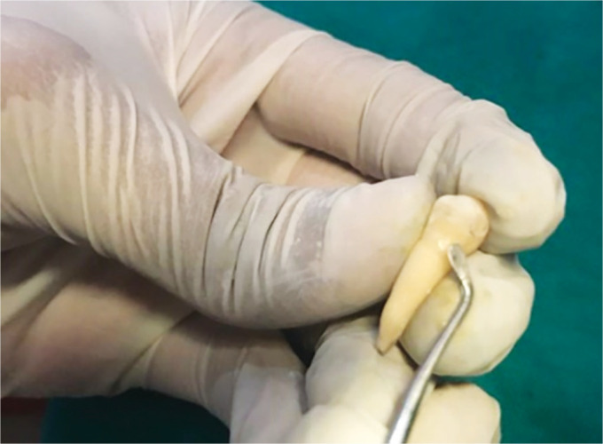
Bulk placement technique
Group II (Horizontal Incremental Technique)
The composite restorative material was placed to fill half of the cavity depth and was light-cured for 40 seconds; second increment was placed to fill the cavity up to the cavosurface margin of the cavity and was light-cured for 40 seconds (Fig. 2).
Fig. 2.
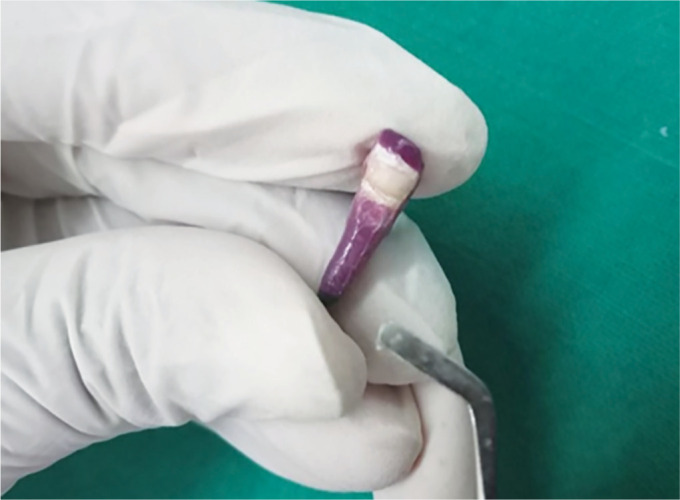
Horizontal incremental technique
Group III (Split Incremental Technique)
The composite restorative material was placed to fill half of the cavity depth and two diagonal cuts were made to split the first uncured increment into four triangular-shaped portions using a blunt probe up to the entire depth of the cavity and were light-cured for 40 seconds. Then second increment was placed into the diagonal cuts and light-cured followed by the third increment that was placed horizontally up to the cavosurface margin to fill the rest of the cavity and light-cured for 40 seconds (Fig. 3).
Fig. 3.
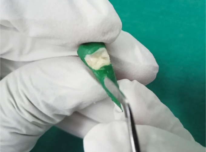
Split incremental technique
Group IV (Mat Incremental Technique)
The composite restorative material was placed to fill half of the cavity depth, and one mesiodistal and two occlusal-gingival cuts were made to split the first uncured increment into six square-shaped portions using a blunt probe up to the entire depth of the cavity and were light-cured for 40 seconds. Then the horizontal and vertical cuts in the form of Mat were filled with restorative composite and light-cured followed by the third increment that was placed horizontally up to cavosurface margin to fill the rest of the cavity and light-cured for 40 seconds (Fig. 4).
Fig. 4.
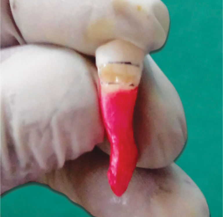
Mat incremental technique
Dye Immersion
After restoration of all the samples with composite (spectrum), they were stored separately in distilled water.
Thermocycling
The samples were thermocycled after which they were immersed in 2% methylene blue for 24 hours, and then were rinsed with water and air-dried. A diamond disc at slow speed in a micromotor straight hand piece was used to section the sample longitudinally in a buccolingual direction.
Microleakage Evaluation
All the groups were assessed under a stereomicroscope for microleakage. Data were collected, tabulated, and sent for statistical analysis.
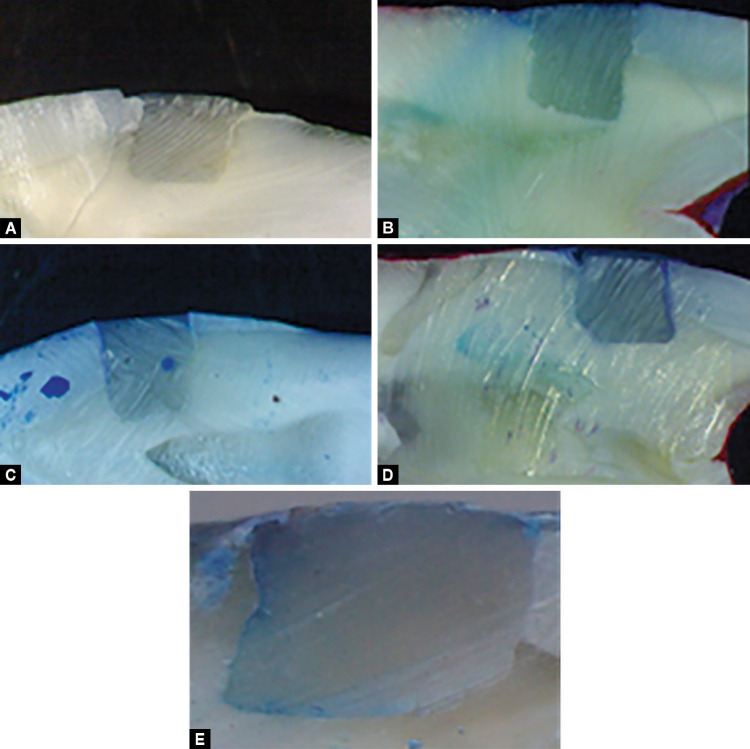
The scoring criteria for the microleakage assessment were followed according to Vinay and Shivanna (2010).
0 = No dye penetration
1 = Dye penetration up to one-third cavity depth
2 = Dye penetration up to two-third cavity depth
3 = Dye penetration up to full depth of cavity
4 = Dye penetration onto axial wall of cavity (Fig. 5)
Figs 5A to E.
(A) Score 0; (B) Score 1; (C) Score 2; (D) Score 3; (E) Score 4
RESULTS
The data were statistically analyzed using the one-way ANOVA and Tukey's test and the following results were obtained:
The mean value of the microleakage was 2.6 for group I (bulk fill technique), 1.4 for group II (horizontal incremental technique), 0.8 for group III, and 0.5 for group IV. It was noted that Group I (bulk fill technique) has highest mean value of 2.6, while group IV (Mat incremental technique) has lowest mean value of microleakage, i.e., 0.5 (Table 1).
Table 1.
Mean values of microleakage
| Groups | N (sample size) | Mean | Standard deviation | Minimum | Maximum |
|---|---|---|---|---|---|
| Group I (bulk placement technique) | 10 | 2.6 | 1.17378779 | 1 | 4 |
| Group II (horizontal incremental technique) | 10 | 1.4 | 0.84327404 | 0 | 3 |
| Group III (split incremental technique) | 10 | 0.8 | 0.63245553 | 0 | 2 |
| Group IV (Mat incremental technique) | 10 | 0.5 | 0.52704627 | 0 | 1 |
Intercomparison of various groups was done using Tukey's (two-sided post hoc) tests. All intercomparisons between the mean microleakage values of various groups were found to be highly significant, except between group III (split incremental technique) and IV (Mat incremental technique) where the results were non-significant at p value < 0.05 (Table 2).
Table 2.
Intercomparison between various groups
| (I) group | (J) group | Mean difference (I–J) | Std. error | Sig. | 95% confidence interval | |
|---|---|---|---|---|---|---|
| Lower bound | Upper bound | |||||
| I | II | 1.677 | 0.391 | 0.03* | 0.62 | 2.73 |
| III | 1.055 | 0.380 | 0.04* | 0.03 | 2.08 | |
| IV | 1.955 | 0.380 | 0.000* | 0.93 | 2.98 | |
| II | I | −1.677 | 0.391 | 0.002* | −2.73 | −0.62 |
| III | −0.622 | 0.399 | 0.004* | −1.70 | 0.45 | |
| IV | 0.278 | 0.399 | 0.007* | −0.80 | 1.35 | |
| III | I | −1.055 | 0.380 | 0.04* | −2.08 | −0.03 |
| II | 0.622 | 0.399 | 0.002* | −0.45 | 1.70 | |
| IV | 0.900 | 0.389 | 0.113** | −0.15 | 1.95 | |
| IV | I | −1.955 | 0.380 | 0.000* | −2.98 | −0.93 |
| II | −0.278 | 0.399 | 0.005* | −1.35 | 0.80 | |
| III | −0.900 | 0.389 | 0.113** | −1.95 | 0.15 | |
DISCUSSION
Various methods are there to decrease polymerization shrinkage such as reducing filler content of the composite material, adopting layering placement techniques, and decreasing the configuration factor (C-factor).
The ratio of bonded to unbonded surfaces of composite restoration is known as C-factor. If number of bonded surface is increased, it will lead to higher C-factor and greater contraction stress on adhesive bond, which results in potential for bond disruption from polymerization effects. On the other hand, if number of unbounded surface is increased, it leads to a low C-factor, which minimizes the polymerization shrinkage.3
The mean values of the microleakage for group I (bulk fill technique), group II (horizontal incremental technique), group III (split incremental technique), and group IV (Mat horizontal incremental technique) were 2.6, 1.4, 0.8, and 0.5, respectively. Result showed that group I, bulk placement technique, showed highest microleakage followed by the horizontal incremental technique, the split horizontal incremental technique, and the Mat incremental technique.
In the present study, the bulk fill technique showed highest microleakage due to the increased polymerization shrinkage, which results due to the great volume of composite and decreased effectiveness of polymerization at deeper portions of the composite. When single increment is used, the polymerization of resin composite would be within five bonding surfaces while at the upper surface, free shrinkage would be there producing a very high level of stress between the bonded surfaces. In this technique, high internal stress is generated in the material and loss of marginal integrity occurs as the larger volume of the composite is polymerized and there is decrease in effectiveness of polymerization at deeper portion of the composite, which results in higher polymerization shrinkage. Therefore, studies show that this technique is less advantageous in terms of polymerization shrinkage. Similar study was also done by Katona on the comparison of the composite restoration technique who concluded that the bulk fill technique appeared to be better as it requires less time but the bulk placement technique may cause deterioration of shape and esthetics of restoration.4
The horizontal incremental technique showed significantly less microleakage than the bulk fill technique. This could be due to the reduced volume of the resin in each increment; thus, the stress generated on the cavity walls is much less and smaller. Increments of composite have shown more uniform and efficient polymerization throughout the entire thickness because of the less bonded to unbonded surface ratio. This incremental technique influences the configuration factor and the extent of the polymerization shrinkage is much less as compared to the bulk fill technique. It is because when composite is placed in increments, polymerization shrinkage occurs in each increment. Less tensile force is created by shrinkage of the thin layer of composite than created by contraction of a composite bulk that fills the whole cavity. The C-factor is also significantly lowered, which further decreases the stress associated with the polymerization shrinkage. Similar study was done by Idriss on the shrinkage stress generated during resin composite application in which it was concluded that the technique of placement and choice of material are important determining factors in microleakage.5
The split horizontal incremental technique showed lower microleakage than the bulk fill technique and the horizontal incremental technique as the diagonal cutting of the flat composite increment into two triangular composite portions created new unbonded composite surfaces, which act as the additional reservoir for flow or plastic deformation during light polymerization. This eventually preserves the interfacial bond and marginal integrity of the restoration. Similar study was done by Damineni et al. on the placement techniques efficacy in marginal sealing of extended class V cavities and it was found that the incremental technique show less microleakage than the bulk placement.6
Polymerization shrinkage is further relieved by splitting the large horizontal increment in the occlusal and proximal cavities into smaller triangular flat portions prior to photocuring. The smaller increment size and the lower C-factor minimize most of the shrinkage stresses by means of the flow surfaces, rather than the bonded interfaces that would increase the cuspal deformation. Al-Zahawi et al. stated the sequence of composite filling in two diagonal cuts would prevent the split increment portions of the composite resin from contacting two opposing cavity walls simultaneously, which will cause the negative effects of polymerization shrinkage stresses on the cavity walls and adhesive interface to be minimized and permit the shrinkage stress to be relieved by free flow of the composite surface at the diagonal cuts, thus reducing the adverse effects of generated polymerization shrinkage stresses.7
The Mat horizontal incremental technique showed the lowest microleakage as the Mat cuts composite increment into six square-shaped composite portions creating new unbonded composite surfaces, thereby the C-factor is reduced further resulting in less polymerization shrinkage due to less stress developed at the bonded cavity walls and margins. The Mat horizontal incremental technique can be used for restoring class V cavities with composite resin and especially when gingival margins extend beyond the CEJ. Such restoration would help in preserving the gingival marginal seal and microleakage of the composite restoration. It was observed that the splitting of the restoration showed significantly lower value of microleakage due to the fact that in this technique the less polymerization shrinkage stress developed at the bonded cavity walls and margins, as the restricted shrinkage is converted to unrestricted shrinkage with subsequent preservation of the gingival margin integrity.
All intergroup comparisons were found to be highly significant except when split incremental and Mat incremental techniques were compared.
Small portions of increments with a low C-factor would minimize the shrinkage stress by the free composite surface flowing at the cuts and not at the bonded interfaces, reducing the adverse effects of the polymerization shrinkage stresses. In addition, it minimizes the detrimental effects of the polymerization shrinkage stress at the adhesive interface and cavity walls by reducing the c-factor ratio. The proposed technique would benefit less experienced general dentist and those who work in busy dental offices at government hospitals, as it would encourage them to satisfy the esthetic dental needs of patients and provide high-quality posterior composite restorations.
Therefore, from the above study it is found that the Mat horizontal incremental technique and the split incremental technique have comparatively lower microleakage among all groups. As in both the techniques primary increment has been splitted into smaller portions. Hence, it can be concluded that the split incremental technique and the Mat horizontal incremental technique could be effective placement techniques for composite restoration. Further clinical trials are required to authenticate the results.
CONCLUSION
Within the limitations of this in vitro study, we can conclude the following:
The microleakage was seen in all the placement techniques, i.e., bulk placement technique, horizontal incremental placement technique, split incremental technique, and Mat horizontal placement technique.
The mean microleakage of the bulk placement technique was statistically higher than all other placement techniques.
The mean microleakage of the Mat incremental technique was statistically lower than horizontal incremental and bulk placement techniques.
The mean microleakage of the Mat incremental technique was comparable with that of the split horizontal incremental technique.
Thus, the Mat incremental placement technique is recommended to be used while doing composite restoration to improve the longevity of restoration.
Footnotes
Source of support: Nil
Conflict of interest: None
REFERENCES
- 1.Terry D, Karl L. Composite resin restoration. A simplified approach. Priv Dent. 2008;2(1):24–38. [Google Scholar]
- 2.Narene AK, Veniashok B, Subhiya A, et al. Polymerisation shrinkage in resin composites - A review. Middle East J Scient Res. 2014;21(1):107–112. [Google Scholar]
- 3.Kumar JS, Jailakshmi S. Bond failure and its prevention in composite restoration – A review. J Pharmaceut Sci Res. 2016;8(7):627–631. [Google Scholar]
- 4.Katona A, Barrack I. Comparison of composite restoration techniques. Interdisciplin Descrip Comp Syst. 2016;14(1):101–115. doi: 10.7906/indecs.14.1.10. DOI: [DOI] [Google Scholar]
- 5.Idriss S, Abduljabbar T, Habib C, et al. Factors associated with microleakage in class II resin composite restorations. Oper Dent. 2007;32(1):60–66. doi: 10.2341/06-16. DOI: [DOI] [PubMed] [Google Scholar]
- 6.Damineni R, Abhilash,, Shailendra M, et al. Evaluation of polymerization shrinkage of light cured composite resins. Advance Hum Biol. 2014;4(3):26–30. [Google Scholar]
- 7.Al-Zahawi AR, Abdul-Rahman MS, Ahmed SM. Effect of two different composites on gingival microleakage of class ii restoration using four different placement techniques (an in vitro study). Int J Recent Adv Multidisciplin Res. 2015;2(9):727–731. [Google Scholar]


