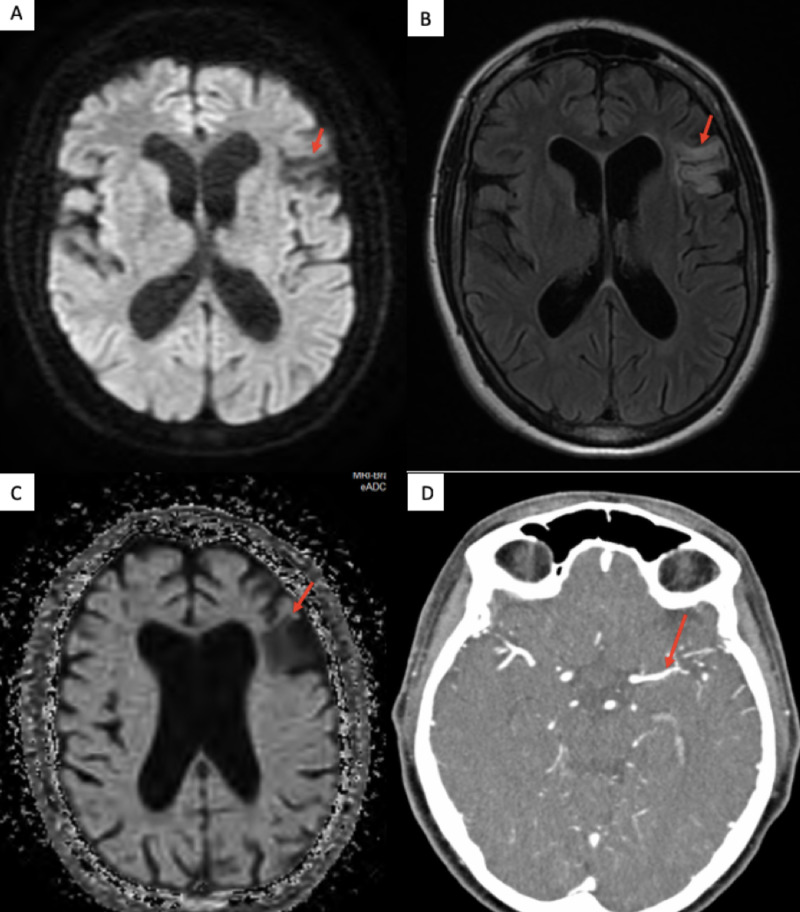
Figure 2. MRI of the brain showing (A) a hypo-intense signal in the left frontal lobe on diffusion-weighted imaging sequence, (B) a hyper-intense signal in the left frontal lobe on T2 flair sequence, (C) a hypo-intense signal on apparent diffusion coefficient sequence, and (D) a mild narrowing left MCA on CT angiography.
Abbreviations: MRI, magnetic resonance imaging; MCA, middle cerebral artery; CT, computed tomography

