

Abstract
Objective
To report 3 patients infected with severe acute respiratory syndrome coronavirus 2 (SARS-CoV-2) who developed generalized myoclonus.
Methods
Patient data were obtained from medical records from the University Hospital “12 de Octubre,” Madrid, Spain.
Results
Three patients (2 men and 1 woman, aged 63–88 years) presented with mild hypersomnia and generalized myoclonus following the onset of the so-called inflammatory phase of coronavirus disease 2019 (COVID-19). All of them had presented previously with anosmia. Myoclonus was generalized with both positive and negative jerks, predominantly involving the facial, trapezius, sternocleidomastoid, and upper extremities muscles. These myoclonic jerks occurred spontaneously and were extremely sensitive to multisensory stimuli (auditive and tactile) or voluntary movements, with an exaggerated startle response. Other causes of myoclonus were ruled out, and none of the patients had undergone respiratory arrest or significant prolonged hypoxia. All of them improved, at least partially, with immunotherapy.
Conclusions
Our 3 cases highlight the occurrence of myoclonus during the COVID-19 pandemic as a post- or para-infectious immune-mediated disorder. However, we cannot rule out that SARS-CoV-2 may spread transneuronally to first- and second-order structures connected with the olfactory bulb. Further investigation is required to clarify the full clinical spectrum of neurologic symptoms and optimal treatment.
The new severe acute respiratory syndrome coronavirus 2 (SARS-CoV-2) has spread rapidly worldwide. The spectrum of coronavirus disease 2019 (COVID-19) ranges from asymptomatic infection to severe respiratory failure, with fever, cough, and fatigue being the most common clinical symptoms.1 Other features, including neurologic manifestations (e.g., myalgia, headache, anosmia, and ageusia), have also been reported.1,2 Neurologists are now starting to see more severe neurologic manifestations associated with COVID-19,3–6 including acute cerebrovascular diseases, skeletal muscle injury, encephalopathy, prominent agitation and confusion, corticospinal tract signs, Guillain-Barré syndrome, Miller Fisher syndrome, and polyneuritis cranialis. The exact pathogenesis of COVID-19-induced neurologic manifestations remains largely unknown. Myoclonus has been reported in other viral infections,7 but not yet associated with SARS-CoV-2 infection.
We report 3 patients who developed generalized myoclonus as a predominant phenomenon during SARS-CoV-2 infection.
Methods
Data were obtained from patients admitted to University Hospital “12 de Octubre,” Madrid, Spain.
Standard protocol approvals, registrations, and patient consents
Written informed consent was obtained from the patients (consent for research).
Data availability
The data supporting the findings of this study are available within the article.
Results
Patient 1
A 63-year-old man was admitted to the emergency department because of a 7-day history of fever and anosmia. On day 8 after onset of symptoms, he noticed shortness of breath. One day later, he developed sudden onset of jerky movements in his face and limbs, which prevented him from eating and speaking properly. His medical history was remarkable for generalized anxiety disorder.
Except for a basal oxygen saturation of 93%, his vital signs were normal. A chest X-ray revealed bilateral diffuse patchy interstitial infiltrates consistent with COVID-19. The initial blood workup revealed elevated C-reactive protein, elevated D-dimers, and lymphopenia. He was diagnosed with COVID-19. Nasopharyngeal swab test for SARS-CoV-2 by qualitative real-time reverse-transcriptase polymerase chain reaction (rRT-PCR) assay could not be performed due to its scarcity in Spain at that time.
By day 10 after onset of symptoms, myoclonus worsened, preventing the patient from moving, speaking, or swallowing. He was intubated and admitted to the intensive care unit owing to myoclonic storm. The neurologic examination at that time revealed mild somnolence with otherwise normal higher mental functions. He had no opsoclonus or other abnormalities in the cranial nerves. He showed generalized myoclonus, with both positive and negative jerks. They predominantly involved the facial, trapezius, sternocleidomastoid, and upper extremities muscles, worsened with voluntary movements, and augmented markedly with auditory and tactile stimuli. An exaggerated startle response was noticed (video 1). There was no motor or sensory deficit. Tone, deep tendon reflexes, and plantar responses were normal. Gait was greatly hindered by the movements.
Video shows generalized myoclonus with both positive and negative jerks, predominantly involving the facial, trapezius, sternocleidomastoid, and upper extremities muscles. The myoclonic jerks were extremely sensitive to multisensory stimuli or voluntary movements, with an exaggerated startle response. Methylprednisolone seemed to improve slightly myoclonus. However, myoclonic jerks worsened again, and the patient had to be reintubated and started on plasmapheresis showing clinical improvement.Download Supplementary Video 1 (19.8MB, mov) via http://dx.doi.org/10.1212/009829_Video_1
Myoclonus only disappeared with deep propofol sedation but tended to reappear as it was withdrawn. Symptomatic treatment with levetiracetam, valproic acid, and clonazepam were initiated with scarce improvement. An extensive laboratory workup, including CSF analysis and cranial MRI, revealed no abnormalities. EEG showed no epileptiform discharges during myoclonus. The main findings of complementary tests are shown in the table.
Table.
Complementary tests


We assumed the myoclonus to be an immune-mediated manifestation of the inflammatory phase of COVID-19. We started immunotherapy (methylprednisolone 1,000 mg/24 hours for 5 days), and the myoclonus seemed to improve slightly (video 1). The patient then could be extubated, without supplemental oxygen needs. Two days later, the myoclonus worsened again, and the patient had to be reintubated and started on plasmapheresis. The patient received a total of 5 daily plasma exchange treatments, showing clinical improvement after completion of therapy (video 1).
Patient 2
An 88-year-old woman was admitted to the emergency department because of a 2-day history of anosmia, fever, and shortness of breath. Chest-X-ray revealed bilateral pneumonia. Her medical history was remarkable for arterial hypertension, hypothyroidism, a nonfunctioning pituitary adenoma, and mild cognitive decline with no history of abnormal movement disorders. The patient's nasopharyngeal swab test for SARS-CoV-2 by qualitative rRT-PCR assay was positive.
By the third week since the onset of symptoms, the patient started having mild hypersomnia and similar jerking movements to patient 1, but milder. Brain CT, EEG, and extensive laboratory workup were unrevealing (table). The myoclonus disappeared with methylprednisolone pulses (250 mg/24 hours for 3 days).
Patient 3
A 76-year-old man with no medical comorbidities had fever, malaise, cough, anosmia, ageusia, and myalgia for 10 days. On day 11 from the onset of symptoms, he developed jerky movements in his face and limbs, mild somnolence, and dyspnea. Myoclonic jerks were milder, but similar to those in patient 1. He was diagnosed with SARS-CoV-2 pneumonia based on the clinical features, blood analysis, radiologic picture, and epidemiologic context. Nasopharyngeal swab test for SARS-CoV-2 by rRT-PCR assay was not performed due to its scarcity in Spain at that time. Brain MRI and EEG were unrevealing. The patient experienced no benefit from clonazepam and levetiracetam. He received methylprednisolone pulses (250 mg/24 hours for 3 days) to treat COVID-19 pneumonia. Two weeks later, the patient started experiencing a spontaneous progressive improvement.
Other patients
We have encountered other patients with COVID-19 who also presented with myoclonus. Regarding these other cases, myoclonus could also be explained by drugs, metabolic disturbances, or severe hypoxia. However, as a preliminary impression, there seems to be an increasing, unexpected amount of severe myoclonus among some of the hundreds of patients admitted to our hospital with COVID-19 infection.
Discussion
To our knowledge, these are the first reported cases of generalized myoclonus in COVID-19. After a thorough study, no other potential causes for this phenomenon were identified. No serious metabolic disturbances were registered, and none of the patients underwent severe hypoxia. No causative drugs were administered, and we did not find autoantibodies that could account for a known immune-mediated disorder. CSF analysis was performed in patient 1 and revealed no relevant abnormalities, and neuroimaging studies were normal in all of the patients.
Jerking movements of our patients could be attributable to brainstem myoclonus, a type of myoclonus originating from the lower brainstem, which is characterized by initial muscle activation in muscles innervated in the lower brainstem followed by a sequence of both higher brainstem innervated muscles and muscles innervated further down the spinal cord.8 Worsening of myoclonus with movements, tactile stimuli, and specially the exaggerated startle response also support this hypothesis.8 EEG studies performed ruled out an epileptic myoclonus, but further neurophysiologic (i.e., EMG) tests could not be performed due to logistic limitations in the setting of the current pandemic.
Regarding myoclonus pathophysiology in COVID-19, we raise 2 possibilities. First, a post- or para-infectious immune-mediated mechanism is possible. Consistent with this hypothesis, postinfectious myoclonus presenting shortly after the start of infections caused by other viruses (occasionally in the form of opsoclonus-myoclonus,9 but also as isolated myoclonus)10 has been reported. Furthermore, our patients started presenting myoclonus following the onset of the so-called inflammatory phase of COVID-19.11 Second, several reports have proved the neuroinvasive potential of SARS-CoV in humans.12–15 Experimental studies have demonstrated that SARS-COV,16 as other respiratory viruses, such as H1N1 influenza virus,17,18 when inoculated intranasally, could spread transneuronally to first- and second-order structures connected with the olfactory bulb. Certain neuronal populations with sleep–wake regulatory functions in the hypothalamus as well as noradrenergic neurons in the locus coeruleus and serotonergic neurons in the dorsal raphe were infected in those models in the absence of encephalitis.16–18 As our 3 patients consecutively presented hyposmia, hypersomnia, and generalized myoclonus, we cautiously hypothesize that SARS-CoV-2 could affect these structures (olfactory bulb and brainstem) sequentially. Furthermore, normal brain MRI and lack of pleocytosis in the CSF cannot rule out this mechanism, because no histologic lesions were observed surrounding infected neurons in the aforementioned studies.16–18
All our patients received a short cycle of high-dose methylprednisolone and one of them received plasma exchange sessions. As not only neuronal death has been described in animal models,16,17 but also noncytolytic effects on neurons,18 whether clinical improvement is related to the treatment administered or is related to a natural and self-limited evolution should be addressed in future studies.
We recognize that the main limitations of each of the cases was the absence of EMG and in 2 of them the absence of nasopharyngeal swab test for SARS-CoV-2 by rRT-PCR assay. The reason for this was the extreme circumstances in our hospitals at the peak of this pandemic.
We describe 3 patients with COVID-19 with generalized myoclonus who had good outcomes. The acute onset of myoclonus in the context of the inflammatory response to SARS-CoV-2 infection, together with the similarities with other viral-triggered myoclonus, and the improvement after immunotherapy leads us to hypothesize that this could have a post- or para-infectious immune-mediated mechanism. However, we cannot rule out that SARS-CoV-2 may spread transneuronally to first- and second-order structures connected with the olfactory bulb, which would be supported by the sequential appearance of hyposmia, hypersomnia, and generalized myoclonus. Further investigation is required to clarify the full clinical spectrum of neurologic symptoms and its optimal treatment.
Acknowledgment
Dr. Julián Benito-León is supported by the NIH, Bethesda, MD (National Institute of Neurological Disorders and Stroke #R01 NS39422), European Commission (grant ICT-2011-287739, NeuroTREMOR), the Ministry of Economy and Competitiveness (grant RTC-2015-3967-1, NetMD—platform for the tracking of movement disorder), and the Spanish Health Research Agency (grant FIS PI12/01602 and grant FIS PI16/00451).
Glossary
- COVID-19
coronavirus disease 2019
- rRT-PCR
real-time reverse-transcriptase polymerase chain reaction
- SARS-CoV-2
severe acute respiratory syndrome coronavirus 2
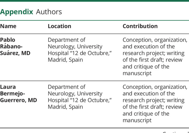
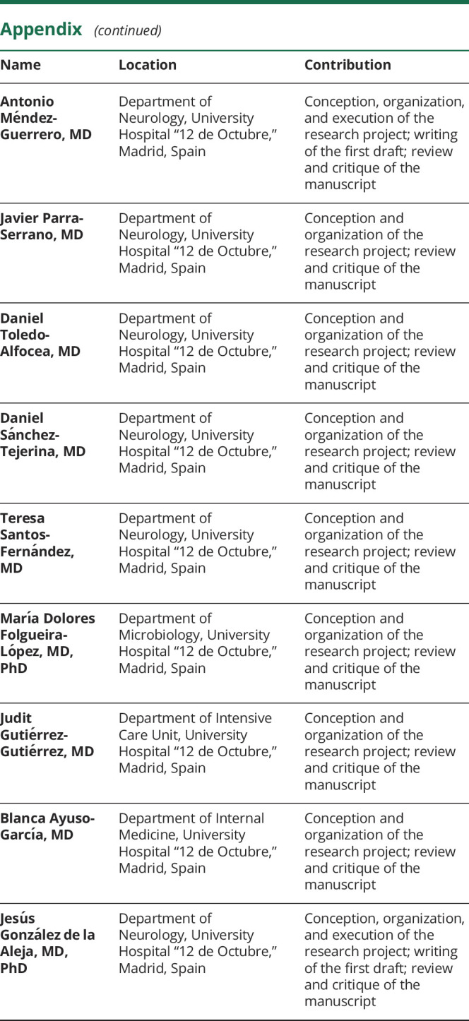
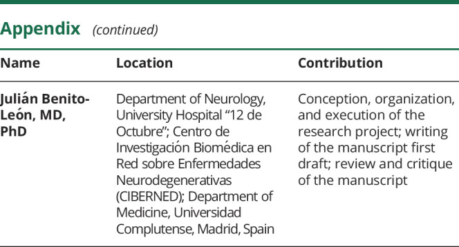
Appendix. Authors
Footnotes
COVID-19 Resources: NPub.org/COVID19
Study funding
This research was supported by FEDER funds.
Disclosure
The authors report no disclosures relevant to the manuscript. Go to Neurology.org/N for full disclosures.
References
- 1.Guan WJ, Ni ZY, Hu Y, et al. Clinical characteristics of coronavirus disease 2019 in China. N Engl J Med 2020;382:1708–1720. [DOI] [PMC free article] [PubMed] [Google Scholar]
- 2.Vaira LA, Salzano G, Deiana G, De Riu G. Anosmia and ageusia: common findings in COVID-19 patients. Laryngoscope Epub 2020 Apr 1. [DOI] [PMC free article] [PubMed]
- 3.Mao L, Jin H, Wang M, et al. Neurologic manifestations of hospitalized patients with coronavirus disease 2019 in Wuhan, China. JAMA Neurol Epub 2020 Apr 10. [DOI] [PMC free article] [PubMed]
- 4.Helms J, Kremer S, Merdji H, et al. Neurologic features in severe SARS-CoV-2 infection. N Engl J Med Epub 2020 Apr 15. [DOI] [PMC free article] [PubMed]
- 5.Toscano G, Palmerini F, Ravaglia S, et al. Guillain-Barre syndrome associated with SARS-CoV-2. N Engl J Med Epub 2020 Apr 17. [DOI] [PMC free article] [PubMed]
- 6.Gutierrez-Ortiz C, Mendez A, Rodrigo-Rey S, et al. Miller Fisher syndrome and polyneuritis cranialis in COVID-19. Neurology Epub 2020 Apr 17. [DOI] [PubMed]
- 7.Delucchi V, Pavlidis E, Piccolo B, Pisani F. Febrile and postinfectious myoclonus: case reports and review of the literature. Neuropediatrics 2015;46:26–32. [DOI] [PubMed] [Google Scholar]
- 8.Beudel M, Elting JWJ, Uyttenboogaart M, van den Broek MWC, Tijssen MAJ. Reticular myoclonus: it really comes from the brainstem! Mov Disord Clin Pract 2014;1:258–260. [DOI] [PMC free article] [PubMed] [Google Scholar]
- 9.Blackburn DJ, Forbes M, Unwin Z, Hoggard N, Hadjivassiliou M, Sarrigiannis PG. Exaggerated startle in post-infectious opsoclonus myoclonus syndrome. Clin Neurophysiol 2018;129:1372–1373. [DOI] [PubMed] [Google Scholar]
- 10.Bhatia K, Thompson PD, Marsden CD. “Isolated” postinfectious myoclonus. J Neurol Neurosurg Psychiatry 1992;55:1089–1091. [DOI] [PMC free article] [PubMed] [Google Scholar]
- 11.Zhou G, Chen S, Chen Z. Advances in COVID-19: the virus, the pathogenesis, and evidence-based control and therapeutic strategies. Front Med 2020;14:117–125. [DOI] [PMC free article] [PubMed] [Google Scholar]
- 12.Gu J, Gong E, Zhang B, et al. Multiple organ infection and the pathogenesis of SARS. J Exp Med 2005;202:415–424. [DOI] [PMC free article] [PubMed] [Google Scholar]
- 13.Xu J, Zhong S, Liu J, et al. Detection of severe acute respiratory syndrome coronavirus in the brain: potential role of the chemokine mig in pathogenesis. Clin Infect Dis 2005;41:1089–1096. [DOI] [PMC free article] [PubMed] [Google Scholar]
- 14.Ding Y, He L, Zhang Q, et al. Organ distribution of severe acute respiratory syndrome (SARS) associated coronavirus (SARS-CoV) in SARS patients: implications for pathogenesis and virus transmission pathways. J Pathol 2004;203:622–630. [DOI] [PMC free article] [PubMed] [Google Scholar]
- 15.Li YC, Bai WZ, Hashikawa T. The neuroinvasive potential of SARS-CoV2 may play a role in the respiratory failure of COVID-19 patients. J Med Virol Epub 2020 Feb 27. [DOI] [PMC free article] [PubMed]
- 16.Netland J, Meyerholz DK, Moore S, Cassell M, Perlman S. Severe acute respiratory syndrome coronavirus infection causes neuronal death in the absence of encephalitis in mice transgenic for human ACE2. J Virol 2008;82:7264–7275. [DOI] [PMC free article] [PubMed] [Google Scholar]
- 17.de Wit E, Siegers JY, Cronin JM, et al. 1918 H1N1 influenza virus replicates and induces proinflammatory cytokine responses in extrarespiratory tissues of ferrets. J Infect Dis 2018;217:1237–1246. [DOI] [PMC free article] [PubMed] [Google Scholar]
- 18.Tesoriero C, Codita A, Zhang MD, et al. H1N1 influenza virus induces narcolepsy-like sleep disruption and targets sleep-wake regulatory neurons in mice. Proc Natl Acad Sci USA 2016;113:E368–E377. [DOI] [PMC free article] [PubMed] [Google Scholar]
Associated Data
This section collects any data citations, data availability statements, or supplementary materials included in this article.
Supplementary Materials
Video shows generalized myoclonus with both positive and negative jerks, predominantly involving the facial, trapezius, sternocleidomastoid, and upper extremities muscles. The myoclonic jerks were extremely sensitive to multisensory stimuli or voluntary movements, with an exaggerated startle response. Methylprednisolone seemed to improve slightly myoclonus. However, myoclonic jerks worsened again, and the patient had to be reintubated and started on plasmapheresis showing clinical improvement.Download Supplementary Video 1 (19.8MB, mov) via http://dx.doi.org/10.1212/009829_Video_1
Data Availability Statement
The data supporting the findings of this study are available within the article.