

A 64-year-old man with cerebral autosomal dominant arteriopathy with subcortical infarcts and leukoencephalopathy (CADASIL) (heterozygous, c.544C>T; p.Arg182Cys) underwent an intracranial high-resolution black-blood protocoled vessel wall MRI (vwMRI). Intramural patchy gadolinium enhancement in the subcortical and leptomeningeal arteries and veins was noted, consistent with the histopathologic findings of CADASIL (figures 1 and 2).1,2 We hypothesize that vwMRI allowed for an in vivo view of the vasculopathy intrinsic to CADASIL. Pending investigation of larger cohorts, this imaging technique may provide a novel mechanism for understanding CADASIL progression and pathogenesis, as well as potentially serving as a biomarker in future disease modification trials and aiding in the differential diagnosis for interpreting clinicians.
Figure 1. Intracranial vessel wall MRI of cerebral autosomal dominant arteriopathy with subcortical infarcts and leukoencephalopathy.
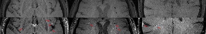
Top row showing T1 precontrast axial images compared with bottom row showing T1 postcontrast mural enhancement of the subcortical and leptomeningeal arteries. Note cortical venous mural enhancement in the bottom right pane. Arrows indicate intramural enhancement.
Figure 2. Baseline white matter burden.

Axial fluid-attenuated inversion recovery sequences show T2 subcortical white matter hyperintensities consistent with cerebral autosomal dominant arteriopathy with subcortical infarcts and leukoencephalopathy. Images provided for comparison.
Appendix. Authors
Study funding
No targeted funding reported.
Disclosure
The authors report no relevant disclosures. Go to Neurology.org/N for full disclosures.
References
- 1.Coupland K, Lendahl U, Karlström H. Role of NOTCH3 mutations in the cerebral small vessel disease cerebral autosomal dominant arteriopathy with subcortical infarcts and leukoencephalopathy. Stroke 2018;49:2793–2800. [DOI] [PubMed] [Google Scholar]
- 2.Rafalowska J, Fidzianska A, Dziewulska D, Podlecka A, Szpak GM, Kwiecinski H. CADASIL or CADVaSIL? Neuropathology 2004;24:16–20. [DOI] [PubMed] [Google Scholar]