Abstract
Background
Versican (VCAN), a significant protein of extracellular matrix (ECM), is capable of accumulating in tumor stroma and critically impacts malignant transforming process and tumor progressing process. Promoted VCAN expression was identified in numerous malignant tumors and showed relationships to cancer relapse and ineffective breast, prostate, and many other cancer types of patients. Nevertheless, the molecular capability and prognosis importance exhibited by VCAN are infrequently presented in gastric cancer (GC).
Methods
According to 5 GC tissues and corresponding general tissues, mRNA expression profiles were taken here. VCAN expression in tissues was confirmed by quantitative reverse transcription polymerase reaction (qRT-PCR). The effect generated by VCAN expression on cell proliferating, invading and migrating processes was assessed in vitro with knockdown and overexpression strategies. Moreover, the relationships between immune response and VCAN expression in GC were assessed with the use of the software online.
Results
There are 181 genes up-regulated and 530 genes down-regulated in GC. According to pathway study, the mentioned differently expressed mRNAs showed correlations with a number of vital physiological processes, cellular components, molecular functions and critical cancer signal pathways. VCAN was reported to be noticeably promoted in GC tissues and related to individual cancer age, race, and stages. VCAN was up-regulated in 16 GC tissues compared to adjacent non-tumorous tissue specimens via qRT-PCR. GC patients exhibiting higher VCAN expression had less post-progression survival (PPS), first progression (FP) and overall survival (OS). Experimental processes in vitro revealed VCAN knockdown hindered, proliferated, invaded, and migrated levels of GC cells, whereas overexpression of VCAN played the opposite effect. Immune factors may interact with VCAN mRNA in GC, and VCAN was found noticeably linked with regulatory T cells (Tregs).
Conclusion
According to the mentioned results, VCAN critically impacts GC progression. Accordingly, VCAN is likely to be a potentially feasible prognosis marking element and a prominent cancer drug for GC patients.
Keywords: VCAN, gastric cancer, prognosis, invasion, immune
Introduction
Gastric cancer (GC), one of the most common malignant tumors worldwide, has a relatively poor prognosis and severely jeopardizes human health. As suggested from World cancer statistics, in 2012, there were about 952,000 cases of GC globally, and the total number of deaths due to GC was about 723,000.1 Radical gastrectomy is still the main method to treat GC, but the diagnostic rate of early GC is less than 10%, which is commonly identified in the late stage of cancer or metastatic GC. The surgical patients were primarily diagnosed in the middle and late stage, and nearly 50–70% of patients with advanced GC experienced postoperative recurrence, with a low 5-year survival rate.2 Though numerous oncogenes and tumor suppressor genes have been demonstrated to critically impact the occurrence and development of GC, and some molecular targeted drugs were employed, no drugs have been suitable for large-scale clinical application for specific targeted therapy of GC.3 Accordingly, it is particularly urgent to explore the molecular mechanism of GC development and seek reliable biomarkers as the basis for early diagnosis and reliable therapeutic targets, as an attempt to expand the treatment methods for advanced GC and facilitate the survival of GC patients.
Versican (VCAN), a chondroitin sulfate proteoglycan, refers to a major part of the extracellular matrix (ECM), providing a hydrated and loose matrix in critical events in progression and disease.4,5 VCAN refers to a sophisticated molecule covering glycosaminoglycan side chains and modular core protein domains, and has a range of synthetic procedures and processes to regulate these elements. Likewise, there are a range of VCAN spatiotemporal expressions in a variety of cells and in many different developmental and pathological periods.6 Studies have reported that VCAN impacts cell adhering, proliferating, migrating and angiogenic processes, thereby critically impacting tissue morphogenesis and maintenance.7 Moreover, VCAN facilitates many pathological steps, covering neurite results, central nervous mechanism damage, hair follicle circulation, tendon remodeling and atherosclerotic vascular disease.8,9
Promoted VCAN expression was identified in numerous malignant tumors and shows relationships to poor patient outcomes and cancer relapse in cancers.10–12 According to 5 GC tissues and corresponding general tissues, mRNA expression profiles were taken here. VCAN expression in tissues was confirmed by quantitative reverse transcription polymerase reaction (qRT-PCR). The effect generated by VCAN expression on cell proliferating, invading and migrating processes was assessed in vitro with knockdown and overexpression strategies. Moreover, the relationships between immune response and VCAN expression in GC were assessed with the use of the software online.
Materials and Methods
Ethics Statement
All experimental processes with animal and human specimens were checked and authorized by the Ethics Committee of Nanjing Medical University (No.2019669). All participants provided written informed consent for the specimens, and the study was conducted following associated guideline and regulation.
mRNA Microarray and Patient Sample Collection
Five GC tissues and corresponding adjacent non-tumorous tissue samples (normal mucosa) were harvested in total from GC cases experiencing surgery treatment during January 2017 and December 2018 (supplementary Table 1). From 5 GC tissues and corresponding normal tissues, CapitalBio Technology Human mRNA Array was employed for detecting diverse mRNAs. Differentially expressed mRNAs were analyzed with the R language package limma. Heatmap and Volcano plot of the identified differentially expressed mRNAs were generated with the R language package pheatmap and ggplot2. GO is a database established by the Gene Ontology Consortium, and the analytical method is KOBAS.
Patients and Clinical Tissue Samples
A total of 16 gastric cancer tissues and corresponding adjacent non-tumorous tissue samples (normal mucosa) were collected from GC patients who adopted surgery treatment during January 2017 and December 2019 from Nanjing first Hospital. All tumors were staged accurately according to the tumor-node-metastasis (TNM) staging system of the International Union Against Cancer (v.8; 2016). The patients received neither radiotherapy nor chemotherapy before the operation.
VCAN Expression Level and Survival Analysis
To detect VCAN expression in different tumor tissues and relevant para-cancer tissues, we used the TCGA portal (www.tcgaportal.org) and GEPIA2 (http://gepia2.cancer-pku.cn/#index). UALCAN (http://ualcan.path.uab.edu/) refers to a comprehensive and interactive web resource to delve into cancer OMICS data. It was used here to conduct subgroup analysis of VCAN expression in GC. Next, Kaplan–Meier Plotter (http://kmplot.com/analysis/index.php?p=background) was used to compare correlations between VCAN expression and overall survival (OS), first progression (FP) and post-progression survival (PPS). The Kaplan–Meier survival plot was used to compare the two groups of cases, and hazard rates with log rank p-values and 95% confidence intervals were acquired.
Tools for VCAN Location
The Human Protein Atlas (https://www.proteinatlas.org/), compiling numerous reports and tissue, cell and pathology atlas forms, and gene data in cells and tissues, was utilized for obtaining VCAN mRNA expression in human tissues and location in cells.
Tool for Immune-Related Analysis
TISIDB (http://cis.hku.hk/TISIDB/index.php), a web portal for tumor and immune system interactions, integrating a range of heterogeneous data, was adopted for delving into the Spearman correlations between VCAN and immunomodulator expression.
RNA Extraction and qRT-PCR
Overall RNA was isolated by Trizol solution (thermo, USA) following the producer’s instruction. The expressions of genes were detected by qRT-PCR with TaKaRa®qPCR SYBR Green Master Mix kit (DaLian, China). Glyceraldehyde-3-phosphate dehydrogenase (GAPDH) acted to be the internal control. With the -ΔCt approach, the results were assessed. VCAN primer pair included: 5ʹ- AGGATACAGCGGAGACCAGT - 3ʹ (Forward) and 5ʹ- GAAGGCAGAGGCACCTGAAT - 3ʹ (Reverse).
Cells Culture and Transfection
MGC-803 and SGC-7901 cells (purchased from Shanghai Institutes for Biological Sciences, China) was cultured with RPMI 1640 medium (BI, USA) covering 10% fetal bovine serum (FBS) (Gibco, USA) at 37°C in a 5% CO2 chamber with penicillin (100 IU/mL) and streptomycin (100 mg/mL). Hongxin Company (Nanjing, China) generated small interfering RNA against VCAN (si-VCAN) and non-targeting control siRNA (si-NC). Transfections were performed with the solution RNAi FectINTM (Cat.G073, Abcam, Canada) and the Opti-MEM (Gibco, USA). Cells were gained to delve into the data for 48 h post-transfection. The target sequence of si-VCAN was as follows: si-RNA1:5′- GGAAAUGGAAGAUGGGCUATT −3′; si-RNA2: 5′- GGAAAAGAUUUGAAAGAGATT −3′; si-RNA3: 5′- GGAUAGGCCUCAAUGACAATT-3′. si-RNA3 was used for subsequent experiments.
Plasmids Construction and Transfection
The VCAN overexpression plasmid pcDNA3-VCAN and control plasmid vector pcDNA3 (Vector map and the sequence of VCAN were shown in supplementary file) were obtained from Hongxin Biological Technology Co., Ltd. (Jiangsu, China). The overexpression Plasmid pcDNA3-VCAN, which used pcDNA3.1(+) as vector, containing a full-length of human VCAN structural cDNA gene, was subjected to the double digestion by Hind III and EcoR I. About 1 × 10⁶ cells were seeded in 6-well plates and cultured for 24 h. Cells were transfected with 2 μg plasmid using DNAFectINTM Plus (Cat.G2500, Abcam, Canada) in a serum-free medium in accordance with the manufacturer’s instructions. After 12–16 h, serum-free medium was changed to a complete medium containing 10% FBS. Then, the transfected cells could be used for further experiments. The transfection efficiency was more than 85%.
Cell Proliferation Experiments
In the clone formation experiment, transfected cells underwent the seeding process in 6-well plates at 1000 cells per well density and then the culture in RPMI 1640 medium covering 10% FBS. Ten days later, the cells underwent the fixing process with methanol and then the staining process with GIMSA. Lastly, colonies underwent the imaging and counting processes. For Cell Counting kit-8 (CCK-8) assay, MGC-803 and SGC-7901 cells were first transfected and incubated at 37 °C. Next, CCK-8 solution (Biosharp, China) was introduced to respective well and incubated for 2h. The absorbance was measured at 0, 24, 48 and 72h time points at 450 nm. All experimental processes were performed in triplicate.
Transwell Migration and Invasion Assays
Following the producer’s directive, MGC-803 and SGC-7901 cells underwent the seeding process in upper chambers with 200 μL of serum-free RPMI 1640 medium. The transwell chamber (Corning, NY, USA) was paved with matrigel mix (BD Biosciences, San Jose, CA, USA) to achieve invasion assays and with no use of matrigel mix to achieve migration assays. RPMI 1640 medium and 10% FBS were introduced to the bottom chamber as a GC cell chemoattractant. After being incubated for 24h, the upper chambers underwent the fixing process and then the staining process by crystal violet (Kaigen, Nanjing, China) for 15 min. To achieve the visualizing process, the cell lines underwent the photographing and counting process in five fields.
Wound Healing Assay
MGC-803 and SGC-7901 cells underwent the transfection after the seeding process on 6-well culture plates. With a standard 20μL pipette tip, artificial linear wounds were eliminated on the fused cell monolayer. Free-floating cells and debris isolated in the well bottom were slowly removed. The medium was introduced, and the plate underwent the incubation at 37 °C. The width of the scratch gap was recorded using an inverted microscope and underwent the photographing process at 0, 24 and 48 h. Respective experimental process was in triplicate for the differences between the original wound width and width of the quantitative cell migrating process.
Statistical Analysis
The analyses were mainly performed with SPSS 25.0 (IBM, SPSS, and Chicago, IL, USA) and GraphPad Prism 8 and p-value <0.05 was distinguished for exhibiting statistics-related significance. Comparison of continuous data was analyzed with an independent t-test between the two groups; however, by the chi-square test, this study delved into categorical data. Kaplan–Meier approach was largely exploited for assessing the survival rate and studied with log rank test. The figures show the corresponding significant levels.
Results
Expression Profiles in GC and Pathway Analysis
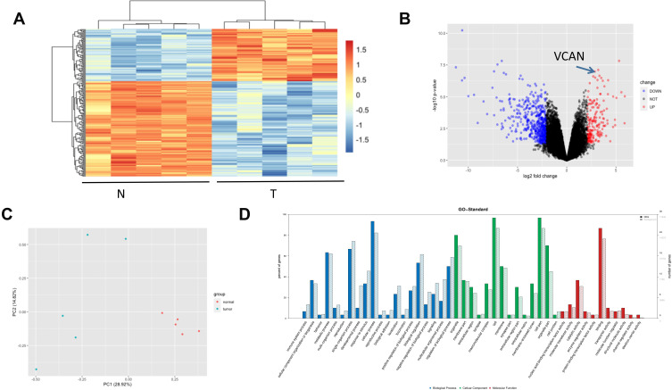
According to 5 GC tissues and relevant adjacent non-tumorous tissue specimens, this study carried out high-throughput human mRNA microarray with samples. Heatmaps (Figure 1A), Volcano plots (Figure 1B) and principal component analysis (PCA) (Figure 1C) present the data. GO analysis suggests that the mentioned differentially expressed mRNAs are relevant to a number of vital physiological processes, cellular components, and molecular functions (Figure 1D).
Figure 1.
Profiling of mRNAs in GC tissues and normal tissues. (A) Heat map shows the up-regulated and down-regulated mRNAs in 5 GC tissues and normal tissues. (T for GC, and N for normal tissues). (B) Volcano plot shows the up-regulated and down-regulated mRNAs. Higher expression levels are indicated by “red”, lower expression levels are indicated by “green”, and no significant difference is indicated by “black”. (C) PCA analysis shows the profiling of mRNAs in GC tissues and normal tissues. (D) GO analysis of mRNAs in GC tissues and normal tissues.
VCAN is Over-Expressed in GC and Has Remarkable Clinical Significance
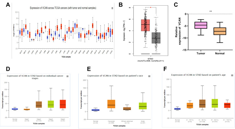
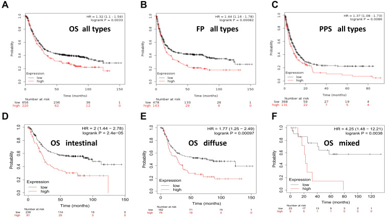
The expression of VCAN in normal and tumor tissues was first detected with TCGA portal, GEPIA2 and UALCAN. The results showed that VCAN was differently expressed in most human normal and tumor tissues (Figure 2A and B). Next, VCAN expression in 16 pairs of GC tissues and adjacent non-tumorous tissue specimens was confirmed by qRT-PCR. Results showed that VCAN was up-regulated in GC tissues compared to adjacent non-tumorous tissues (Figure 2C). The results of subgroup analysis indicated that the expression of VCAN mRNA in GC patients was noticeably associated with individual cancer stages, race, and age (Figure 2D–F). To delve into the prognostic potential of VCAN in GC, Kaplan–Meier Plotter was used. As presented in Figure 3A–C, that higher VCAN expression was associated with shorter OS, FP and PPS compared to lower expression. Sub-analysis showed that whether it is intestinal, diffuse, and mixed pathological type, the expression of VCAN was negatively linked with OS (Figure 3D–F).
Figure 2.
Clinical role of VCAN in GC. (A) VCAN expressed in most human cancers. (B) VCAN expression is higher in GC tissues than that in normal tissues through GEPIA2. (C) VCAN expression is confirmed higher in GC tissues than that in normal tissues by qRT-PCR. (D–F) VCAN expression is correlated with stages,race, and age in the GC patients. *P<0.05; **P<0.01.
Figure 3.
Prognosis value of VCAN in GC patients. (A–C) VCAN expression is correlated with poor OS, FP and PPS in the GC patients. (OS, overall survival; FP, first progression; PPS, post-progression survival). (D–F) Sub-analysis of intestinal, diffuse, and mixed pathological type between the expression of VCAN and OS.
VCAN Plays a Promoting Role in GC Cells in vitro
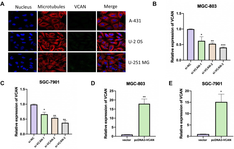
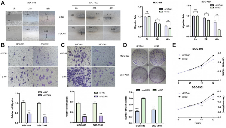
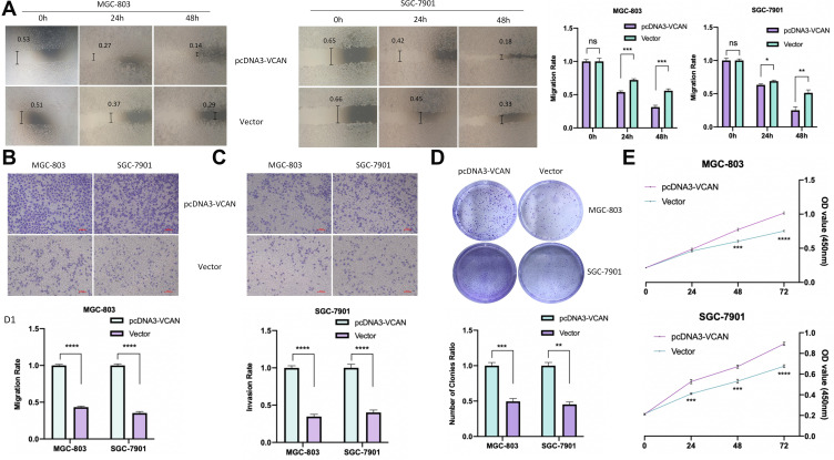
The Human Protein Atlas database showed that in A-431, U-2 OS and U-251 MG cell lines, VCAN located in the extracellular matrix (Figure 4A). For the exploration of the physiological characteristics role of VCAN in tumor, VCAN expression was effectively knocked down or overexpressed in GC cells (Figure 4B–E). The results of a scratch-wound assay demonstrated that suppression of VCAN exhibited a notably lower scratch closure rate than identified in controls in GC cell lines (Figure 5A).In comparison with control ones seen from transwell assay in the confluent monolayer of the cultured GC cell lines, the suppression of VCAN by si-VCAN achieved a lower relative migration and invasion rate (Figure 5B and C). Plate cloning experiment and CCK-8 assays showed that VCAN knockdown noticeably inhibited the proliferation of MGC-803 and SGC-7901 cell lines compared to control group (Figure 5D and E). Overexpression of VCAN plays the opposite effect (Figure 6A–E). The mentioned findings demonstrated that inhibition of VCAN can slow the progression of GC in vitro covering proliferation, invasion and migration.
Figure 4.
VCAN location and models establishment. (A) VCAN located in the membrane. (B and C) VCAN was effectively reduced using the si-VCAN in MGC-803 and SGC-7901 cells. (D and E) VCAN was effectively over-expressed in MGC-803 and SGC-7901 cells.*P<0.05; **P<0.01; ***P<0.001.
Figure 5.
Knockdown of VCAN could inhibit the proliferation of GC cell. (A) Scratch assay of knockdown of VCAN. (B) Knockdown of VCAN could inhibit the migration of GC cell. (C) Knockdown of VCAN could inhibit the invasion of GC cell. (D) Plate cloning experiment of knockdown of VCAN. (E) CCK8 results showed that suppression of VCAN by si-VCAN exhibited a slower proliferation. *P<0.05; **P<0.01; ***P<0.001; ****P<0.0001.
Figure 6.
Overexpression of VCAN could promote the proliferation of GC cell. (A) Scratch assay of overexpression of VCAN. (B) Overexpression of VCAN could promote the migration of GC cell. (C) Overexpression of VCAN could promote the invasion of GC cell. (D) Plate cloning experiment of overexpression of VCAN. (E) CCK8 results showed that Overexpression of VCAN exhibited a higher proliferation.*P<0.05; **P<0.01; ***P<0.001; ****P<0.0001.
VCAN Expression is Correlated with Immune System
More and more researches have proved that immune system is highly related to tumor process. We investigated the relationship between VCAN expression and immune factors. Results showed that VCAN was noticeably linked with Regulatory T cells (Tregs) (Figure 7A). Some immunosuppressive membrane proteins covering T-cell immunoreceptor with Ig and ITIM domains (TIGIT), cytotoxic T lymphocyte-associated antigen-4 (CTLA4), Indoleamine 2.3-dioxygenase 1(IDO1), inducible costimulatory molecule (ICOS), show evident relationships to VCAN expression by filtering: p < 0.05 as well as |±rho| ≥ 0.1 were listed (Figure 7B–E). However, programmed cell death protein 1 (PDCD1) showed no association (Figure 7F). The specific molecular mechanism needs further study.
Figure 7.
Correlation between VCAN expression and immune system (A) VCAN was significantly linked with Tregs. (B–F) Immunosuppressive membrane proteins IDO1, TIGIT, CTLA4, ICOS have significant correlation with VCAN expression while PDCD1 not.
Discussion
In the present study, our comprehensive approaches with mRNA microarray identified a list of genes with differential expression in GC tumor tissues compared to non-tumor tissues. We found that VCAN was noticeably up-regulated in GC tissues and associated with cancer stages, race, and age. GC patients with higher VCAN expression displayed worse prognosis. This is not the first study to study the expression of VCAN in GC clinical specimens. Li et al assessed the associations between clinical variables and VCAN. The Gene Expression Omnibus and the Human Protein Atlas were employed to achieve subsequent verification. Gene set enrichment study (GSEA) was conducted with The Cancer Genome Atlas dataset. According to their study, significant VCAN expression showed relationships to high stage and T classification in GC. The area under the ROC curve reached 0.853. Compared with patients exhibiting low VCAN expression, cases exhibiting high VCAN expression achieved worse prognosis. According to multiple-variate study, VCAN independently reduces overall survival in both groups. GSEA found pathways participating in chemokine signaling, T cell receptor signaling, Wnt signaling, ECM-receptor interaction, and cancer as differentially up-regulated in GCs exhibiting large VCAN expression.13 According to Jiang et al, the VCAN gene was noticeably associated with overall survival and disease-free survival in GC.14 A literature search found that VCAN is closely related to clinicopathology in other cancer clinical samples not only in GC. Lu et al found that high expression of VCAN is likely to predict poor survival results in Pancreatic ductal adenocarcinoma.15 The elevated expression of VCAN has also been reported noticeably correlated with metastasis and worse 5-year overall survival after radical nephrectomy in clear cell renal cell carcinoma.16 All of the mentioned evidence indicates that VCAN is likely to be a potential target for cancer diagnosis, treatment, and prognostic prediction.
According to in vitro experiments, VCAN knockdown hindered proliferated, invaded, and migrated levels of GC cells while overexpression of VCAN played the opposite effect. According to existing research, VCAN is anti-adhesive, and this activity appears to reside in the G1 domain of VCAN.17–19 Nevertheless, the carboxy-terminal domain of VCAN shows interaction with the β1 integrin of glioma cells, which can activate focal adhesion kinase (FAK), facilitate cell adhesion and avoid apoptosis in the mentioned cell.20 Moreover, VCAN can bind to adhesion molecules onto inflammatory leukocytes surface. For instance, VCAN can bind to L and P selectins via specific over-sulfated sequences in the chondroitin sulfate chains.21 VCAN also impacts cell proliferation. For instance, mitogens, eg, platelet-derived growth factor (PDGF), promote VCAN expression in arterial smooth muscle cells and facilitate the expansion of the pericellular ECM that is required for the proliferation and migration of the mentioned cells.22 Thus, VCAN expression shows relationships to a proliferative cell phenotype and is often found in tissues exhibiting elevated proliferation, such as in development and in a variety of tumors covering GC. Yang et al reported that functional evaluations showed that silencing VCAN by shRNA noticeably hindered cell migration and invasion capacity, whereas increased VCAN by overexpressing NPM1-mA enhanced migration and invasion ability of leukemia cells.23 Note that in GC tissues and cell lines, lncRNA VCAN antisense RNA 1 (VCAN-AS1) showed evident promotion, while its rise showed relationships to clinical results of GC cases. Moreover, silencing VCAN-AS1 represses cell proliferating, migrating, and invading processes, whereas it facilitates apoptosis. Furthermore, VCAN-AS1 can regulate TP53 expression in a negative manner through the competitive bind to eIF4A3.10 Circ_VCAN was found noticeably upregulated in radioresistant glioma tissues compared with radiosensitive tissues, and that circ_VCAN expression was negatively correlated with miR-1183 expression in glioma tissues. Circ_VCAN expression was decreased and miR-1183 expression rose in U87 and U251 cells after irradiation. Both knockdown of circ_VCAN and treatment with miR-1183 mimics inhibited proliferating, migrating, and invading processes, and facilitated apoptosis of the U251 and U87 cells irradiated. Moreover, luciferase reporter assays revealed that circ_VCAN might function as a sponge for miR-1183.24 The above results indicate that different transcript forms of VCAN have powerful functions for cancer progression, especially in their ability for adhering and proliferating cancer cells.
Tumor infiltrative forkhead box P3-positive (FOXP3+) regulatory T cells (Tregs) critically impact GC immune microenvironment.25 Tregs refer to immunosuppressive lymphocytes adversely affecting the regulation of the immune system through the regulation of the active immune function of effector T cells (Teffs). The extensive increase in FOXP3+ Tregs makes effector T cells’ immune function ineffective in the microenvironment of tumor.26 As revealed from the assessment here, VCAN expression shows high relationships to Tregs and some immunosuppressive membrane proteins covering TIGIT, CTLA4, IDO1, ICOS have a significant correlation with VCAN expression. We searched the literature and found that there are some reports on the research of VCAN and immunity. For example, Hope et al reported that VCAN strongly correlated with CD8+ T cell infiltration in colorectal cancer, regardless of mismatch repair status. Tumors exhibiting low total VCAN and active VCAN proteolysis showed relationships to obvious CD8+ T cell infiltration. Tumor-intrinsic WNT pathway activation showed relationships to CD8+ T cell excluding process and VCAN gaining process. Besides controlling VCAN levels in the tumor site, VCAN proteolysis can synthesize bioactive fragments with novel functions.27 Chang et al indicated that type I interferon signaling regulates VCAN expression, and VCAN is necessary for type I interferon production. Macrophage-derived VCAN refers to a molecule that can regulate immune with anti-inflammatory properties in acute pulmonary inflammation.28 According to Chelsea Hope et al, human myeloma tumors with CD8+ infiltration/aggregates received VCAN proteolysis at a site assessed for synthesizing a glycosaminoglycan-bereft N-terminal fragment, versikine myeloma-relevant macrophages (MAMs), instead of tumor cells, chiefly produced V1-VCAN, the precursor to versikine, but stromal cell-derived ADAMTS1 was the VCAN-degrading protease exhibiting the highest expression. Different from intact VCAN, versikine-induced Il-6 production showed part reliance on Tlr2. In a model of macrophage-myeloma cell crosstalk, versikine triggered components of “T-cell inflammation,” covering T-cell chemoattractant CCL2 and IRF8-dependent type I interferon transcriptional signatures. For this reason, the interplay between stromal cells and myeloid cells in the myeloma microenvironment produces versikine, an emerging bioactive damage-associated molecular pattern probably facilitating immune sensing of myeloma tumors and modulating the tolerogenic consequences of intact VCAN accumulating process.29 Accordingly, therapeutic VCAN administration is likely to facilitate cancer immunotherapy.
Conclusion
The mentioned results suggest that VCAN is critical to GC progression. Accordingly, VCAN is likely to be a potentially feasible prognosis marking element and a prominent cancer drug for GC patients.
Funding Statement
This research was supported by Huai’an Natural Science Research Program HAB201805 for Xiaowei Wang.
Disclosure
The authors report no conflicts of interest in this work.
References
- 1.Ferlay J, Soerjomataram I, Dikshit R, et al. Cancer incidence and mortality worldwide: sources, methods and major patterns in GLOBOCAN 2012. Int J Cancer. 2015;136(5):E359–86. doi: 10.1002/ijc.29210 [DOI] [PubMed] [Google Scholar]
- 2.Sukri A, Hanafiah A, Mohamad Zin N, Kosai NR. Epidemiology and role of Helicobacter pylori virulence factors in gastric cancer carcinogenesis. APMIS. 2020;128(2):150–161. doi: 10.1111/apm.13034 [DOI] [PubMed] [Google Scholar]
- 3.Palle J, Rochand A, Pernot S, Gallois C, Taïeb J, Zaanan A. Human epidermal growth factor receptor 2 (HER2) in advanced gastric cancer: current knowledge and future perspectives. Drugs. 2020;80(4):401–415. doi: 10.1007/s40265-020-01272-5 [DOI] [PubMed] [Google Scholar]
- 4.Wu YJ, La Pierre DP, Wu J, Yee AJ, Yang BB. The interaction of versican with its binding partners. Cell Res. 2005;15(7):483–494. doi: 10.1038/sj.cr.7290318 [DOI] [PubMed] [Google Scholar]
- 5.Kinsella MG, Bressler SL, Wight TN. The regulated synthesis of versican, decorin, and biglycan: extracellular matrix proteoglycans that influence cellular phenotype. Crit Rev Eukaryot Gene Expr. 2004;14(3):203–234. doi: 10.1615/CritRevEukaryotGeneExpr.v14.i3.40 [DOI] [PubMed] [Google Scholar]
- 6.Wight TN. Versican: a versatile extracellular matrix proteoglycan in cell biology. Curr Opin Cell Biol. 2002;14(5):617–623. doi: 10.1016/S0955-0674(02)00375-7 [DOI] [PubMed] [Google Scholar]
- 7.Rahmani M, Wong BW, Ang L, et al. Versican: signaling to transcriptional control pathways. Can J Physiol Pharmacol. 2006;84(1):77–92. doi: 10.1139/y05-154 [DOI] [PubMed] [Google Scholar]
- 8.Du WW, Yang W, Yee AJ. Roles of versican in cancer biology–tumorigenesis, progression and metastasis. Histol Histopathol. 2013;28(6):701–713. doi: 10.14670/HH-28.701 [DOI] [PubMed] [Google Scholar]
- 9.Shen Q, Chen M, Zhao X, Liu Y, Ren X, Zhang L. Versican expression level in cumulus cells is associated with human oocyte developmental competence. Syst Biol Reprod Med. 2020;6:1–9. [DOI] [PubMed] [Google Scholar]
- 10.Feng L, Li J, Li F, et al. Long noncoding RNA VCAN-AS1 contributes to the progression of gastric cancer via regulating p53 expression. J Cell Physiol. 2020;235(5):4388–4398. doi: 10.1002/jcp.29315 [DOI] [PubMed] [Google Scholar]
- 11.Kulbe H, Otto R, Darb-Esfahani S, et al. Discovery and validation of novel biomarkers for detection of epithelial ovarian cancer. Cells. 2019;8(7):713. doi: 10.3390/cells8070713 [DOI] [PMC free article] [PubMed] [Google Scholar]
- 12.Liu X, Han C, Liao X, et al. Genetic variants in the exon region of versican predict survival of patients with resected early-stage hepatitis B virus-associated hepatocellular carcinoma. Cancer Manag Res. 2018;10:1027–1036. doi: 10.2147/CMAR.S161906 [DOI] [PMC free article] [PubMed] [Google Scholar]
- 13.Li W, Han F, Fu M, Wang Z. High expression of VCAN is an independent predictor of poor prognosis in gastric cancer. J Int Med Res. 2020;48(1):300060519891271. [DOI] [PMC free article] [PubMed] [Google Scholar]
- 14.Jiang K, Liu H, Xie D, Xiao Q. Differentially expressed genes ASPN, COL1A1, FN1, VCAN and MUC5AC are potential prognostic biomarkers for gastric cancer. Oncol Lett. 2019;17(3):3191–3202. doi: 10.3892/ol.2019.9952 [DOI] [PMC free article] [PubMed] [Google Scholar]
- 15.Lu Y, Li C, Chen H, Zhong W. Identification of hub genes and analysis of prognostic values in pancreatic ductal adenocarcinoma by integrated bioinformatics methods. Mol Biol Rep. 2018;45(6):1799–1807. doi: 10.1007/s11033-018-4325-2 [DOI] [PubMed] [Google Scholar]
- 16.Mitsui Y, Shiina H, Kato T, et al. Versican promotes tumor progression, metastasis and predicts poor prognosis in renal carcinoma. Mol Cancer Res. 2017;15(7):884–895. doi: 10.1158/1541-7786.MCR-16-0444 [DOI] [PMC free article] [PubMed] [Google Scholar]
- 17.Yamagata M, Suzuki S, Akiyama SK, Yamada KM, Kimata K. Regulation of cell-substrate adhesion by proteoglycans immobilized on extracellular substrates. J Biol Chem. 1989;264(14):8012–8018. [PubMed] [Google Scholar]
- 18.Yamagata M, Kimata K. Repression of a malignant cell-substratum adhesion phenotype by inhibiting the production of the anti-adhesive proteoglycan, PG-M/versican. J Cell Sci. 1994;107(Pt 9):2581–2590. [DOI] [PubMed] [Google Scholar]
- 19.Wu Y, Chen L, Zheng PS, Yang BB. beta 1-integrin-mediated glioma cell adhesion and free radical-induced apoptosis are regulated by binding to a C-terminal domain of PG-M/versican. J Biol Chem. 2002;277(14):12294–12301. doi: 10.1074/jbc.M110748200 [DOI] [PubMed] [Google Scholar]
- 20.Kawashima H, Hirose M, Hirose J, Nagakubo D, Plaas AH, Miyasaka M. Binding of a large chondroitin sulfate/dermatan sulfate proteoglycan, versican, to L-selectin, P-selectin, and CD44. J Biol Chem. 2000;275(45):35448–35456. doi: 10.1074/jbc.M003387200 [DOI] [PubMed] [Google Scholar]
- 21.Kawashima H, Atarashi K, Hirose M, et al. Oversulfated chondroitin/dermatan sulfates containing GlcAbeta1/IdoAalpha1-3GalNAc(4,6-O-disulfate) interact with L- and P-selectin and chemokines. J Biol Chem. 2002;277(15):12921–12930. doi: 10.1074/jbc.M200396200 [DOI] [PubMed] [Google Scholar]
- 22.Evanko SP, Johnson PY, Braun KR, Underhill CB, Dudhia J, Wight TN. Platelet-derived growth factor stimulates the formation of versican-hyaluronan aggregates and pericellular matrix expansion in arterial smooth muscle cells. Arch Biochem Biophys. 2001;394(1):29–38. doi: 10.1006/abbi.2001.2507 [DOI] [PubMed] [Google Scholar]
- 23.Yang L, Wang L, Yang Z, et al. Up-regulation of EMT-related gene VCAN by NPM1 mutant-driven TGF-β/cPML signalling promotes leukemia cell invasion. J Cancer. 2019;10(26):6570–6583. doi: 10.7150/jca.30223 [DOI] [PMC free article] [PubMed] [Google Scholar]
- 24.Zhu C, Mao X, Zhao H. The circ_VCAN with radioresistance contributes to the carcinogenesis of glioma by regulating microRNA-1183. Medicine (Baltimore). 2020;99(8):e19171. doi: 10.1097/MD.0000000000019171 [DOI] [PMC free article] [PubMed] [Google Scholar]
- 25.Sakaguchi S, Yamaguchi T, Nomura T, Ono M. Regulatory T cells and immune tolerance. Cell. 2008;133(5):775–787. doi: 10.1016/j.cell.2008.05.009 [DOI] [PubMed] [Google Scholar]
- 26.Fontenot JD, Gavin MA, Rudensky AY. Foxp3 programs the development and function of CD4+CD25+ regulatory T cells. Nat Immunol. 2003;4(4):330–336. doi: 10.1038/ni904 [DOI] [PubMed] [Google Scholar]
- 27.Hope C, Emmerich PB, Papadas A, et al. Versican-derived matrikines regulate Batf3-dendritic cell differentiation and promote T cell infiltration in colorectal cancer. J Immunol. 2017;199(5):1933–1941. doi: 10.4049/jimmunol.1700529 [DOI] [PMC free article] [PubMed] [Google Scholar]
- 28.Chang MY, Kang I, Gale M, et al. Versican is produced by Trif- and type I interferon-dependent signaling in macrophages and contributes to fine control of innate immunity in lungs. Am J Physiol Lung Cell Mol Physiol. 2017;313(6):L1069–L1086. doi: 10.1152/ajplung.00353.2017 [DOI] [PMC free article] [PubMed] [Google Scholar]
- 29.Hope C, Foulcer S, Jagodinsky J, et al. Immunoregulatory roles of versican proteolysis in the myeloma microenvironment. Blood. 2016;128(5):680–685. doi: 10.1182/blood-2016-03-705780 [DOI] [PMC free article] [PubMed] [Google Scholar]


