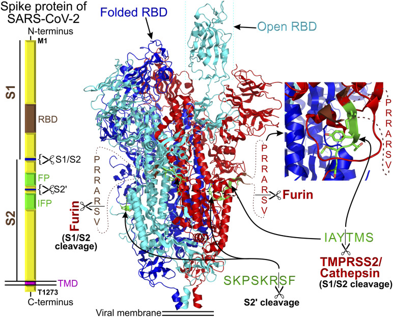
Figure 3.
Block diagram of the homotrimeric SARS-CoV-2 spike protein assembly. “RBD” stands for receptor binding domain, “FP” stands for fusion peptide, and the IPF block depicts the location of internal fusion peptide. S1 and S2 are two segments of SARS-CoV-2 ectodomain that can be cleaved with the indicated endopeptidases. The cryo-EM structure (15) of the spike protein is shown in the center of the figure. The proteolytic sites are shown in green. In the structure, the residues preceding and following the furin cleavage site are colored in brown. The inset shows a magnifying view of the TMPRSS2/cathepsin cleavage site. The structure of the homotrimeric SARS-CoV-2 spike protein complex was replotted from pdb ID: 6VYB (15). Each spike protein in the homotrimer is color coded for better identification.

