Abstract
Background
Coronary artery calcium (CAC) predicts coronary heart disease events and is important for individualized cardiac risk assessment. This report assesses the interscan variability of CT for coronary calcium quantification using image acquisition with standard and reduced radiation dose protocols and whether the use of reduced radiation dose acquisition with iterative reconstruction (IR; “reduced-dose/IR ”) allows for similar image quality and reproducibility when compared to standard radiation dose acquisition with filtered back projection (FBP; “standard-dose/FBP”) on 320-detector row computed tomography (320-CT).
Methods
200 consecutive patients (60 ± 9 years, 59% male) prospectively underwent two standard- and two reduced-dose acquisitions (800 total scans, 1600 reconstructions) using 320 slice CT and 120 kV tube voltage. Automated tube current modulation was used and for reduced-dose scans, prescribed tube current was lowered by 70%. Image noise and Agatston scores were determined and compared.
Results
Regarding stratification by Agatston score categories (0, 1–10, 11–100, 101–400,> 400), reduceddose/IR versus standard-dose/FBP had excellent agreement at 89% (95% CI: 86–92%) with kappa 0.86 (95% CI: 0.81–0.90). Standard-dose/FBP rescan agreement was 93% (95% CI: 89–96%) with kappa=0.91 (95% CI:0.86–0.95) while reduced-dose/IR rescan agreement was similar at 91% (95% CI: 87–94%) with kappa 0.88 (95% CI: 0.83–0.93). Image noise was significantly higher but clinically acceptable for reduced-dose/IR (18 Hounsfield Unit [HU] mean) compared to standard-dose/FBP (16 HU; p<0.0001). Median radiation exposure was 74% lower for reduced- (0.37 mSv) versus standard-dose (1.4 mSv) acquisitions.
Conclusion
Rescan agreement was excellent for reduced-dose image acquisition with iterative reconstruction and standard-dose acquisition with filtered back projection for the quantification of coronary calcium by CT. These methods make it possible to reduce radiation exposure by 74%.
Keywords: Cardiac Computed Tomography, Coronary Artery Calcium, Agatston Score, Radiation Reduction, Iterative Reconstruction
Introduction
The presence of coronary artery calcium (CAC) by non-contrast Cardiac CT is a well-established predictor of coronary heart disease events and may be used for individualized cardiac risk assessment (1–3). Interscan variability in the acquisition of CAC imaging may affect the proper clinical risk stratification of patients (4,5). The recent introduction of iterative reconstruction (IR) reduces image noise and hence permits the use of acquisition protocols with lower radiation exposure for CT angiography, but has not been prospectively validated against conventional filtered back projection (FBP) on a 320-detector row CT scanner (6–11).
This study assesses the reproducibility of standard- and reduced-radiation dose acquisition protocols, the latter combined with the use of iterative reconstruction, for CAC quantification. The aim was to investigate and whether CAC acquisition at reduced radiation dose reconstructed with IR (“reduced-dose/IR”) provides similar reproducibility compared to CAC acquisition at standard radiation dose reconstructed with FBP (“standard-dose/FBP”).
Technical Methods
The study was approved by the Institutional Review Board (IRB) and Radiation Safety Committee of the National Institutes of Health and National Heart, Lung, and Blood Institute (URL: https://clinicaltrials.gov/ct2/show/NCT01621594. Unique identifier: NCT01621594).
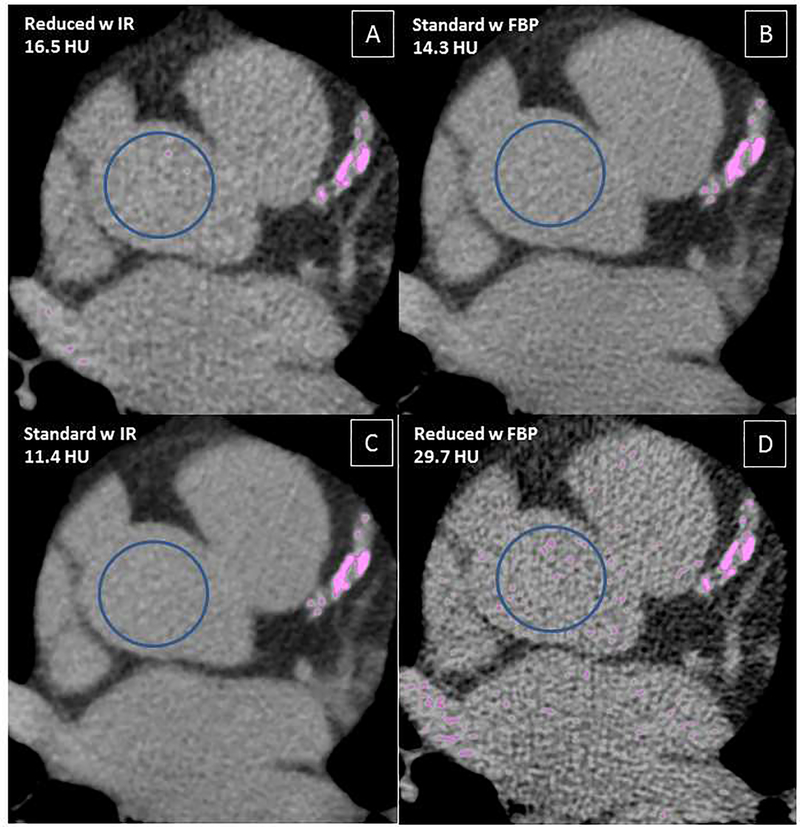
200 consecutive patients prospectively underwent non-enhanced CT for coronary calcium quantification twice at a standard radiation dose and twice at a reduced radiation dose in randomized order (Figure 1). Each scan underwent reconstruction with both FBP and IR (AIDR3D Standard, Toshiba Medical Systems, Otawara, Japan). Standard-dose/FBP was the reference standard. Patient characteristics were prospectively obtained.
Figure 1:
Example of CTs with a region of interest (ROI) in the ascending aorta measuring the image noise as the standard deviation (SD) of the ROI in Hounsfield Units (HU) in acquisitions with Iterative Reconstruction (IR) and Filtered Back Projection (FBP):
A: Reduced-dose acquisition with iterative reconstruction; B: Standard-dose acquisition with filtered back projection; C: Standard-dose acquisition with w iterative reconstruction; D: Reduced-dose acquisition with filtered back projection
CT imaging was performed using a prospectively ECG-triggered axialacquisition protocol on a 320 × 0.5mm detector row CT (AquilionONE ViSION, Toshiba, Japan) with a gantry rotation time of 275ms, 0.5mm slice thickness and tube voltage of 120 kV. Data were reconstructed with 3mm slice thickness and no interslice gap or overlap (12). Tube current was modulated through automated exposure control (Sure Exposure 3D, Toshiba, Japan).
CAC quantification used the Agatston approach and Society of Cardiovascular Computed Tomography (SCCT) standard methodology (12–16). Reduced- versus standard-dose scans were interpreted in random order in separate sessions by an experienced cardiologist. To quantitatively compare attenuation and image noise between the four reconstructed data sets, standard deviation (SD) of the region-of-interest (ROI) measurements were obtained in the ascending aorta (Figure 1).
Statistical analysis
Data are presented as mean ± SD or frequency (percentage) for patient characteristics with mean and median with 5th and 95th percentile for coronary artery calcium scores.
For each imaging method (standard or reduced radiation, reconstruction with FBP or IR), we assessed the intra-method scan reproducibility in multiple ways: by Bland-Altman plots of the difference of the two scans vs. the average of the two scans (17), reproducibility of categorizing scans into the following ranges: 0, 1–10, 11–100, 101–400, > 400, and absolute scan differences We computed the agreement percentage with a bootstrap 95% confidence interval and simple kappa statistic corresponding to the five categories. As in Sevrukov et al., we obtained 95% repeatability bounds for absolute difference of two scans as (18). To reduce outlier impact, these regressions excluded 2 subjects (1%) with standard-dose/FBP Agatston scores > 2000.
Technical Results
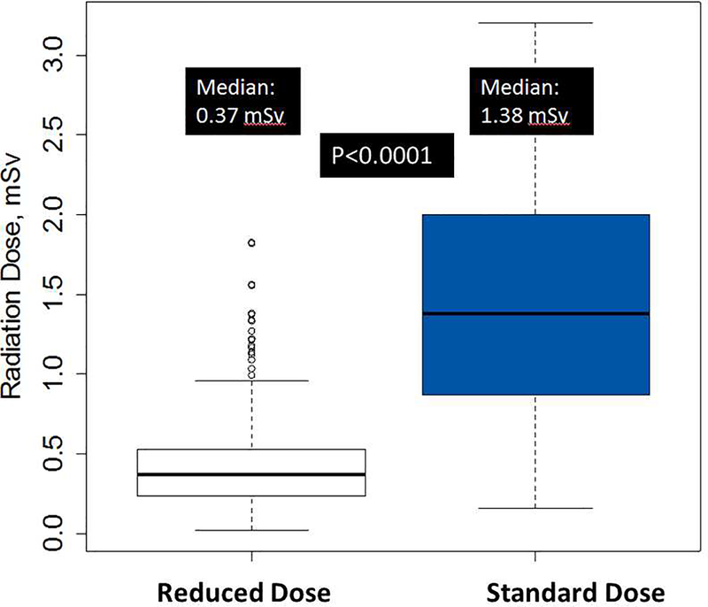
Scan parameters and radiation dose are listed in Table 1. The median (5th–95th percentiles) radiation exposure was 74% (51%−76%) lower for low versus standard dose scans corresponding to overall medians of 0.37 mSv (5th, 95th: 0.15, 1.2) and for standard dose was 1.4 mSv (5th, 95th: 0.46, 3.2; p<0.0001).
Table 1:
Scan Parameters and Radiation Dose
| N=200 | Standard Dose 1 | Standard Dose 2 | Reduced Dose 1 | Reduced Dose 2 |
|---|---|---|---|---|
| Current ± SD, mA | 389.8 ± 202.2 | 390.6 ± 203.3 | 120.5 ± 82.1 | 120.5 ± 85.2 |
| Z-Coverage ± SD, mm | 117 ± 7 | 117 ± 7 | 117 ± 7 | 117 ± 7 |
| Scans at 120mm Z-coverage, n (%) | 168 (84%) | 168 (84%) | 168 (84%) | 168 (84%) |
| DLP, mGy * cm Median (5th, 95th) | 26.3 (16.8, 84.1) | 26.3 (16.8, 84.1) | 26.4 (17.1, 38.0) | 26.4 (17.1, 38.0) |
| Effective Dose, mSv Median (5th, 95th) | 1.4 (0.46, 3.2) | 1.4 (0.46, 3.2) | 0.37 (0.15, 1.2) | 0.37 (0.15, 1.2) |
| Heart Rate ± SD, beats per minute | 58 ± 8 | 58 ± 8 | 58 ± 8 | 58 ± 8 |
Abbreviations: mA = milliampere; mm = millimeters; DLP = Dose Length Product; mSv = millisievert; SD = Standard Deviation
Quantitatively examining image noise, the median value for standard-dose/FBP was 15.6 HU (5th–95th percentiles: 11.3–22.8 HU). Reduced-dose/IR image noise was 18.1 HU (13.9–22.2 HU, p<0.00001), but qualitatively clinically acceptable.
A majority of patients (n=124, 62%) had CAC (Agatston score > 0) detected on standard-dose/FBP scanning. The CAC for the cohort encompassed a wide range of standard FBP Agatston scores (0–4715), but 95% of scores were ≤ 1147. Baseline characteristics of the patient population (n=200) were representative of a wide range of cardiovascular risk (Table 2).
Table 2:
Baseline Characteristics (N=200)
| Age, years ± SD | 60 ± 9 years |
| Male, n (%) | 118 (59%) |
| Body Mass Index, kg/m2 ± SD | 28 ± 5.4 |
| Ethnicity | |
| White, n (%) | 144 (72%) |
| Black, n (%) | 30 (15%) |
| Asian, n (%) | 15 (7.5%) |
| Hispanic, n (%) | 11 (5.5%) |
| CAD Risk Factors | |
| Hypertension, n (%) | 95 (48%) |
| Diabetes Mellitus, n (%) | 29 (15%) |
| Hyperlipidemia, n (%) | 92 (46%) |
| Family History of CAD, n (%) | 35 (23%) |
| Current Smoker, n (%) | 14 (7%) |
| Former Smoker, n (%) | 34 (17%) |
| Any Risk Factor for CAD | 115 (76%) |
Abbreviations: SD = Standard Deviation; CAD = Coronary Artery Disease; ACE-I = Angiotensin converting enzyme-inhibitor; ARB = Angiotensin Receptor Blocker
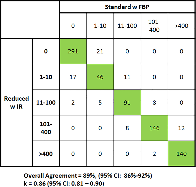
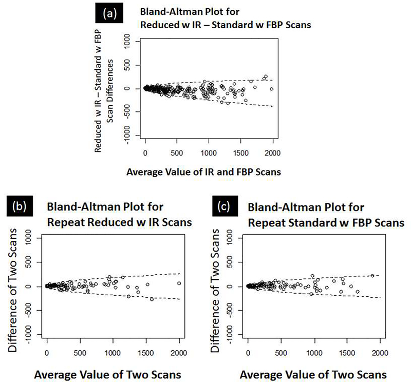
Reduced-dose/IR Agatston scores were classified within the same Agatston group as standard-dose/FBP scores in 89% of cases (714/800) with a 95% CI of 86–92% (Figure 2). This corresponded to a kappa = 0.86 (95% CI of 0.81 – 0.90). For the 79 patients with zero CAC on both reduced-dose/IR scans or both standard-dose/FBP scans, 71/79 (90%) had a zero calcium score on all standard radiation dose and reduced radiation dose scans. By Bland-Altman analysis, the absolute differences for reduced-dose/IR and standard-dose/FBP were nominal at low values and increased across higher CAC scores (Figure 3(a)).
Figure 2:
Overall Agreement of standard-dose acquisition with filtered back projection (FBP) vs. reduced-dose acquisition with iterative reconstruction by standard Agatston categories. With n=200 patients and 4 measurements per patient, there were 8 possible reconstruction and dose combinations. This resulted in n=800 distinct acquisitions and n=1600 total reconstructions. In this specific comparison, Agatston scores of low-dose acquisition with iterative reconstruction were classified within the same category as standard-dose acquisition with filtered back projection in 714/800 cases (89%, 95% CI 86–92%).
Figure 3:
(a) Difference between reduced-dose/IR – standard-dose/FBP Agatston Scores: Bland-Altman plot of difference between reduced-dose/IR and standard-dose/FBP combinations with upper and lower 95% confidence bounds shown. The difference in reduced-dose/IR and standard-dose/FBP was small at low values (<400) and increased as the mean scores increased. The 95% repeatability bounds for the reduced-dose/IR – standard-dose/FBP scan differences are −0.05 · average value .
(b) Repeatability of reduced-dose/IR and (c) standard-dose/FBP calcium scores: The variability for both reduced-dose/IR and standard-dose/FBP was small at low values (<400) and increased as the average scan scores increased. Superimposed on the Bland-Altman plots are the 95% repeatability bounds for the scan differences. For reduced-dose/IR, the 95% bounds are . For standard-dose/FBP, the 95% bounds are .
There was very good rescan agreement for repeat scans with respect to the Agatston categories. For reduced-dose/IR, the agreement was 91% (95% CI: 87–94%) with kappa=0.87 (95% CI:0.83–0.93), for standard-dose/FBP the agreement was 93% (95% CI: 89–96%) with kappa=0.91 (95% CI:0.86–0.95), for standard-dose/IR the agreement was 92% (95% CI: 87–94%) with kappa=0.89 (95% CI: 0.84 – 0.94), and for reduced-dose/ FBP the agreement was 90% (95% CI: 86–94%) with kappa = 0.88 (95% CI: 0.82 – 0.93). By Bland-Altman methods, the absolute differences of both reduced-dose/IR and standard-dose/FBP rescan values were nominal at small values and increased across increasing scores (Figure 3 (b) and (c)).
Discussion
This study is the largest prospective, in-vivo study to evaluate interscan variability and reduced radiation dose CAC scoring on a 320-detector row CT scanner. The use of iterative reconstruction in coronary calcium imaging by CT has evolved from anthropomorphic phantom studies to application in patients at standard radiation dose to assess image noise improvement and most recently reduced radiation dose(12,19–26). The results in our study compare favorably to smaller studies evaluating reduced radiation dose acquisition protocols in combination with IR by Hecht et al. and by Matsuura et al. who tested the use of a hybrid IR algorithm based on Poisson denoising algorithm (iDose, Phillips, Best, Netherlands) in 102 consecutive patients and 77 patients, respectively (25,27,28). Willemink et al. evaluated IR in 30 patients at four dose levels and found CAC reclassifications rates to remain within 15% at 20% of the routine radiation dose(29).
With regard to rescan variability, several reported factors include heart rate, calcification density and different reconstruction algorithms (30,31). Our findings demonstrate that IR rescan differences are similar to prior studies. Detrano et al. examined the Multi-Ethnic Study of Atherosclerosis (MESA) cohort using electron-beam computed tomography (EBCT) and multi-detector row CT (MDCT) and found high concordance (96%, k=0.92) between EBCT and MDCT, but with a rescan variability of about 20% (5). Later, Ghadri, et al. showed that inter-scan variability was high between 64-slice MDCT and 64-slice dual source CT with a coefficient of variation of 15% (4). Most recently, Willemink et al. have shown differences in Agatston classification of up to 6.5% when CAC was performed by testing CAC in cadaveric hearts on 4 different platforms(32).
Several limitations for this study are to be acknowledged. This study was a single-center trial using one single platform. The use of 2 standard-dose and 2 reduced-dose acquisitions increased radiation exposure to patients, though overall radiation dose delivered was within an accepted limit as specified by both the IRB and NIH Radiation Safety Committee. The 74% radiation dose reduction we used may have been conservative and an even greater radiation dose reduction may be achievable without a significant change in risk prognostication.
In conclusion, reduced-dose image acquisition in combination with iterative reconstruction, when compared to standard-dose image acquisition with filtered back projection, achieves a median radiation dose of 0.37 mSv, resulting in comparable image quality, rescan agreement and risk classification while providing 74% radiation dose reduction.
Figure 4: Radiation Exposure for Reduced vs. Standard Dose Scans:
As shown in the following box and whisker plots, median radiation exposure for reduced dose was median 0.37 mSv (5th, 95th: 0.15, 1.17) and for standard dose was median 1.38 mSv (5th, 95th: 0.46, 3.18). For reduced-dose scans, the outliers represent patients with high BMI (36–45 kg/m2) where the automatic exposure control determined to use high tube current. For standard-dose scans, the scanner reached maximal x-ray tube output so there are no outliers beyond the 1.5 * interquartile range. The median radiation reduction was 74% for reduced-dose vs. standard-dose scans (p<0.0001).
Acknowledgements
The authors wish to acknowledge the work and dedication of Shirley Rollison who performed all of the CT scans.
Funding: This research was supported by the Intramural Research Program of the National Institutes of Health and National Heart, Lung, and Blood Institute.
ABBREVIATIONS LIST
- CAC
Coronary Artery Calcium
- CHD
Coronary Heart Disease
- IR
Iterative Reconstruction
- CT
Computed Tomography
- FBP
Filtered Back Projection
- IRB
Institutional Review Board
- SCCT
Society of Cardiovascular Computed Tomography
- mSv
MilliSievert
- EBCT
Electron Beam Computed Tomography
- MDCT
Multidetector Computed Tomography
Footnotes
Conflict of Interest: None
Disclosures: Arai AE, Chen MY – Institutional Research Agreement with Toshiba Medical Research.
Clinical Trial Registration: URL: https://clinicaltrials.gov/ct2/show/NCT01621594. Unique identifier: NCT01621594
Publisher's Disclaimer: This is a PDF file of an unedited manuscript that has been accepted for publication. As a service to our customers we are providing this early version of the manuscript. The manuscript will undergo copyediting, typesetting, and review of the resulting proof before it is published in its final citable form. Please note that during the production process errors may be discovered which could affect the content, and all legal disclaimers that apply to the journal pertain.
References
- 1.Detrano R, Guerci AD, Carr JJ et al. Coronary calcium as a predictor of coronary events in four racial or ethnic groups. N Engl J Med 2008;358:1336–45. [DOI] [PubMed] [Google Scholar]
- 2.Budoff MJ, Shaw LJ, Liu ST et al. Long-term prognosis associated with coronary calcification: observations from a registry of 25,253 patients. J Am Coll Cardiol 2007;49:1860–70. [DOI] [PubMed] [Google Scholar]
- 3.Chang SM, Nabi F, Xu J et al. Value of CACS Compared With ETT and Myocardial Perfusion Imaging for Predicting Long-Term Cardiac Outcome in Asymptomatic and Symptomatic Patients at Low Risk for Coronary Disease: Clinical Implications in a Multimodality Imaging World. JACC Cardiovasc Imaging 2015;8:134–44. [DOI] [PubMed] [Google Scholar]
- 4.Ghadri JR, Goetti R, Fiechter M et al. Inter-scan variability of coronary artery calcium scoring assessed on 64-multidetector computed tomography vs. dual-source computed tomography: a head-to-head comparison. Eur Heart J 2011;32:1865–74. [DOI] [PubMed] [Google Scholar]
- 5.Detrano RC, Anderson M, Nelson J et al. Coronary calcium measurements: effect of CT scanner type and calcium measure on rescan reproducibility--MESA study. Radiology 2005;236:477–84. [DOI] [PubMed] [Google Scholar]
- 6.Yin WH, Lu B, Li N et al. Iterative reconstruction to preserve image quality and diagnostic accuracy at reduced radiation dose in coronary CT angiography: an intraindividual comparison. JACC Cardiovasc Imaging 2013;6:1239–49. [DOI] [PubMed] [Google Scholar]
- 7.Chen MY, Steigner ML, Leung SW et al. Simulated 50 % radiation dose reduction in coronary CT angiography using adaptive iterative dose reduction in three-dimensions (AIDR3D). Int J Cardiovasc Imaging 2013;29:1167–75. [DOI] [PMC free article] [PubMed] [Google Scholar]
- 8.Stehli J, Fuchs TA, Bull S et al. Accuracy of coronary CT angiography using a submillisievert fraction of radiation exposure: comparison with invasive coronary angiography. J Am Coll Cardiol 2014;64:772–80. [DOI] [PubMed] [Google Scholar]
- 9.Layritz C, Schmid J, Achenbach S et al. Accuracy of prospectively ECG-triggered very low-dose coronary dual-source CT angiography using iterative reconstruction for the detection of coronary artery stenosis: comparison with invasive catheterization. Eur Heart J Cardiovasc Imaging 2014;15:1238–45. [DOI] [PubMed] [Google Scholar]
- 10.Utsunomiya D, Weigold WG, Weissman G, Taylor AJ. Effect of hybrid iterative reconstruction technique on quantitative and qualitative image analysis at 256-slice prospective gating cardiac CT. Eur Radiol 2012;22:1287–94. [DOI] [PubMed] [Google Scholar]
- 11.Oda S, Weissman G, Vembar M, Weigold WG. Iterative model reconstruction: improved image quality of low-tube-voltage prospective ECG-gated coronary CT angiography images at 256-slice CT. Eur J Radiol 2014;83:1408–15. [DOI] [PubMed] [Google Scholar]
- 12.Blobel J, Mews J, Schuijf JD, Overlaet W. Determining the radiation dose reduction potential for coronary calcium scanning with computed tomography: an anthropomorphic phantom study comparing filtered backprojection and the adaptive iterative dose reduction algorithm for image reconstruction. Invest Radiol 2013;48:857–62. [DOI] [PubMed] [Google Scholar]
- 13.Halliburton SS, Abbara S, Chen MY et al. SCCT guidelines on radiation dose and doseoptimization strategies in cardiovascular CT. J Cardiovasc Comput Tomogr 2011;5:198–224. [DOI] [PMC free article] [PubMed] [Google Scholar]
- 14.Voros S, Rivera JJ, Berman DS et al. Guideline for minimizing radiation exposure during acquisition of coronary artery calcium scans with the use of multidetector computed tomography: a report by the Society for Atherosclerosis Imaging and Prevention Tomographic Imaging and Prevention Councils in collaboration with the Society of Cardiovascular Computed Tomography. J Cardiovasc Comput Tomogr 2011;5:75–83. [DOI] [PubMed] [Google Scholar]
- 15.Agatston AS, Janowitz WR, Hildner FJ, Zusmer NR, Viamonte M Jr., Detrano R. Quantification of coronary artery calcium using ultrafast computed tomography. J Am Coll Cardiol 1990;15:827–32. [DOI] [PubMed] [Google Scholar]
- 16.Callister TQ, Cooil B, Raya SP, Lippolis NJ, Russo DJ, Raggi P. Coronary artery disease: improved reproducibility of calcium scoring with an electron-beam CT volumetric method. Radiology 1998;208:807–14. [DOI] [PubMed] [Google Scholar]
- 17.Bland JM, Altman DG. Measuring agreement in method comparison studies. Stat Methods Med Res 1999;8:135–60. [DOI] [PubMed] [Google Scholar]
- 18.Sevrukov AB, Bland JM, Kondos GT. Serial electron beam CT measurements of coronary artery calcium: Has your patient’s calcium score actually changed? AJR Am J Roentgenol 2005;185:1546–53. [DOI] [PubMed] [Google Scholar]
- 19.Gebhard C, Fiechter M, Fuchs TA et al. Coronary artery calcium scoring: Influence of adaptive statistical iterative reconstruction using 64-MDCT. Int J Cardiol 2013;167:2932–7. [DOI] [PubMed] [Google Scholar]
- 20.van Osch JA, Mouden M, van Dalen JA et al. Influence of iterative image reconstruction on CTbased calcium score measurements. Int J Cardiovasc Imaging 2014;30:961–7. [DOI] [PubMed] [Google Scholar]
- 21.Takahashi M, Kimura F, Umezawa T, Watanabe Y, Ogawa H. Comparison of adaptive statistical iterative and filtered back projection reconstruction techniques in quantifying coronary calcium. J Cardiovasc Comput Tomogr 2015. [DOI] [PubMed] [Google Scholar]
- 22.Kurata A, Dharampal A, Dedic A et al. Impact of iterative reconstruction on CT coronary calcium quantification. Eur Radiol 2013;23:3246–52. [DOI] [PubMed] [Google Scholar]
- 23.Szilveszter B, Elzomor H, Karolyi M et al. The effect of iterative model reconstruction on coronary artery calcium quantification. Int J Cardiovasc Imaging 2015. [DOI] [PubMed] [Google Scholar]
- 24.Murazaki H, Funama Y, Hatemura M, Fujioka C, Tomiguchi S. [Quantitative evaluation of calcium (content) in the coronary artery using hybrid iterative reconstruction (iDose) algorithm on lowdose 64-detector CT: comparison of iDose and filtered back projection]. Nihon Hoshasen Gijutsu Gakkai Zasshi 2011;67:360–6. [DOI] [PubMed] [Google Scholar]
- 25.Baron KB, Choi AD, Chen MY. Low Radiation Dose Calcium Scoring: Evidence and Techniques. Curr Cardiovasc Imaging Rep 2016;9:12. [DOI] [PMC free article] [PubMed] [Google Scholar]
- 26.Tatsugami F, Higaki T, Fukumoto W et al. Radiation dose reduction for coronary artery calcium scoring at 320-detector CT with adaptive iterative dose reduction 3D. Int J Cardiovasc Imaging 2015;31:1045–52. [DOI] [PubMed] [Google Scholar]
- 27.Hecht HS, de Siqueira ME, Cham M et al. Low- vs. standard-dose coronary artery calcium scanning. Eur Heart J Cardiovasc Imaging 2014. [DOI] [PubMed] [Google Scholar]
- 28.Matsuura N, Urashima M, Fukumoto W et al. Radiation dose reduction at coronary artery calcium scoring by using a low tube current technique and hybrid iterative reconstruction. J Comput Assist Tomogr 2015;39:119–24. [DOI] [PubMed] [Google Scholar]
- 29.Willemink MJ, den Harder AM, Foppen W et al. Finding the optimal dose reduction and iterative reconstruction level for coronary calcium scoring. J Cardiovasc Comput Tomogr 2015. [DOI] [PubMed] [Google Scholar]
- 30.Hong C, Bae KT, Pilgram TK. Coronary artery calcium: accuracy and reproducibility of measurements with multi-detector row CT--assessment of effects of different thresholds and quantification methods. Radiology 2003;227:795–801. [DOI] [PubMed] [Google Scholar]
- 31.Lu B, Budoff MJ, Zhuang N et al. Causes of interscan variability of coronary artery calcium measurements at electron-beam CT. Acad Radiol 2002;9:654–61. [DOI] [PubMed] [Google Scholar]
- 32.Willemink MJ, Vliegenthart R, Takx RA et al. Coronary artery calcification scoring with state-of-the-art CT scanners from different vendors has substantial effect on risk classification. Radiology 2014;273:695–702. [DOI] [PubMed] [Google Scholar]


