

Abstract
Aneurysmal bone cysts are benign, rare bony tumours frequently observed among children and young adults principally located in the long bones, pelvis, and spine and rarely in other anatomical district such as the hand. We report the case of a 12-year-old girl with an aneurysmal bone cyst, in active stage, involving the still-open epiphysis of the fourth metacarpal of the right hand, which was in a first time treated by curettage, and 3 months later, occurring a recurrence, by a radically excision of the bone and reconstruction with a graft from the iliac crest. At 10-year follow-up the patient had good cosmetic results and a functioning hand. We also performed a systematic Literature review in order to retrieve the key information regarding: the diagnosis, the clinical features and the treatment.
Key words: Aneurysmal bone cyst, metacarpal, bone graft reconstruction, hand’s tumour
Introduction
Aneurysmal bone cysts (ABC) are benign, rare bony tumours that constitute only 1-2% of all bone tumours, described firstly by Jaffe and Lichtenstein in 1942.1 ABC can be primary or can arise from a preexistent lesion however the etiology remains unknown. ABC are most common in the youth, principally located in the long bones, without epiphysis involvement, pelvis and spine. The localization in other anatomical district such as the hand.2 Histologically, ABC appear as multicystic, lytic lesion with cavernous spaces stuffed with blood. The walls of cysts contain fibroblasts and thin strips of bone. The tumors are separated from the surrounding tissue by a thin layer of periosteal new bone.3 The most common treatment of an aneurysmal bone cyst is surgical curettage of the lesion, sometimes filling of the cavity with a bone graft and intraoperative adjuvant therapy may be required. Usually the prognosis following treatment is satisfactory. However, a recurrence rate was reported in the first 2 years after treatment from 10 to 59%, especially in young patients due to skeletal immaturity.4 The recurrence rate also depends on the histopathological pattern of the lesion. Preoperative staging and stagedependent surgical procedures are essential for treatment of ABC and the risk of local recurrence is linked to aggressiveness of the primary lesion and to efficacy of the surgery. The aim of the study is to describe a case of a patient with IV metacarpal bon ABC. We also performed a systematic review of the literature in order to retrieve the key information regarding: the diagnosis, the clinical features and the treatment.
Material and Methods
Search Criteria
The study was conducted in accordance with the Preferred Reporting Items for Systematic Reviews and Meta-Analyses (PRISMA) guidelines (Figure 1). A systematic review of the literature indexed in PubMed, MEDLINE, Cochrane Library and Scopus databases, using as search-terms “Aneurysmal”, “Aneurysmatic”, “bone”, “cyst”, “hand” and their MeSH terms combinations (using Boolean operator AND, OR) was performed from 1950 to March 2020. The research was repeated until March 6, 2020.
Inclusion and Exclusion Criteria
The inclusion criteria of the review were the presence in the evaluated manuscript of: demographic features, symptoms, diagnostic settings, treatment, possible complications and outcomes in patients with ABC of the hand. Only article written in English and available abstract were included. Were excluded from the review: surgical technique reports, expert opinions, studies on animals, unpublished reports, cadaver or in vitro investigations, book chapters, abstracts from scientific meetings.
Data Collection
Two independent reviewers (A.P and R.V.) separately conducted the described search by title and abstract. If the articles met inclusion criteria following a title and abstract screened, the full text was obtained and reviewed. Any discordance was solved by consensus with a third author (R.D.V.). From each included article were extracted: age and gender of the patients, location of the ABC, type of surgical treatment performed, risk factors, complications related to the treatment performed and duration of follow-up. Numbers software (Apple Inc., Cupertino, CA) was used to tabulate the obtained data.
Statistical Analysis
Categorical variables are presented as frequency and percentages. Continuous variables are presented as means and standard deviation.
Case Report
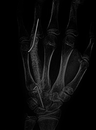
A 12-year-old girl, with history of pain in her right hand since 2 months, was visited at our Emergency Unit. On physical examination there was a slight swelling in the dorsal region of the hand, painful to acupressure. The performed radiographs showed a cystic lesion with expanded cortical near the distal region of the fourth metacarpal, close to the still-open epiphysis (Figure 1a). A MRI was requested. Meanwhile the patient was treated in another hospital by simple surgical curettage. In following three months, she had pain and worsening of dorsal hand swelling. The radiographs and MRI showed a cystic lesion with expanded cortical involving all the fourth metacarpal, also the growth plate (Figure 2). The cortical bone was expanded and thin, which made it impossible to remove the tumour by curettage and filling the cavity with bone graft. Instead the entire diaphysis, including the tumour, was removed and a 7 cm bicortical iliac crest graft, oversizing metacarpal dimension, was harvested and fixed with Kirschner wires to the proximal phalanx and then left in place for six weeks (Figure 3). The hand was also immobilized in a short arm cast for the first 4 weeks. Follow-up radiographs showed that the bone graft healed in the correct position. Macroscopical pattern and microscopy of the tumour showed the typical features of the aneurysmal bone cyst with thin strips of bone and fibroblasts surrounded by blood vessels. At ten year follow up range of movement was satisfactory (Figure 4) only the strength in the operated hand was less than in the other hand but she didn’t have functional limitation and referred a normal life. The radiographs are shown in Figure 4.
Systematic literature review
Patients features and demographical data
Only a few cases of hand’s ABC were reported in the Literature. A total of 744 reports, excluding duplicates, were independently screened, thereby 40 were finally included in our review (Figure 1). Our review showed 83 cases, including our patients, of hand’s ABC in the Literature. (4-55) The mean age of included patients was 18.6 (+/- 10.6) years; 50 patients (60%) were paediatric (< of 18 years) the Male/Female ratio was 1.1; the mean follows up time was 52.1 months (+/-52.2). Demographic and clinical features are summarized in Table 1.1-51
Symptoms onset, localization, risk factors and diagnosis
All patients had an onset symptom. Presentation symptoms were: hand’s swelling in 70 patients (84%), hand’s pain in 60 cases (72.3%), pathologic fracture of the involved bone in one case (1.2%). In 47 cases (56.4%) pain and swelling were associated.
Hand’s ABC seems to be prevalent in the metacarpal bones (47 patients, 56.4%), followed by the proximal phalanx (19 patients, 23.8%), the middle phalanx (6 patients, 7.2%), the capitate (3 patients 3.6%), the distal phalanx (3 patients, 3.6%), the lunate (2 cases, 2.4%), the hamate (in one case 1.2%), and the trapezium (in one case 1.2%). In one case the ABC was localized in a sesamoid bone of proximal interphalangeal joint of the index. In 14 patients (16.8%) a traumatic injury of the affected hand was reported. In all case except one (98.8%) a histological diagnosis was made.
Treatment and outcomes and complication
Eighty-two patients (98.8%) were surgically treated. The chosen surgical approaches were: tumour resection and autologous bone graft in 37 cases (44.4%), curettage of the lesion and autologous bone graft in 26 cases (31.2%), curettage of the cyst in 6 cases (7.2%), curettage and bipolar cauterization in 6 cases (7.2%), tumour excision in 6 cases (7.2%), amputation in 2 patients (2.4%) and conservative treatment only in one case.
Figure 1.

The PRISMA flow-chart.
Figure 2.

a) Rx pattern before simple curettage. b) Rx pattern three month later simple curettage.
Concerning surgical outcome: in 68 patients (81.6%) the first surgery was curative without signs of recurrence. Recurrence of the disease was found in 15 patients (18%), therefore they underwent reoperation. In 8 of this patients, tumour resection and autologous bone graft was used as rescue surgery. In 4 patients after recurrence a new curettage of the lesion and autologous bone graft was performed. In one patient after recurrence, cryotherapy and curettage was used as rescue surgery. In one patient the reoperation consisted in the amputation of affected finger. In one patient the reoperation consisted in curettage and bipolar cauterization of the lesion.
About complication, in 15 patients (18%) was found a limitation of range of motion (ROM); therefore, in 5 of these patients was necessary surgical debridement and tenolysis. In 3 paediatric patients (3.6%) a premature physeal closure was found.
Discussion
The origin of the term “aneurysmal bone cyst” derived from two cases of unicameral bone cysts reported by Jaffe and Lichtenstein in 1942.1 In that report, they noted two large “peculiar blood-containing cysts,” which they described as “aneurysmal bone cyst”. Jaffe argued that aneurysmal bone cysts could be the result of an hemorrhagic “blowout” in a preexisting lesion, which may be destroyed in the process.1,2 Lichtenstein instead proposed a vascular origin, without specifying whether this lesion was a localized venous thrombosis or an congenital arteriovenous malformation.3,4 Although many hypotheses have been developed over the years, today the nature of ABC is unclear. Many authors defined aneurysmal bone cysts as a secondary evolution of a pre-existing lesion.2,3 Other authors proposed two different aetiologies characterizing the lesion as either primary or secondary to a known precursor.45 Most cases are found among children and young adults, in fact the majority of patients with aneurysmal bone cysts are younger than age 20 years. These lesions are principally located in the long bones, pelvis, and spine. Most rarely it is observed in the hand.4,19,22
The natural history of ABC is characterized by four radiologic stages: initial, active, stabilization, and healing. In the initial phase, the lesion is composed of a well-defined area of osteolysis. During the growth phase the lesion grows exponentially leading to the “destruction” of the bone and to the typical “blown-out” radiological appearance. Then follows a period of stabilization defined on the X-ray as having a “soap bubble appearance” (which is caused by the maturation of the bony shell). Final healing results in progressive calcification and ossification, with the lesion transformed into a dense bony mass.46
There has been no agreement on a definitive or ideal treatment in the entire scientific literature which is why many different treatment options are used.
Figure 3.
Rx pattern of reconstruction.
Figure 4.

a) Rx at ten years follow up. b) Flexion of fingers at ten years follow up.
Table 1.
Demographic and clinical features.
| Study | Case | Sex | Age (Year) | Risk Factors | Symptoms | Localization | Complication | Surgical treatment | Donor site | Outcome | Follow Up (month) |
|---|---|---|---|---|---|---|---|---|---|---|---|
| Mason et al.. 1958 | 1 | M | 9 | _ | P, Sw | PP III F | CABG | Iliac crest | Cured | 33 | |
| Harto-Garofalidis et al. 1967 | 2 | M | 17 | _ | P, Sw | PP I F | _ | TRABG | Tibia | Cured | _ |
| Chari et al. 1971 | 3 | F | 16 | _ | P, Sw | IV MTB | Limitation of ROM | TRABG | Tibia | Cured | 10 |
| Burkhalter et al. 1978 | 4 | F | 22 | P, Sw | IV MTB | _ | TRABG | Iliac crest | Cured | 12 | |
| 5 | M | 8 | Trauma | P, Sw | IV MTB | Limitation of ROM | TRABG | Fibula | Cured | 18 | |
| 6 | M | 10 | P, Sw | III MTB Limitation | Limitation of ROM | 1° Curettage 2° TRABG | Fibula | Reoperation/Cured | 72 | ||
| Fuhs et al. 1979 | 7 | M | 17 | Trauma | P, Sw | DP IV F | Limitation of ROM, Recurrence | Amputation | _ | Amputation | 12 |
| 8 | M | 20 | Trauma | P, Sw | I MTB | _ | TRABG | Iliac crest | Cured | _ | |
| Chalmers et al. 1981 | 9 | M | 36 | _ | P, Sw | PP V F | Limitation of ROM | CABG | _ | Reoperation/Cured | _ |
| 10 | F | 13 | P, Sw | PP II F | Recurrence | CABG | _ | Cured | _ | ||
| 11 | M | 16 | _ | P, Sw | PP I F | _ | Curettage | _ | Cured | _ | |
| Barbieri et al. 1984 | 12 | F | 35 | _ | P, Sw | IV MTB | _ | F trasposition | _ | Cured | 24 |
| 13 | F | 11 | _ | P, Sw | II MTB | Limitation of ROM | TRABG | _ | Cured | 48 | |
| 14 | F | 30 | _ | P, Sw | IV MTB | _ | CABG | _ | Cured | 6 | |
| Lin et al. 1984 | 15 | M | 16 | _ | P, Sw | Hamate | _ | TE | _ | Cured | 15 |
| Frassica et al. 1988 | 16 | F | 13 | Trauma | P, Sw | PP V F | _ | CABG | _ | Reoperation/Cured | 244 |
| 17 | M | 20 | _ | P, Sw | Trapezium | Recurrence | TE | _ | Cured | 176 | |
| 18 | F | 49 | _ | P | PP I F | _ | CABG | _ | Reoperation/Cured | 254 | |
| 19 | F | 36 | _ | P, Sw | V MTB | Recurrence | TRABG | _ | Cured | 132 | |
| 20 | M | 28 | _ | P, Sw | III MTB | _ | CABG | _ | Cured | 55 | |
| 21 | M | 55 | _ | P | DP V F | _ | CABG | _ | Cured | 59 | |
| 22 | M | 14 | _ | PF | I MTB | Recurrence | Curettage | _ | Reoperation/Amputation | 42 | |
| 23 | F | 14 | _ | P | IV MTB | _ | CABG | _ | Cured | 43 | |
| 24 | F | 28 | _ | P | I MTB | _ | CABG | _ | Cured | 32 | |
| 25 | F | 16 | _ | P | V MTB | Recurrence | 1° CABG 2° TRABG | _ | Reoperation/ Cured | 27 | |
| Kotwal et al. 1988 | 26 | F | 5 | _ | P, Sw | III MTB | _ | TRABG | Fibula | Cured | 48 |
| Kozlowski et al. 1988 | 27 | M | 8 | Previous surgery CABG | P, Sw | III MTB | _ | CABG | _ | _ | _ |
| 28 | M | 9 | Trauma | P, Sw | DP III F | _ | Curettage | _ | _ | _ | |
| 29 | F | 10 | _ | P, Sw | II MTB | _ | TRABG | _ | _ | _ | |
| 30 | M | 11 | Trauma | P, Sw | MP III F | _ | CABG | _ | _ | _ | |
| 31 | F | 13 | _ | P, Sw | II MTB | _ | CABG | _ | _ | _ | |
| Milliez et al. 1988 | 32 | F | 10 | _ | P, Sw | MP III F | Recurrence | CABG | Iliac crest | Reoperation/Cured | 6 |
| Dossing et al. 1990 | 33 | F | 40 | Trauma | P, Sw | MP I F | Recurrence | 1° CABG 2° TRABG | _ | Reoperation/Cured | 18 |
| Mortensen et al. 1990 | 34 | M | 6 | _ | P, Sw | PP III F | _ | TRABG | Ulna | Cured | 72 |
| Rao et al. 1993 | 35 | M | 31 | _ | P | I MTB | Recurrence | 1° CABG 2° TRABG | IV MTB | Reoperation/Cured | 44 |
| 36 | F | 11 | _ | Sw | MP II F | Recurrence | 1° CABG 2° TRABG | PP II F of the foot | Reoperation/Cured | 6 | |
| Borrelli et al. 1994 | 37 | M | 10 | _ | Sw | III MTB | _ | TRABG | Fibula | Cured | 30 |
| Mankin et al. 1995 | 38 | F | 40 | _ | P | Lunate | Limitation of ROM | TE, SCA | _ | Cured | 6 |
| Platt et al. 1995 | 39 | F | 14 | Trauma | P | Capitate | _ | No | _ | Cured | 16 |
| Apaydin et al. 1996 | 40 | M | 32 | _ | P, Sw | I MTB | _ | TRABG | Iliac crest | Cured | 18 |
| Sakka et al. 1997 | 41 | M | 8 | Trauma | P, Sw | DP I F | _ | Curettage | _ | Cured | 36 |
| Athanasian et al. 1999 | 42 | F | 14 | Trauma | P, Sw | PP III F | Recurrence | 1° CABG 2° Criosurgery | Iliac crest | Reoperation/Cured | 60 |
| Sproule et al. 2002 | 43 | M | 8 | Trauma | P, Sw | PP I F | Limitation of ROM | CABG | _ | Cured | 12 |
| Gundes et al. 2005 | 44 | M | 25 | _ | P, Sw | IV MTB | Limitation of ROM | TRABG | Fibula | Cured | 12 |
| Havulinna et al. 2005 | 45 | M | 34 | _ | Sw | Sesamoid II F | _ | TE | _ | Cured | 24 |
| Basarir et al. 2006 | 46 | M | 18 | _ | P, Sw | V MTB | _ | CABG | Iliac crest | Cured | 18 |
| 47 | F | 22 | _ | P, Sw | PP IV F | Recurrence | 1° CABG 2° TRABG | Iliac crest | Reoperation/Cured | 22 | |
| 48 | M | 16 | Trauma | P, Sw | IV MTB | _ | TRABG | Iliac crest | Cured | 14 | |
| 49 | F | 52 | Trauma | P, Sw | II MTB | Recurrence | 1° CABG 2° TRABG | Iliac crest | Reoperation/Cured | 125 | |
| Sakamoto et al. 2006 | 50 | M | 15 | _ | P | Capitate | _ | Curettage | _ | Cured | 48 |
| Sahu et al. 2008 | 51 | F | 12 | _ | Sw | I MTB | _ | TE | _ | Cured | 24 |
| Ozyurek et al. 2009 | 52 | M | 21 | _ | P, Sw | V MTB | _ | TRABG | Iliac crest | Cured | 36 |
| Jafari et al. 2011 | 53 | M | 16 | _ | Sw | I MTB | Limitation of ROM | TRABG | Fibula | Cured | _ |
| 54 | F | 6 | _ | Sw | III MTB | _ | TRABG | Fibula | Cured | _ | |
| 55 | M | 17 | _ | Sw | I MTB | _ | TRABG | Fibula | Cured | _ | |
| 56 | M | 27 | _ | Sw | PP V F | Graft resorption | TRABG | Iliac crest | Cured | _ | |
| 57 | F | 15 | _ | Sw | III MTB | _ | TRABG | Iliac crest | Cured | _ | |
| 58 | F | 10 | _ | Sw | II MTB | _ | TRABG | Fibula | Cured | _ | |
| 59 | M | 9 | _ | Sw | V MTB | _ | TRABG | Fibula | Cured | _ | |
| 60 | F | 23 | _ | Sw | II MTB | Recurrence | TRABG | Iliac crest | Reoperation/Cured | 18 | |
| 61 | F | 15 | _ | Sw | V MTB | Limitation of ROM | TRABG | Fibula | Cured | _ | |
| 62 | F | 16 | _ | Sw | III MTB | Limitation of ROM | TRABG | Fibula | Cured | _ | |
| 63 | M | 17 | _ | Sw | PP II F | _ | TRABG | Iliac crest | Cured | _ | |
| 64 | F | 20 | _ | Sw | PP IV F | _ | TRABG | Iliac crest | Cured | _ | |
| 65 | M | 20 | _ | P | Capitate | _ | CABG | Iliac crest | Cured | 24 | |
| 66 | M | 15 | Trauma | P, Sw | III MTB | _ | TRABG | Radius | Cured | 144 | |
| 67 | F | 23 | _ | P | Lunate | _ | TE | _ | Cured | 72 | |
| 68 | M | 6 | _ | Sw | III MTB | _ | TRABG | Fibula | Cured | 24 | |
| 69 | M | 11 | _ | Sw | MP III F | Asimmetry, PPC | CC | _ | Cured | 36 | |
| 70 | F | 12 | _ | Sw | IV MTB | PPC | CC | _ | Cured | 36 | |
| 71 | M | 6 | _ | Sw | PP IV F | Recurrence, PPC, Limitation of ROM | 1°CABG 2°CC | _ | Reoperation/Cured | 36 | |
| 72 | F | 20 | _ | Sw | PP III F | _ | CC | _ | Cured | 36 | |
| 73 | M | 28 | _ | Sw | MP V F | _ | CC | _ | Cured | 36 | |
| 74 | F | 22 | _ | Sw | PP IV F | _ | CC | _ | Cured | 36 | |
| 75 | M | 29 | _ | P, Sw | V MTB | _ | TRABG | VI MTB | Cured | 85 | |
| 76 | F | 14 | _ | P, Sw | III MTB | _ | TRABG | VI MTB | Cured | 84 | |
| 77 | F | 15 | _ | P, Sw | III MTB | _ | TRABG | IV MTB | Cured | 84 | |
| 78 | M | 23 | _ | P, Sw | III MTB | _ | TRABG | III MTB | Cured | 90 | |
| 79 | M | 19 | _ | P | Pisiform | _ | TE | _ | Cured | _ | |
| 80 | F | 12 | _ | P, Sw | III MTB | _ | CABG | Iliac crest | Cured | 6’ | |
| 81 | M | 21 | _ | P, Sw | IV MTB | Limitation of ROM | TR Bone Allograft | IV MTB | Cured | 96 | |
| 82 | M | 2 | _ | P, Sw | PP III F | _ | CABG | _ | Cured | 60 | |
| 83 | F | 12 | _ | P, Sw | IV MTB | Limitation of ROM | 1° Curettage 2° TRABG | Iliac crest | Reoperation/Cured | 120 |
CABG: Curettage and autologous bone graft; CC: Curettage and cauterization; Dp: distal phalanx; F: finger; MCB: metacarpal bone;MP: middle phalanx; MTB Metatarsal bone; P: pain; PF: pathologic fracture; PP: proximal phalanx; PPC: premature physeal closure; ROM: Range of Motion; SCA: scapho-capitate arthrodesis; Sw: swelling. TE: tumor excision; TRABG: Tumor resection, curettage and autologous bone graft.
Conventional treatments of an ABC, partly similar to simple cysts treatment,47,48 were represented by: the surgical removal of the entire lesion (en-block or piece-meal) or curettage of the lesion, with or without bone graft, application of liquid nitrogen or by reconstruction using an intramedullary rod, bone auto-graft, polymethylmethacrylate with Steinmann pins, Masquelet technique, implantation of allograft chips.5-44,49 The efficacy of these methods, specifically of intramedullary nailing and bicortical or tricortical bone grafts from the iliac crest, had already been demonstrated in other studies.50,51
Due to its rarity in the hand, no evidence-based treatment regimen has been established, however, from our systematic review it emerged that a radical treatment with tumour resection and bone autograft was associated with the best result and with the lowest number of recurrences. This treatment can be used also as a rescue treatment in the event of disease recurrence.
Our case shows that good results can be achieved although the tumour involved the growth plate and had destroyed the entire diaphysis of the metacarpal and also shows that a bicortical iliac crest graft can transform into an almost normal-looking bone.
Conclusions
Although most authors recommend a less radical approach, a wide resection and a cortical bone graft is indicated in cases when the articular surface or growth plate is involved or when full bone invasion occurred.
Funding Statement
Funding: No funding was riceived for this work
References
- 1.Jaffe HL, Lichtenstein L. Solitary unicameral bone cyst with emphasis on the roentgen picture the pathologic appearance, and the pathogenosis. Arch Surg 1942;44:1004-25 [Google Scholar]
- 2.Mcndenhall WM, Ziotecki RA, Gibbs CP, et al. Aneurysmal bone cyst. Am J Clin Oncol 2006;29:311-5. [DOI] [PubMed] [Google Scholar]
- 3.Vergel De Dios AM, Bond JR, Shives TC, et al. Aneurysmal bone cyst. A clinicopathologic study of 238 cases. Cancer 1992;69;2921-31. [DOI] [PubMed] [Google Scholar]
- 4.Freiberg AA, Loder RT, Heidelberger KP, Hensinger RN. Aneurysmal bone cysts in young children. J Pediatr Orthop 1994;14:86-91. [DOI] [PubMed] [Google Scholar]
- 5.Mason ML, Wheelock MC. Aneurysmal bone cyst of the hand. Q Bull Northwest Univ Med Sch 1958;32:268-71 [PMC free article] [PubMed] [Google Scholar]
- 6.Harto-Garofalidis G, Rigopoulos C, Fragiadakis E. Aneurysmal bone cyst of the proximal phalanx of the thumb: successful replacement by tibial autograft. A case report. Clin Orthop Relat Res 1967;54:125-31. [PubMed] [Google Scholar]
- 7.Chari PR, Rao KM. Aneurysmal bone cyst of the fourth metacarpal bone. Aust N Z J Surg 1971;41:166-9. [PubMed] [Google Scholar]
- 8.Burkhalter WE, Schroeder FC, Eversmann WW. Aneurysmal bone cysts occurring in the metacarpals: a report of three cases. J Hand Surg Am 1978;3:579-84 [DOI] [PubMed] [Google Scholar]
- 9.Fuhs SE, Herndon JH. Aneurysmal bone cyst involving the hand: a review and report of two cases. J Hand Surg Am 1979;4:152-9. [DOI] [PubMed] [Google Scholar]
- 10.Chalmers J. Aneurysmal bone cysts of the phalanges. A report of three cases. Hand 1981;13:296-300. [DOI] [PubMed] [Google Scholar]
- 11.Barbieri CH. Aneurysmal bone cyst of the hand. An unusual situation. J Hand Surg Br 1984;9:89-92. [DOI] [PubMed] [Google Scholar]
- 12.Lin E, Engel J, Bubis JJ, Herman O. Aneurysmal bone cyst of the hamate bone. J Hand Surg Am 1984;9:847-50. [DOI] [PubMed] [Google Scholar]
- 13.Frassica FJ, Amadio PC, Wold LE, Beabout JW. Aneurysmal bone cyst: clinicopathologic features and treatment of ten cases involving the hand. J Hand Surg Am 1988;13:676-83. [DOI] [PubMed] [Google Scholar]
- 14.Kotwal PP, Jayaswal A, Singh MK, Dave PK. Aneurysmal bone cyst in the metacarpal of a child: a case report. J Hand Surg Br 1988;13:479-80. [DOI] [PubMed] [Google Scholar]
- 15.Kozlowski K, Azouz EM, Campbell J, et al. Primary bone tumours of the hand. Report of 21 cases. Pediatr Radiol 1988;18:140-8. [DOI] [PubMed] [Google Scholar]
- 16.Milliez PY, Thomine JM. Rare benign bone tumors and dystrophy in the hand. Review of literature and report of four cases. Ann Chir Main 1988;7:189-201. [DOI] [PubMed] [Google Scholar]
- 17.Døssing KV. Aneurysmal bone cyst of the hand. An unusual location in the first phalanges of the first finger. Case report. Scand J Plast Reconstr Surg Hand Surg 1990;24:173-5. [DOI] [PubMed] [Google Scholar]
- 18.Mortensen NH, Kuur E. Aneurysmal bone cyst of the proximal phalanx. J Hand Surg Br 1990;15:482-3. [DOI] [PubMed] [Google Scholar]
- 19.Rao GS, Keogh P, Webster H, et al. Aneurysmal bone cysts in the hand treated by free non-vascular transfer of metatarsal or proximal phalanx from the foot. J Hand Surg Br 1993;18:736-41. [DOI] [PubMed] [Google Scholar]
- 20.Borrelli J, McCormack RR. Aneurysmal bone cyst involving the long finger metacarpal in a child: a case report. J Hand Surg Am 1994;19:800-2 [DOI] [PubMed] [Google Scholar]
- 21.Mankin KP, Bischoff RJ, Gelberman RH, Rosenberg AE. Aneurysmal bone cyst involving the lunate. J Hand Surg Br 1995;20:12-5. [DOI] [PubMed] [Google Scholar]
- 22.Platt AJ, Klugman DJ. Aneurysmal bone cyst of the capitate. J Hand Surg Br. 1995;20:8-11. [DOI] [PubMed] [Google Scholar]
- 23.Apaydin A, Ozkaynak C, Yilmaz S, et al. Aneurysmal bone cyst of metacarpal. Skeletal Radiol 1996;25:76-8. [DOI] [PubMed] [Google Scholar]
- 24.Sakka SA, Lock M. Aneurysmal bone cyst of the terminal phalanx of the thumb in a child. Arch Orthop Trauma Surg 1997;116:119-20 [DOI] [PubMed] [Google Scholar]
- 25.Athanasian EA, McCormack RR. Recurrent aneurysmal bone cyst of the proximal phalanx treated with cryosurgery: a case report. J Hand Surg Am 1999;24:405-12. [DOI] [PubMed] [Google Scholar]
- 26.Sproule JA, Salmo E, Mortimer G, O'Sullivan M. Aneursymal bone cyst of the proximal phalanx of the thumb in a child. Hand Surg 2002;7:147-50. [DOI] [PubMed] [Google Scholar]
- 27.Gudemez E, Eksioglu F. Aneurysmal bone cyst of the thumb metacarpal: enblock resection and free toe phalanx transplantation. Orthopedics 2003;26:1229-30. [DOI] [PubMed] [Google Scholar]
- 28.Havulinna J, Parkkinen J, Laitinen M. Aneurysmal bone cyst of the index sesamoid. J Hand Surg Am 2005;30:1091-3. [DOI] [PubMed] [Google Scholar]
- 29.Başarir K, Saglik Y, Yildiz Y, Tezen E. Aneurysmal bone cyst of the hand: a report of four cases. Hand Surg 2006;11:35-41. [DOI] [PubMed] [Google Scholar]
- 30.Sakamoto A, Tanaka K, Matsuda S, et al. Aneurysmal bone cyst of the capitate: case report and a review emphasizing local recurrence. Fukuoka Igaku Zasshi 2006;97:302-7. [PubMed] [Google Scholar]
- 31.Tuzuner T, Subasi M, Alper M. The trapezium: a new location for an aneurysmal bone cyst. J Hand Surg Br 2006;31:347-8 [DOI] [PubMed] [Google Scholar]
- 32.Sahu A, Gujral SS, Gaur S. Extraosseous aneurysmal cyst in hand: a case report. Cases J 2008;24;1:268. [DOI] [PMC free article] [PubMed] [Google Scholar]
- 33.Ozyurek S, Rodop O, Kose O, et al. Aneurysmal bone cyst of the fifth metacarpal. Orthopedics 2009;32. [DOI] [PubMed] [Google Scholar]
- 34.Jafari D, Jamshidi K, Najdmazhar F, et al. Expansile aneurysmal bone cyst in the tubular bones of the hand treated with en bloc excision and autograft reconstruction: a report of 12 cases. J Hand Surg Eur 2011;36:648-55. [DOI] [PubMed] [Google Scholar]
- 35.Moussallem CD, Arnalsteen DM, Khlifi H, et al. Aneurysmal bone cyst of the lunate: case report. J Hand Surg Am 2011;36:106-9. [DOI] [PubMed] [Google Scholar]
- 36.Kieseritzky J, Widenfalk B. Treatment of a large aneurysmal bone cyst in a 15- year-old boy using a corticospongial bone graft and a 12-year follow-up after operation. J Plast Surg Hand Surg 2012;46:132-6. [DOI] [PubMed] [Google Scholar]
- 37.Rajappa S, Kumar MM. Aneurysmal bone cyst of lunate treated by excision without intercarpal fusion: a case report. J Hand Surg Eur 2013;38:1007-8. [DOI] [PubMed] [Google Scholar]
- 38.Singh P, Kumar R. Aneurysmal Bone Cyst In Metacarpal of a Child. J Orthop Case Rep 2013;3:3-6. [DOI] [PMC free article] [PubMed] [Google Scholar]
- 39.Al-Qattan MM. Bipolar electric cauterization as adjuvant treatment after curettage of aneurysmal bone cysts of the hand. Ann Plast Surg 2014;72:38-40. [DOI] [PubMed] [Google Scholar]
- 40.Pallapati SC, Thomas BP, Anderson GA. En bloc Excision and Matched Metatarsal Transfer for Expansive Benign Osteolytic Lesions of the Metacarpal. J Hand Surg Am 2016;41:e417-e423. [DOI] [PubMed] [Google Scholar]
- 41.Zancolli EP, Ranson JM, Thirkannad SM. Aneurysmal Bone Cyst Involving the Pisiform: A Case Report. Hand 2017;12:NP55-7. [DOI] [PMC free article] [PubMed] [Google Scholar]
- 42.Nanda SN, Tripathi S, Shiraz SM, Warrier S. Aneurysmal Bone Cyst of 3rd Metacarpal, Management and Followup: A Case Report. J Orthop Case Rep 2018;8:9-12. [DOI] [PMC free article] [PubMed] [Google Scholar]
- 43.Zaidenberg EE, Farias Cisneros E, Miller R, Zaidenberg CR. Metacarpal Aneurysmal Bone Cyst En Bloc Resection Reconstructed With Osteoarticular Allograft. J Hand Surg Am 2019;44:425.e1-5 [DOI] [PubMed] [Google Scholar]
- 44.Dhamangaonkar AC, Sathe A, Banerjee S. An Unusual Case of Aneurysmal Bone Cyst of Proximal Phalanx in a 2 Year Old Child. J Hand Surg Asian Pac 2020;25:114-8. [DOI] [PubMed] [Google Scholar]
- 45.Martinez v, Sissons HA. Anourysmal bone cyst: a review of 123 cases including primary lesions and those secondary to other bone pathology. Cancer 1988;61:2291-304 [DOI] [PubMed] [Google Scholar]
- 46.Dabska M, Buraczowski J. Aneurysmal bone cyst: pathology, clinical course and radiologic appearance. Cancer 1969;23:371-88. [DOI] [PubMed] [Google Scholar]
- 47.Perisano C, Rosa MA, Donati F, et al. Treatment options of simple bone cysts: the role of bone substitutes, growth factors and literature review. J Biol Regul Homeost Agents 2016;30:159-64. [PubMed] [Google Scholar]
- 48.Cerciello S, Careri S, D’Adamio S, et al. Aneurysmal bone cysts. Minerva Ortop Traumatol 2017;68:220-6. [Google Scholar]
- 49.Careri S, Vitiello R, Oliva MS, et al. Masquelet technique and osteomyelitis: innovations and literature review. Eur Rev Med Pharmacol Sci 2019;23:210-6. [DOI] [PubMed] [Google Scholar]
- 50.De Vitis R, Passiatore M, Cilli V, et al. Intramedullary nailing for treatment of forearm non-union: Is it useful? - A case series. J Orthop 2020;10;20:97-104. [DOI] [PMC free article] [PubMed] [Google Scholar]
- 51.De Vitis R, Passiatore M, Perna A, et al. Modified Matti-Russe technique using a "butterfly bone graft" for treatment of scaphoid non-union. J Orthop 2019;27;19:63-6. [DOI] [PMC free article] [PubMed] [Google Scholar]