Alzheimer's disease (AD) is a major cause of dementia. The molecular basis of disease progression is unknown. We hypothesized that the identified AD genome‐wide association study (GWAS) genes would regulate this process. We used CRISPR‐mediated knockout of these genes to test their effects in the cell model systems of tau propagation. GWAS gene knockouts had no effect in these models of tau pathology.
![]()
Keywords: Alzheimer's disease, Alzheimer's risk factors, GWAS, Tau prion mechanism, Tau propagation
Abstract
Alzheimer's disease (AD) afflicts millions of people worldwide and is caused by accumulated amyloid beta and tau pathology. Progression of tau pathology in AD may utilize prion mechanisms of propagation in which pathological tau aggregates released from one cell are taken up by neighboring or connected cells and act as templates for their own replication, a process termed ‘seeding’. We have used HEK293T cells to model various aspects of pathological tau propagation, including uptake of tau aggregates, induced seeding by exogenous aggregates, seeding caused by Lipofectamine‐mediated delivery to the cell interior, and stable maintenance of aggregates in dividing cells. The factors that regulate these processes are not well understood, and we hypothesized that AD risk modifier genes might play a role. We identified 22 genes strongly linked to AD via meta‐analysis of genome‐wide association study (GWAS). We used CRISPR/Cas9 to individually knock out each gene in HEK293T cells and verified disruption using genomic sequencing. We then tested the effect of gene knockout in tau aggregate uptake, naked and Lipofectamine‐mediated seeding, and aggregate maintenance in these cultured cell lines. GWAS gene knockouts had no effect in these models of tau pathology. With obvious caveats due to the model systems used, these results imply that the 22 AD risk modifier genes are unlikely to directly modulate tau uptake, seeding, or aggregate maintenance in a cell‐autonomous fashion.
Abbreviations
- AD
Alzheimer's disease
- FRET
fluorescence resonance energy transfer
- GeCKO
genome‐scale CRISPR knockout
- gRNA
guide RNA
- GWAS
genome‐wide association study
- HSPGs
heparan sulfate proteoglycans
- TIDE
tracking of indels by decomposition
Tauopathies are neurodegenerative diseases characterized by accumulation of tau protein in ordered assemblies. Tauopathy progresses according to predictable patterns in patients [1] and has been proposed to involve brain networks [2, 3]. Previous studies have described the diversity of self‐propagating fibrillar conformations in vitro [4], and the ability of tau aggregates to propagate pathology from the outside to the inside of a cell, and between cells [5]. The Tolnay group observed that inoculation of mouse brain with tau aggregates induced local pathology in a transgenic mouse model [6]. These and other findings have led to the idea that tau has properties similar to the prion protein, PrP [7]. PrP forms distinct pathogenic conformers, termed ‘strains’, that create different diseases. Distinct tau strains have been propagated in cultured cells and used to create transmissible tauopathy in mouse models, with faithful, interanimal propagation of defined pathological patterns [7]. Thus, an infectious form of tau can be created in vitro that transmits unique, conformation‐dependent diseases between animals. Consequently, we have referred to tau as a prion. This idea remains controversial [8, 9]. Similar results from multiple groups [10, 11, 12] and the effectiveness of immunotherapies against tau in mouse models [13] have now led to a general recognition that transcellular propagation of pathology could underlie pathogenesis of tauopathies and other amyloidoses. The precise mechanisms are unknown, but clues could come from genes associated with variance in Alzheimer's disease (AD) risk.
Genome‐wide association study (GWAS) has been used to identify risk‐modifying genes in AD [14]. Thousands of individuals with AD have been evaluated, and a relatively small number of genes have been consistently identified (Table 1) [15, 16]. We hypothesized that increased AD risk might derive from increased efficiency of transcellular propagation of tau pathology. This is very labor‐intensive to study in cultured neurons or animal models. Consequently, we have utilized cell‐based assays to study various steps in tau propagation: uptake [17, 18], conversion of intracellular tau to an aggregated state [17, 19] and indefinite propagation of unique strains [7, 20]. These events appear to be relatively well‐conserved between simple cells such as HEK293T, and neurons within a mouse brain. We therefore tested the role of AD GWAS genes through systematic genetic knockout via CRISPR/Cas9 in HEK239T cell models of aggregate uptake, seeding, and maintenance.
Table 1.
General function and chromosomal localization of the identified AD GWAS genes.
| Gene | Chromosome | Function |
|---|---|---|
| APOE | 19 | Lipid metabolism |
| BIN1 | 2 | Endocytosis |
| CLU | 8 | Immune response and complement pathway |
| HLA‐DRB1 | 6 | Immune and inflammatory response |
| HLA‐DRB5 | 6 | Immune and inflammatory response |
| PTK2B | 8 | Cell signaling pathways and ion channel activation |
| INPP5D | 2 | Immune response and myeloid cell proliferation |
| MEF2C | 5 | Immune response and inflammation |
| CD33 | 19 | Immune and complement system |
| PICALM | 11 | Clathrin‐mediated endocytosis |
| SORL1 | 11 | Intracellular sorting and trafficking |
| ABCA7 | 19 | Lipid metabolism |
| FERMT2 | 14 | Focal adhesion assembly, cell shape modulation |
| CASS4 | 20 | Cytoskeletal modulation and axonal transport |
| CD2AP | 6 | Receptor endocytosis and cytokinesis |
| ZCWPW1 | 7 | Epigenetic regulator of gene expression |
| SLC24A4 | 14 | Ion transportation |
| CELF1 | 11 | Adipogenesis and splicing of mRNA |
| CR1 | 1 | Inflammatory response |
| MS4A6A | 11 | Inflammatory response |
| EPHA1 | 7 | Immune and complement system |
| NME8 | 7 | Implicated in ciliary function |
Materials and methods
Generation of CRISPR/Cas9 knockout cells and lentiviral transduction
Two human guide RNA (gRNA) sequences per gene were selected from the optimized GeCKO version 2 [21] or Brunello libraries [22]. DNA oligonucleotides were synthesized integrated DNA technologies and cloned into the lentiCRISPR v2 vector [21] for lentivirus production. Lentivirus was created as described previously [23]. For transduction, a 1 : 30 dilution of virus suspension was added to the cells. After 24 h, infected cells were treated with 1 μg·mL−1 puromycin (Life Technologies, Carlsbad, CA, USA) and cultured for 2 days, followed by passage at 1 : 5 dilution, and a second round of virus and puromycin application. The cells were cultured at least 10 days after the first lentiviral transduction before using them for experiments.
Confirmation of gene editing by TIDE
Two gRNAs for each gene were used to produce knockout cell lines for analysis by tracking of indels by decomposition (TIDE) to confirm the presence of indels in predicted DNA regions, as established by Brinkman et al.[ 24] Genomic DNA was extracted (Qiagen DNeasy Blood & Tissue Kit, Qiagen, Germantown, MD, USA). DNA concentration was determined by spectrophotometer (DeNovix DS‐11 FX+, Wilmington, DE, USA). PCR primers were designed around the region of expected CRISPR/Cas9 cut site according to the protocol for TIDE. PCR was performed using 100 ng of genomic DNA with 2× TaqPro Red Complete Polymerase (Denville Scientific, Metuchen, NJ, USA). PCR conditions were at 95 °C, and then 30× (15 s at 95 °C, 15 s at 60 °C, 1 min at 72 °C) and 10 min at 72 °C. The PCR product was run on a 1% agarose gel to verify the product size and gel‐extracted using the QIAquick Gel Extraction Kit (Qiagen). Purified PCR samples were Sanger‐sequenced at the sequencing core facility at UT Southwestern Medical Center. Sequencing files were used for TIDE. The analysis was performed according to the software instructions. The presence of aberrant sequence signal, R 2‐value, and the knockout efficiency were considered to evaluate the results [24]. One gRNA was selected for each gene based on its gene knockout efficacy (Table 2).
Table 2.
gRNAs used in this study. The knockout efficiency of gRNAs was verified using TIDE. Published expression patterns in HEK293 cells from the Human Protein Atlas are also listed. Normalized expression (NX) is a notation used in Human Protein Atlas to represent transcript expression levels for each gene. Higher NX value represents higher expression of the gene in the cell. ND denotes no detection of the gene transcript.
| Gene | Normalized expression level | gRNA sequence | Knockout efficiency (%) | |
|---|---|---|---|---|
| 1 | APOE | 0.3 | AGCTGCGCCAGCAGACCGAG | 87.9 |
| 2 | BIN1 | 1.2 | TGAGGCAAACAAGATCGCAG | 80.7 |
| 3 | CLU | 17.6 | AATTCAAAATGCTGTCAACG | 93.7 |
| 4 | HLADRB1 | 1.7 | TCTGCAGTAGGTGTCCACCG | 97.5 |
| 5 | HLADRB5 | 1.1 | CAGAGACATCTATAACCAAG | 94.2 |
| 6 | PTK2B | 0.2 | GCAGTACGCCTCGCTCAGGG | 96.2 |
| 7 | INPP5D | ND | CGATCACGTAAATGTCATGG | 90.1 |
| 8 | MEF2C | 2.9 | GGAGGTCGATGTGTTACACC | 90.5 |
| 9 | CD33 | ND | TGGGGTGATTATGAGCACCG | 88.7 |
| 10 | PICALM | 8.2 | TGATATACCAGACCTTTCAC | 80.6 |
| 11 | SORL1 | 0.8 | ACGCTTATGCCCAGTACCTC | 90.0 |
| 12 | ABCA7 | 2.1 | GAGGCCACAGCAATTCGACC | 79.6 |
| 13 | FERMT2 | 4.9 | CATTGGACCTTAGATAAGTA | 90.4 |
| 14 | CASS4 | 0.1 | CATCATGGACTGTGCGCCCA | 94.2 |
| 15 | CD2AP | 13.6 | TACTTCACCTATACCTTCTC | 91.6 |
| 16 | ZCWPW1 | 1.1 | ACTGAAATCTCTTGAGTATG | 89.0 |
| 17 | SLC24A4 | 0.7 | CTCCCGTCCTTGCTGACCCG | 91.8 |
| 18 | CELF1 | 17.7 | CGGGAACTCTTCGAACAGTA | 92.9 |
| 19 | CR1 | ND | GTCAATGCAATGCCCCAGAA | 86.3 |
| 20 | MS4A6A | ND | TATCAATCGCCACAGAGAAA | 88.2 |
| 21 | EPHA1 | ND | GGAGGCTTCCCGCGTCCACG | 84.5 |
| 22 | NME8 | ND | AAAACGAGAAGTCCAGTTAC | 88.3 |
Uptake assay
HEK293T cells were plated at 15 000 cells per well in a 96‐well plate. Fluorescently labeled tau aggregates were sonicated (Qsonica, Newtown, CT, USA) for 30 s at a setting of 65 (corresponding to ~ 80 watts) and were applied to cell media for 4 h as per prior studies [18]. For positive control in uptake inhibition, fibrils were preincubated overnight at 4 °C in media containing heparin at 100 μg·mL−1. Cells were harvested with 0.05% trypsin and suspended in flow cytometry buffer (HBSS plus 1% FBS and 1 mm EDTA) before quantitation by flow cytometry (LSRFortessa SORP; BD Biosciences, San Jose, CA, USA). Aggregate internalization was quantified by measuring median fluorescence intensity (MFI) per cell. Technical triplicates were carried out in each condition, and a minimum of 5000 single cells were analyzed per replicate. We determined the average MFI of the replicates for each condition and normalized to aggregate uptake of control sample. Data analysis was performed using flowjo version 10 software (TreeStar, Inc.) and graphpad prism version 8 for Windows (San Diego, CA, USA).
Seeding assay
A stable monoclonal fluorescence resonance energy transfer (FRET) biosensor cell line overexpressing tau repeat domain (RD) containing a single disease‐associated mutation (P301S) fused to mCerulean3 (C) or mClover3 (CL) (tau RD‐C/CL) was created by selection and amplification of a single cell after viral transduction and culture in puromycin. Biosensor cells were plated at a density of 10 000 cells/well in a 96‐well plate. Recombinant tau fibrils were sonicated for 30 s at a setting of 65. Aggregates were applied to the cells in volumes of 50 μL per well and incubated for an additional 48 h. Tau (50 nm) was added directly to the cells after sonication. Alternatively, Lipofectamine 2000 (Thermo Fisher Scientific, Waltham, MA, USA) was used to transduce tau (5 nm). After 48 h, cells were harvested with 0.05% trypsin, fixed in 2% paraformaldehyde for 10 min, and then resuspended in flow cytometry buffer (HBSS plus 1% FBS and 1 mm EDTA). We quantified FRET as described previously using the LSRFortessa [25] except that we identified single cells that were both mClover‐ and mCerulean‐positive and subsequently quantified FRET‐positive cells within this population. For each data set, three independent experiments with three technical replicates were performed. For each experiment, a minimum of ~ 5000 single cells per replicate were analyzed. Data analysis was performed using flowjo version 10 software and graphpad prism version 8.
Seed maintenance assay
A stable monoclonal LM 39‐9 cell line overexpressing tau RD (P301L/V337M) tagged to cyan and yellow fluorescent proteins was used for the tau seed maintenance experiment. These cells had previously been developed for their ability to stably propagate aggregates that enable detection by FRET, as distinct from the first description of the tau RD(P301L/V337M)‐YFP cells described previously [7, 20]. LM 39‐9 cells were plated at 10 000 cells per well in a 96‐well plate. After transduction with virus encoding appropriate gRNA, cells were maintained for 2 weeks prior to analysis. Cells were harvested with 0.05% trypsin and fixed in 2% paraformaldehyde for 10 min, and then resuspended in flow cytometry buffer (HBSS plus 1% FBS and 1 mm EDTA). The LSRFortessa SORP (BD Biosciences) was used to perform FRET flow cytometry. FRET was quantified as described previously [26] with the following modification: we identified single cells that were YFP‐ and CFP‐positive and subsequently quantified FRET‐positive cells within this population. For each data set, three independent experiments with three technical replicates were performed. For each experiment, a minimum of ~ 5000 single cells per replicate were analyzed. Data analysis was performed using flowjo version 10 software (TreeStar Inc., San Jose, CA, USA) and graphpad prism version 8 for Windows.
Results
Knockout of 22 AD GWAS candidates
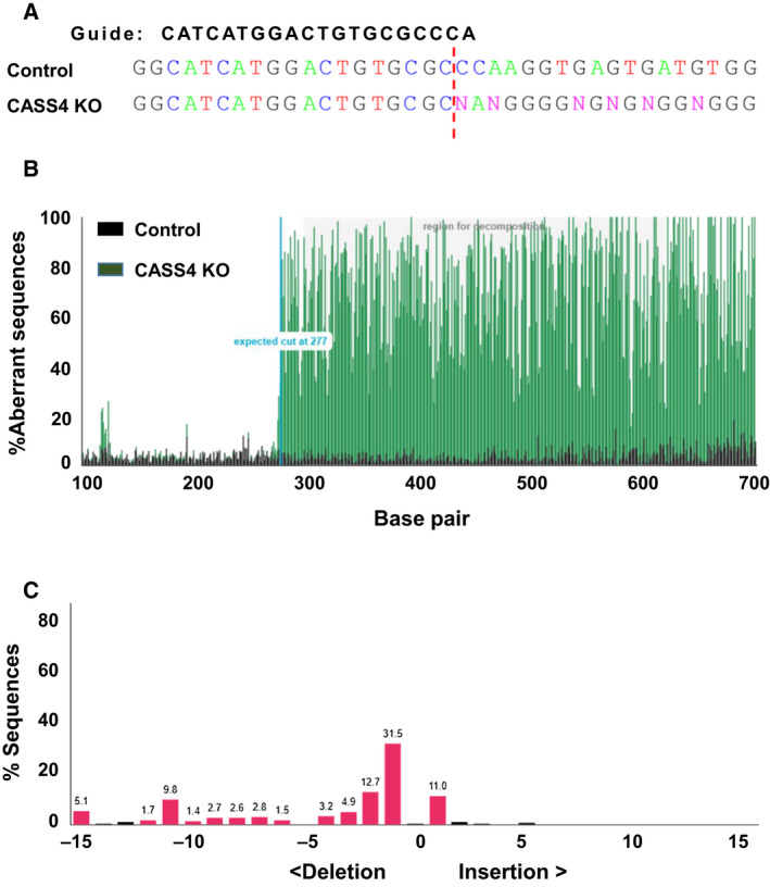
We first identified the candidate genes based on the reported GWAS of AD [14]. Meta‐analysis of different associational studies has confirmed the importance of 22 genes as AD risk modifiers (Table 1) [16]. We targeted each gene individually with gRNAs (Table 2). The gRNAs were cloned into a lentivirus construct [27] and transduced into cultured cells. Cells were cultured for 10 days in the presence of puromycin to select for stable integration of the virus and presumed genetic disruption. We confirmed genetic disruption of each gene using TIDE, a method based on sequencing the target genes to detect disruption of the sequence through insertion/deletion (indel) at the site of gRNA binding [24]. This confirmed high‐frequency indels at each of the genes targeted by our constructs (see Table 2; Fig. 1A–C for an example). We selected the gRNAs with high indel efficiency (> 80%) in TIDE analysis (Table 2) and used those gRNAs for subsequent assays. We noted that six genes are reportedly expressed at very low or undetectable levels in HEK293 cells (Table 2), but nonetheless carried these through in our analyses. Knockout of PICALM, CD2AP, and CELF1 resulted in slow growth rate and some cell death, whereas knockout of FERMT2 resulted in cell growth delay as well as some obvious deformity in cell shape. The cells looked more spherical in shape. Knockout of the other genes did not create any visible phenotype.
Fig. 1.

Analysis of gene editing efficiency by TIDE. (A) Representative image of one of the genes targeted with CRISPR/Cas9. After lentivirus exposure, cells were cultured for 2 weeks in puromycin. DNA was isolated from control (scrambled gRNA treated) and CASS4 KO, and Sanger‐sequenced. The gRNA sequence used for making the knockout is shown in bold. The predicted Cas9 cut site is shown as a red dotted line. The insertions/deletions (N) can be seen in the CASS4 KO. (B) Representative image from TIDE analysis of CASS4. Plot shows the overlay of sequence between scrambled control and CASS4. The increase in aberrant sequence after the expected cut site is evident in the CASS4 KO sample, indicating effective gene disruption. (C) Plots represent the spectrum of indels and their frequencies for CASS4. R 2 = 0.94. The plot was analyzed and derived from the TIDE web tool (https://tide.nki.nl/).
AD GWAS gene disruption does not affect tau uptake
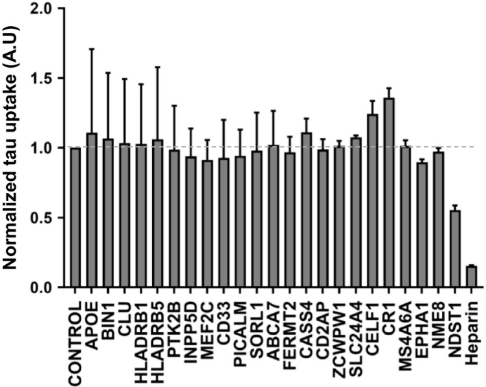
Heparan sulfate proteoglycans (HSPGs) play a critical role in binding tau aggregates, mediating their uptake, and seeding activity [17, 18]. Compounds such as heparin or similar small molecules, which bind tau assemblies and compete for their binding to HSPGs, block tau uptake [18]. We tested the role of GWAS genes by evaluating HEK293T cells in which we had individually disrupted each gene. As a positive control, we knocked out NDST1, which we have previously determined to be required for proper HSPG sulfation and to mediate tau uptake [18]. We prepared full‐length tau(2N4R) fibrils and labeled them using Alexa Fluor 647 via succinimidyl ester amine reaction. We applied labeled tau fibrils to cultured cells for 4 h, followed by washing, trypsin treatment (to digest extracellular tau and release cells from the culture plate), and analysis by flow cytometry according to prior methods [17]. We observed no effect of GWAS gene knockout on tau uptake (Fig. 2), whereas heparin treatment reduced uptake approximately 90%, and NDST1 knockdown reduced uptake approximately 50% (Fig. 2).
Fig. 2.

Knockout of GWAS genes does not modify uptake of tau. GWAS genes were individually targeted in HEK293T cells using CRISPR/Cas9 to create polyclonal knockout cell lines. The cell lines were then tested for internalization of fluorescently labeled tau aggregates by measuring MFI per cell with flow cytometry. None of the gene knockouts changed tau uptake. Data were collected in triplicate and normalized to uptake from control cells treated with scrambled gRNA. The X‐axis indicates the targeted genes, and the Y‐axis indicates tau uptake relative to scrambled gRNA. Heparin and NDST1 were used as positive controls for uptake inhibition. Error bars indicate the SEM. The dotted gray line represents the value of control cells. One‐way ANOVA was used to analyze the result and test for statistically significant differences.
AD GWAS gene disruption does not affect tau seeding
An exogenous tau assembly that gains entry to the cytoplasm acts as a template to convert endogenous tau to a fibrillar form, a process termed ‘seeding’. Seeding is initiated by application of relatively low concentrations of tau assemblies to the cell media. In the absence of additional reagents, these assemblies bind HSPGs, are internalized, and initiate seeding reactions. This type of seeding, which we have termed ‘naked’, is relatively inefficient and typically results in conversion of approximately 1–5% of the cells to an aggregated state. When the tau repeat domain containing a single disease‐associated mutation (P301S) is fused to mClover3 (CL) or mCerulean3 (C) (or a similarly compatible fluorescent protein pair), aggregation enables FRET induced by proximity of the fluorescent proteins. This allows quantitation of seeding activity by flow cytometry.
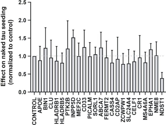
As an alternative, incubation of tau seeds with Lipofectamine 2000 (or a similar reagent) enables transduction of seeds with very high efficiency, approximately 100‐fold more than naked seeding. We expressed tau RD(P301S)‐C/CL in HEK293T cells to form a monoclonal ‘biosensor’ line with high sensitivity to exogenous tau aggregates, similar to a line previously reported [26]. To test the role of GWAS genes in the tau seeding process, we knocked out each in the biosensor cells. These cells were then treated with exogenous tau fibrils alone, or with Lipofectamine 2000 [19]. We measured seeding activity by quantitative flow cytometry. We observed no significant change in naked seeding across all AD GWAS genes (Fig. 3). We observed minor differences in seeding in INPP5D and NME8 knockout cells with Lipofectamine‐mediated seeding (Fig. 4) but that change was not consistent with naked seeding. In conclusion, we observed no consistent significant effect of GWAS gene knockout upon tau seeding.
Fig. 3.

Knockout of AD GWAS genes does not modify naked tau seeding. GWAS genes were individually targeted in HEK293T RD(P301S)‐C/CL biosensor cells using CRISPR/Cas9 to create polyclonal knockout cell lines, which were cultured for 2 weeks in the presence of puromycin. Recombinant tau fibrils were added to those cells to induce seeding. Seeding was quantified using FRET, and the percentage of FRET‐positive cells was normalized to the scrambled gRNA. Data were collected in triplicate. The X‐axis indicates the targeted genes, and the Y‐axis indicates normalized seeding activity. None of the genes modified the seeding efficiency. Heparin and NDST1 were positive controls for uptake inhibition. Error bars indicate the SEM. The dotted gray line represents the value of control cells. One‐way ANOVA was used to analyze the result and test for statistically significant differences.
Fig. 4.

Knockout of AD GWAS genes does not modify Lipofectamine‐mediated tau seeding. AD GWAS genes were individually targeted in HEK293T RD(P301S)‐C/CL biosensor cells using CRISPR/Cas9 to create polyclonal knockout cell lines, which were cultured for 2 weeks in the presence of puromycin. Recombinant tau fibrils were mixed with Lipofectamine 2000 to facilitate direct delivery to the cytoplasm. Seeding was quantified using FRET. Data were collected in triplicate. The X‐axis indicates the targeted genes, and the Y‐axis represents percentage of FRET‐positive cells. No knockout modified seeding efficiency. Error bars indicate the SEM. The dotted gray line represents the value of control cells. One‐way ANOVA was used to analyze the result and test for statistically significant differences.
GWAS gene disruption does not affect tau aggregate maintenance
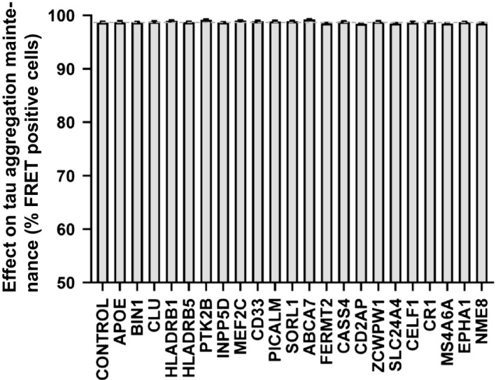
Previous studies have shown that dividing cells propagate tau aggregates of distinct conformation, termed strains, that transmit pathology between animals, and specify unique pathologies [7, 20]. Studies of yeast prions indicate that aggregate propagation requires accessory factors, for example, Hsp104 [28], and thus we hypothesized AD GWAS genes might affect this process. We previously created a cell line that propagated a distinct tau strain, termed LM39‐9. These cells constitutively express aggregates of tau RD containing two disease‐associated mutations (P301L/V337M) that are fused to cyan and yellow fluorescent proteins (which constitute a FRET pair). LM39‐9 cells exhibit high aggregate transmission efficiency (~ 99%), which can be easily tracked over time by flow cytometry. We used lentivirus to individually disrupt each of the AD GWAS genes, cultured the LM39‐9 cells for 2 weeks, and then quantified the percentage of cells containing aggregates using flow cytometry. We observed no loss of aggregation following disruption of any GWAS gene (Fig. 5), indicating none was critical to aggregate maintenance in this cell model.
Fig. 5.

Knockout of AD GWAS genes does not modify tau aggregate maintenance. AD GWAS genes were individually targeted in LM39‐9 cells using CRISPR/Cas9 to create polyclonal knockout cell lines, which were cultured for 2 weeks in the presence of puromycin. The cell lines were tested for the loss of tau aggregates using FRET flow cytometry. The X‐axis indicates the targeted genes, and the Y‐axis represents percentage of FRET‐positive cells. None of the genes inhibited aggregate maintenance within the LM‐39‐9 cell lines. Error bars indicate the SEM. The dotted gray line represents the value of control cells. One‐way ANOVA was used to analyze the result and test for statistically significant differences.
Discussion
The mechanisms that govern prion activities of tau are largely unknown. AD GWAS genes are putative modifiers of pathogenesis and are thus of interest. Consequently, in this work we tested the hypothesis that AD GWAS genes would impact uptake, seeding, or aggregate maintenance of tau, which may play a critical role in progression of disease. We confirmed that each of the 22 gRNAs disrupted their target genes at high frequency. We then studied the effects of the knockouts across a range of putative steps in pathogenesis for which we have previously developed quantitative cell‐based assays. We did not observe any impact on knockout in any of the fundamental events of tau aggregate propagation that we can measure in simple cell systems.
Alzheimer's disease and related dementias involve progressive accumulation of tau assemblies in neurons and glia. If tau propagation underlies these disorders, it is conceivable that many cellular mechanisms could be specific to cells of the brain. In this case, modeling these processes in simple cultured cell systems as we have done might not be particularly productive and this may explain why we did not observe any effect of AD GWAS genes on the components of tau pathology we measured in HEK293T cells. Six of the genes studied (INPP5D, CD33, CR1, MS4A6A, EPHA1, NME8) are reportedly not expressed at high levels in these cells and thus cannot be completely excluded as not important for tau prion propagation (although they are clearly not required for this process to occur in HEK293T cells). Due to the impracticality of optimizing detection methods to measure expression levels of multiple proteins, we confirmed the function of our knockout vectors by sequencing the target genes.
Future studies in neurons, which present additional challenges to the screening studies we have performed here, would give more clarity on the function of these genes in relation to AD pathology. However, the HEK293T models have previously proven very useful in defining modes of cell uptake of pathological tau assemblies, seeding, and strain maintenance that have translated well to primary neurons and mouse models [7, 17, 18]. Similarly, these simple systems have readily propagated unique tau strains derived from recombinant fibrils and human tauopathy brains that can be transmitted and propagated in animal models [7, 29]. We fully recognize that without extension of findings derived from simple systems such as these into animal or even human studies, it will be difficult to know how these simple models reflect actual events in the brain.
The relationship of AD GWAS to AD pathogenesis is complex, as hits may involve genes that are not directly involved in cell‐autonomous aspects of tau propagation. For example, genes associated with microglial function, such as TREM2, would not be expected to score positive in these studies and it is important to note that some of the AD GWAS genes are enriched in microglia. Additionally, while this study was limited to the study of AD GWAS genes related to tau uptake, seeding, and aggregate maintenance, clearly there are many realms of cell biology at play that should be studied further. For example, loss of protein homeostasis could play an important role in age‐related disorders like AD [30], and a recent study showed the importance of protein homeostasis in neurodegenerative diseases [31]. Despite obvious caveats of the present study, specifically the use of an HEK293T cell system for tau uptake, seeding, and aggregate maintenance, we hope this work will be useful for those interested in using reductionist cell models to study the role of genes involved in fundamental events of tau propagation.
Conflict of interest
The authors declare no conflict of interest.
Author contributions
MID and SK designed research; SK performed research; and MID and SK wrote the manuscript.
Acknowledgements
The study was supported by the Rainwater Charitable Foundation and the Crowley Foundation.
Data accessibility
All data generated and analyzed during this study are included in this article.
References
- 1. Lee VM, Goedert M and Trojanowski JQ (2001) Neurodegenerative tauopathies. Annu Rev Neurosci 24, 1121–1159. [DOI] [PubMed] [Google Scholar]
- 2. Braak H and Braak E (1991) Neuropathological stageing of Alzheimer‐related changes. Acta Neuropathol 82, 239–259. [DOI] [PubMed] [Google Scholar]
- 3. Seeley WW, Crawford RK, Zhou J, Miller BL and Greicius MD (2009) Neurodegenerative diseases target large‐scale human brain networks. Neuron 62, 42–52. [DOI] [PMC free article] [PubMed] [Google Scholar]
- 4. Frost B, Ollesch J, Wille H and Diamond MI (2009) Conformational diversity of wild‐type Tau fibrils specified by templated conformation change. J Biol Chem 284, 3546–3551. [DOI] [PMC free article] [PubMed] [Google Scholar]
- 5. Frost B, Jacks RL and Diamond MI (2009) Propagation of tau misfolding from the outside to the inside of a cell. J Biol Chem 284, 12845–12852. [DOI] [PMC free article] [PubMed] [Google Scholar]
- 6. Clavaguera F, Bolmont T, Crowther RA, Abramowski D, Frank S, Probst A, Fraser G, Stalder AK, Beibel M, Staufenbiel M et al (2009) Transmission and spreading of tauopathy in transgenic mouse brain. Nat Cell Biol 11, 909–913. [DOI] [PMC free article] [PubMed] [Google Scholar]
- 7. Sanders DW, Kaufman SK, Devos SL, Sharma AM, Mirbaha H, Li A, Barker SJ, Foley AC, Thorpe JR, Serpell LC et al (2014) Distinct tau prion strains propagate in cells and mice and define different tauopathies. Neuron 82, 1271–1288. [DOI] [PMC free article] [PubMed] [Google Scholar]
- 8. Surmeier DJ, Obeso JA and Halliday GM (2017) Selective neuronal vulnerability in Parkinson disease. Nat Rev Neurosci 18, 101–113. [DOI] [PMC free article] [PubMed] [Google Scholar]
- 9. Walsh DM and Selkoe DJ (2016) A critical appraisal of the pathogenic protein spread hypothesis of neurodegeneration. Nat Rev Neurosci 17, 251–260. [DOI] [PMC free article] [PubMed] [Google Scholar]
- 10. Guo JL and Lee VM‐Y (2011) Seeding of normal Tau by pathological Tau conformers drives pathogenesis of Alzheimer‐like tangles. J Biol Chem 286, 15317–15331. [DOI] [PMC free article] [PubMed] [Google Scholar]
- 11. Iba M, Guo JL, McBride JD, Zhang B, Trojanowski JQ and Lee VM‐Y (2013) Synthetic tau fibrils mediate transmission of neurofibrillary tangles in a transgenic mouse model of Alzheimer's‐like tauopathy. J Neurosci 33, 1024–1037. [DOI] [PMC free article] [PubMed] [Google Scholar]
- 12. Woerman AL, Aoyagi A, Patel S, Kazmi SA, Lobach I, Grinberg LT, McKee AC, Seeley WW, Olson SH, Prusiner SB et al (2016) Tau prions from Alzheimer's disease and chronic traumatic encephalopathy patients propagate in cultured cells. Proc Natl Acad Sci USA 113, E8187–E8196. [DOI] [PMC free article] [PubMed] [Google Scholar]
- 13. Gallardo G and Holtzman DM (2017) Antibody therapeutics targeting Aβ and Tau. Cold Spring Harb Perspect Med 7, a024331. [DOI] [PMC free article] [PubMed] [Google Scholar]
- 14. Cuyvers E and Sleegers K (2016) Genetic variations underlying Alzheimer's disease: evidence from genome‐wide association studies and beyond. Lancet Neurol 15, 857–868. [DOI] [PubMed] [Google Scholar]
- 15. Lambert JC, Ibrahim‐Verbaas CA, Harold D, Naj AC, Sims R, Bellenguez C, DeStafano AL, Bis JC, Beecham GW, Grenier‐Boley B et al (2013) Meta‐analysis of 74,046 individuals identifies 11 new susceptibility loci for Alzheimer's disease. Nat Genet 45, 1452–1458. [DOI] [PMC free article] [PubMed] [Google Scholar]
- 16. Van Cauwenberghe C, Van Broeckhoven C and Sleegers K (2016) The genetic landscape of Alzheimer disease: clinical implications and perspectives. Genet Med 18, 421–430. [DOI] [PMC free article] [PubMed] [Google Scholar]
- 17. Holmes BB, Devos SL, Kfoury N, Li M, Jacks R, Yanamandra K, Ouidja MO, Brodsky FM, Marasa J, Bagchi DP et al (2013) Heparan sulfate proteoglycans mediate internalization and propagation of specific proteopathic seeds. Proc Natl Acad Sci USA 110, E3138–E3147. [DOI] [PMC free article] [PubMed] [Google Scholar]
- 18. Stopschinski BE, Holmes BB, Miller GM, Manon VA, Vaquer‐Alicea J, Prueitt WL, Hsieh‐Wilson LC and Diamond MI (2018) Specific glycosaminoglycan chain length and sulfation patterns are required for cell uptake of tau vs. α‐synuclein and β‐amyloid aggregates. J Biol Chem 293, 10826–10840. [DOI] [PMC free article] [PubMed] [Google Scholar]
- 19. Holmes BB, Furman JL, Mahan TE, Yamasaki TR, Mirbaha H, Eades WC, Belaygorod L, Cairns NJ, Holtzman DM and Diamond MI (2014) Proteopathic tau seeding predicts tauopathy in vivo. Proc Natl Acad Sci USA 111, E4376–E4385. [DOI] [PMC free article] [PubMed] [Google Scholar]
- 20. Kaufman SK, Sanders DW, Thomas TL, Ruchinskas AJ, Vaquer‐Alicea J, Sharma AM, Miller TM and Diamond MI (2016) Tau prion strains dictate patterns of cell pathology, progression rate, and regional vulnerability in vivo. Neuron 92, 796–812. [DOI] [PMC free article] [PubMed] [Google Scholar]
- 21. Sanjana NE, Shalem O and Zhang F (2014) Improved vectors and genome‐wide libraries for CRISPR screening. Nat Methods 11, 783–784. [DOI] [PMC free article] [PubMed] [Google Scholar]
- 22. Doench JG, Fusi N, Sullender M, Hegde M, Vaimberg EW, Donovan KF, Smith I, Tothova Z, Wilen C, Orchard R et al (2016) Optimized sgRNA design to maximize activity and minimize off‐target effects of CRISPR‐Cas9. Nat Biotechnol 34, 184–191. [DOI] [PMC free article] [PubMed] [Google Scholar]
- 23. Araki T, Sasaki Y and Milbrandt J (2004) Increased nuclear NAD biosynthesis and SIRT1 activation prevent axonal degeneration. Science 305, 1010–1013. [DOI] [PubMed] [Google Scholar]
- 24. Brinkman EK, Chen T, Amendola M and van Steensel B (2014) Easy quantitative assessment of genome editing by sequence trace decomposition. Nucleic Acids Res 42, e168. [DOI] [PMC free article] [PubMed] [Google Scholar]
- 25. Furman JL, Holmes BB and Diamond MI (2015) Sensitive detection of proteopathic seeding activity with FRET flow cytometry. J Vis Exp 106, e53205. [DOI] [PMC free article] [PubMed] [Google Scholar]
- 26. Furman JL and Diamond MI (2017) FRET and flow cytometry assays to measure proteopathic seeding activity in biological samples. Methods Mol Biol 1523, 349–359. [DOI] [PubMed] [Google Scholar]
- 27. Shalem O, Sanjana NE, Hartenian E, Shi X, Scott DA, Mikkelsen TS, Heckl D, Ebert BL, Root DE, Doench JG et al (2014) Genome‐scale CRISPR‐Cas9 knockout screening in human cells. Science 343, 84–87. [DOI] [PMC free article] [PubMed] [Google Scholar]
- 28. Chernova TA, Wilkinson KD and Chernoff YO (2017) Prions, chaperones, and proteostasis in yeast. Cold Spring Harb Perspect Biol 9, a023663. [DOI] [PMC free article] [PubMed] [Google Scholar]
- 29. Kaufman SK, Del Tredici K, Thomas TL, Braak H and Diamond MI (2018) Tau seeding activity begins in the transentorhinal/entorhinal regions and anticipates phospho‐tau pathology in Alzheimer's disease and PART. Acta Neuropathol 521, 4339–4311. [DOI] [PMC free article] [PubMed] [Google Scholar]
- 30. Freer R, Sormanni P, Vecchi G, Ciryam P, Dobson CM and Vendruscolo M (2016) A protein homeostasis signature in healthy brains recapitulates tissue vulnerability to Alzheimer's disease. Sci Adv 2, e1600947. [DOI] [PMC free article] [PubMed] [Google Scholar]
- 31. Hongjun F, Possenti A, Freer R, Nakano Y, Hernandez NC, Villegas MT, Cauhy PVM, Lassus BA, Chen S, Fowler SL et al (2019) A tau homeostasis signature is linked with the cellular and regional vulnerability of excitatory neurons to tau pathology. Nat Neurosci 22, 47–56. [DOI] [PMC free article] [PubMed] [Google Scholar]
Associated Data
This section collects any data citations, data availability statements, or supplementary materials included in this article.
Data Availability Statement
All data generated and analyzed during this study are included in this article.


