Abstract
Simple Summary
Yield and quality of goat milk are important indexes for screening dairy goat breeds. Therefore, it is necessary for us to improve the yield and quality of goat milk. In this study, we demonstrated that circRNA8220/miR-8516/STC2 could promote the synthesis of β-casein and triglyceride through PI3K/AKT/mTOR pathway. In addition, we found that circRNA8220/miR-8516/STC2 also promote proliferation via Ras/MEK/ERK pathway in goat mammary epithelial cells (GMECs). These findings contribute to a better understanding of circRNA-controlled breast development and lactation mechanisms and provide new potential insights into the regulation of breast development and milk composition in dairy goats.
Abstract
Circular RNAs (circRNAs), which are considered a large class of endogenous noncoding RNAs, function as regulators in various biological procedures. In this study, the function and molecular mechanisms of circRNA8220 in goat mammary epithelial cells (GMECs) were explored. CircRNA8220 could spong miR-8516 and block the function of miR-8516 by binding to the target site of miR-8516 a negative feedback relationship existed between circRNA8220 and miR-8516. Stanniocalcin 2 (STC2) was a target gene of miR-8516. circRNA8220 could up-regulate the expression of STC2 by sponging miR-8516 in GMECs. circRNA8220/miR-8516/STC2 could promote proliferation and enhance the synthesis of β-casein and triglycerides (TG) via Ras/MEK/ERK and PI3K/AKT/mTOR signaling pathways, respectively.
Keywords: circRNA8220, miR-8516, STC2, β-casein, triglycerides
1. Introduction
Mammary epithelial cells are mammalian secretory cells, which are the basis of lactation in goat mammary gland. The number and activity of mammary epithelial cells are closely related to breast lactation and play an important role in breast development [1]. Hypoplasia of mammary gland leads to decrease of milk production result in economic losses [2]. Therefore, a better understanding of molecular mechanisms underlying the development of mammary epithelial cells for increasing milk production is critical. However, the underlying molecular mechanisms of lactation in dairy goats is not clear.
Circular RNAs (circRNAs), which are RNA molecules that are characterised by covalently closed loop structures, down-regulate gene expression at the transcriptional or post-transcriptional level by binding to miRNAs or other molecules [3]. For example, circRNA_100290 regulates CDK6 expression by sponging miR-29b family members [4]. Hsa_circRNA_103809 promotes the expression of ZNF121 by sponging miR-4302 in lung cancer cells [5]. To date, many functions of circRNA have been found; for example, circRNA_100782 plays a role in proliferation regulation of pancreatic carcinoma [6]. Antagonistic roles of circRNA_100338 and miR-141-3p have been found in the regulation of invasive potential in liver cancer cells [7]. We selected circRNA8220 from circRNA libraries that were constructed by our team through bioinformatics prediction approach. However, the regulatory mechanism of circRNA8220 on milk synthesis and proliferation in goat mammary epithelial cells (GMECs) are still unclear.
miRNAs, which are noncoding single-stranded RNA molecules, regulate gene expression by binding to the 3′-untranslated region (UTRs), 5′-UTRs and even coding sequences of their target mRNAs; then, they affect a series of physiological functions in animals [8,9,10]. Incremental proofs demonstrate that miRNAs have an important regulatory function on milk synthesis [11,12]. The expression of miR-8516 has been found to be significantly different in peak lactation compared with prelactation [13]. However, no research on miR-8516 and effect of miR-8516 is available. Thus, studying the function of target gene of miR-8516 in GMECs is interesting.
Stanniocalcin 2 (STC2), which is a glycoprotein hormone, regulates a series of biological processes in an autocrine or paracrine manner [14], such as cell proliferation [15], tumorigenesis [16] and atherosclerosis [17]. STC2 promotes colorectal cancer development and epithelial–mesenchymal transition process via activating MEK/ERK and PI3K/AKT signaling pathways [16]. However, the function of STC2 in milk synthesis, proliferation and apoptosis of GMECs is unknown.
On the basis of these considerations, we decided to explore the function and mechanism of circRNA8220 and STC2 in GMECs in vitro. Our findings revealed that circ-8220 could bind to miR-8516 via the target site inhibit miR-8516 activity lead to increase of STC2 expression, promote GMECs proliferation and enhance the synthesis of β-casein and triglycerides (TG) via Ras/MEK/ERK and PI3K/AKT/mTOR signaling pathways, respectively. In addition, this study explored the effects of circ-8220 and STC2 on proliferation and lactation in vitro.
2. Materials and Methods
2.1. Animals and Cell Culture
The Guanzhong dairy goats were selected from Longxian Goat Breeding Center near Northwest A&F University of China. Breast tissues were collected periods from three healthy Guanzhong dairy goats (3 year olds) at the peak of lactation by surgery. First of all, one side of the mammary gland of the dairy goat was wiped clean with alcohol then local was performed on the mammary tissue. A 5–6-cm incision was made and the skin pulled back using sterile forceps, exposing the mammary tissue. 1 cm2 samples were taken using a sterile scalpel blade and forceps. In order to stop any external bleeding, pressure was applied with sterile gauze to stop any external bleeding. The incision was closed with 6 to 8 surgical staples (#89063337, Appose ULC Skin Stapler, 35 wide; Henry Schein Inc., Melville, NY, USA). Mammary gland tissue was stored in PBS, added with 100 μg/mL streptomycin and 100 μg/mL penicillin and transferred to the laboratory within 1 h. The GMECs were cultivated in DMEM/F12 medium (Gibco, CA, USA) containing 10% foetal bovine serum, 10 ng/mL epidermal growth factor 1 (EGF-1, Gibco, CA, USA), 5 mg/mL insulin, 100 U/mL streptomycin/mL penicillin and 0.3 mmol/L hydrocortisone at 37 °C in a humidified atmosphere with 5% CO2. Human embryonic kidney cell line (HEK293T) was cultured in high-glucose DMEM (Dulbecco’s Modified Eagle Medium) supplemented with 10% fetal bovine serum, 100 μg/mL penicillin and 100 μg/mL streptomycin at 37 °C and 5% CO2 to proliferate. The GMECs were purified and cultured depending on previous reports [18].
2.2. pcDNA3.1-STC2 Vector Construction
Goat STC2 CDs sequence (XM_005694539.3) was obtained by PCR using total RNA extract from GMECs, inserted into pMD™19-T vector (TaKaRa, Beijing, China) and entirely sequenced. Thereafter, STC2 CD sequence was inserted into pcDNA3.1 vector (Thermo Fisher, Shanghai, China) between the Xho I and Kpn I sites. The insertion of the sequence into pcDNA3.1 vector was checked by entire sequencing. The forward primer of STC2 was (Hind III) 5′-CTTAAGCTTATGTGTGCCGAGCGGCTG-3′ the reverse primer was (Not I) 5′-CGAGCGGCCGCTCACCTCCGGATATCGGAATACTCA-3′.
2.3. pCD5-circRNA8220 Vector Construction
Goat circRNA8220 full length was obtained by PCR and inserted into pMD™19-T vector. Thereafter, the sequence was subcloned into the pCD5-ciR vector (Geneseed, Guangzhou, China) between EcoRI and BamHI sites. The forward primer of circRNA8220 was (EcoRI) 5′-CGGAATTCTAATACTTTCAGCTGGAGCTGATTGGACACAAT-3′ the reverse primer was (BamHI) 5′-CGGGATCCAGTTGTTCTTACCTCTCATCACAGTAGGTGAAC-3′.
2.4. Transfection and RT-qPCR
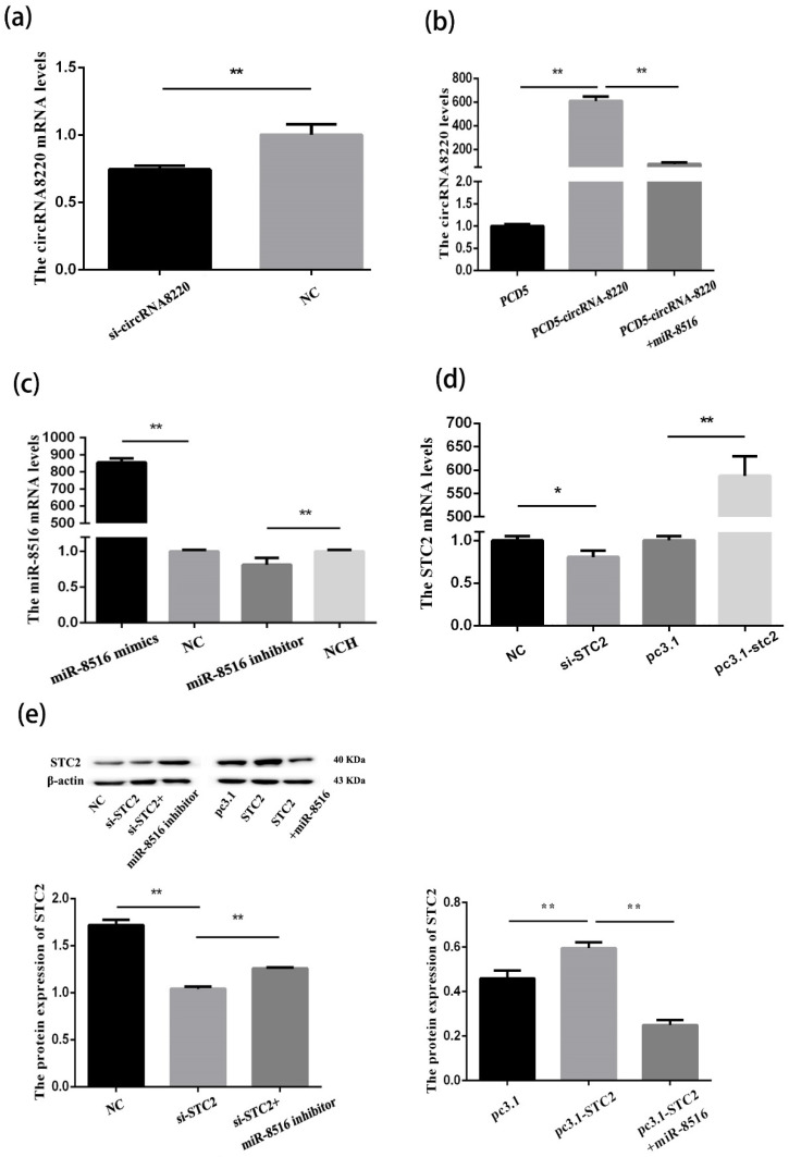
When GMEC density reached 70–80%, pcDNA3.1-STC2 and pCD5-circRNA8220 vectors, STC2-siRNA, circRNA8220-siRNA and miR-8516 mimics/inhibitors and NC and inhibitor NC (GenePharma) were transfected using Lipofectamine™ RNAiMAX Reagent (Invitrogen, CA, USA) following the specifications of the manufacturer. At 24 or 48 h later, total RNA was extracted using Trizol reagent (TaKaRa, Dalin, China) and converted to cDNA using the Prime Script RT reagent kit with gDNA eraser (TaKaRa, Dalin, China) in accordance with the specifications of the manufacturer. The mRNA concentrations of interest were quantified via SYBR Premix Ex Taq II (TaKaRa, Beijing, China). The results were acquired from the CFX Connect Real-Time PCR Detection System (Bio-Rad, CA, USA). The PCR reaction procedure was as follows: 95 °C for 10 min 40 cycles at 94 °C for 15 s, 60 °C for 30 s and 72 °C for 30 s. Gene expression compared with β-actin or U6 mRNA level was calculated by the 2−ΔΔCt method. All primers for RT-qPCR are shown in Appendix A Table A1. The transfection efficiencies of pcDNA3.1-STC2 and pCD5-circRNA8220 vectors and STC2-siRNA, circRNA8220-siRNA and miR-8516 mimics/inhibitors (GenePharma) are shown in Appendix A Figure A1.
2.5. Luciferase Reporter Assay
To explore miR-8516 whether target gene STC2 and circRNA8220, 260 bp sequence of STC2 3′UTR and 344 bp total sequence of circRNA8220 were cloned into the psiCHECK-2 vectors (Addgene, CA, USA), respectively. The mutated plasmids with mutated target site (psiCHECK-2-Mut) were also constructed. The wild-type (psiCHECK-2-WT) or mutated (psiCHECK-2-Mut) plasmids were co-transfected with miR-8516 mimic or inhibitor into 293T cells using Lipofectamine™ RNAiMAX Reagent. At 24 h later, the result was obtained from thermo scientific varioskan flash (Thermo scientific, USA) using the Dual-Glo luciferase system (Promega, USA). The primer sequences are shown in Appendix A Table A1. Each experiment was performed three times in triplicate.
2.6. Cell Apoptosis and Proliferation Assay
The apoptosis ratio of GMECs was detected using Annexin V-FITC PI staining apoptosis assay kit (SeaBiotech, Shanghai, China) by flow cytometry method (FCM) after 24 h transfection following the protocol of the manufacturer. The viability of GMECs was tested by CCK8 assay. A total of 10 μL CCK8 solution (ZETATM life, USA) was put in each well after 24 h transfection. The resultant was then incubated in 96-well plates at 37 °C for 2 h. The results were calculated at 450 nm using an epoch microplate reader (Biotek, Winooski, USA).
To prove the credibility of the proliferation assay results in GMECs, the 5-Ethynyl-2′-deoxyuridine (Edu) assay was implemented in 96-well plates after GMECs were treated for 24 h. The cells were then washed by PBS three times after dyeing with Edu (Ribobio, Guangzhou, China) with a final concentration of 50 μM for 2 h. Thereafter, the cells were dyed with DAPI for 15 min in 37 °C. The result was observed by fluorescence microscopy.
2.7. Detecting the Concentration of β-casein and TG
Supernatant of cell culture medium and the cells were collected after 24 h transfection. The concentrations of β-casein and TG (triglyceride) were detected using Goat β-casein ELISA (Enzyme-linked immunosorbent assay) KIT (MLBIO, Shanghai, China) and Goat TG ELISA KIT (MLBIO, Shanghai, China) in accordance with the instructions of the manufacturer. The results were obtained from an epoch microplate reader (Biotek, Winooski, USA) at 450 nm. Concentrations were calculated in accordance with the standard curve. Variable coefficient values of intra- and inter-assay were less than 15% and 10%, respectively.
2.8. Western Blot
Proteins were extracted from cells by adding RIPA (Radio Immunoprecipitation Assay) lysis buffer (Biotek, Beijing, China) and mixed with phenylmethanesulfonyl fluoride (PMSF, Solarbio, Beijing, China) at 0.1 mg/mL after 48 h post-transfection. The concentration of proteins (GMECs) was detected with the BCA (Bicinchoninic Acid) protein assay kit (Vazyme Biotech, Nanjing, China) approximately 30 μg total protein was added a 12% SDS–PAGE (polyacrylamide gelelectrophoresis). The separated proteins were transferred onto polyvinylidene difluoride membrane (PVDF, Merck Millipore, MA, USA). The polyvinylidene difluoride membrane was immersed in 10% skimmed milk powder and diluted with Tris-buffered saline inclusive of 0.1% Tween 20 (pH 7.6) for 2.5 h at room temperature. The membrane was immersed in corresponding primary antibodies overnight at 4 °C (Table 1). In the next step, the membrane was incubated in suitable HRP (horseradish peroxidases)-conjugated secondary antibodies against mouse, rabbit at 4 °C for 2 h. Proteins were visualised using ECL prime western blotting detection reagent (Amersham, GE Healthcare Lifesciences) by gel documentation system (Biospectrum 410, UVP). Proteins were quantified by the Quantity One program (Bio-Rad, CA, USA).
Table 1.
Antibody used in this study.
| Name | Manufacturer | Product Number |
|---|---|---|
| β-Actin | Beyotime, Shanghai, China | AA128 |
| STC2 | GeneTex, Alton Pkwylrvine, USA | GTX82231 |
| Ras | Gene Tex, America | GTX132480 |
| p-MEK1 (Ser298) | Abways, Shanghai, China | CY5854 |
| MEK1 | Abways, Shanghai, China | CY5168 |
| p-ERK1/2 (Thr202/Tyr204) | Abways Shanghai, China | CY5277 |
| ERK1/2 | Abways, Shanghai, China | CY5487 |
| p-PI3K (Tyr607) | Abways, Shanghai, China | CY6427 |
| PI3K | Abways, Shanghai, China | CY6915 |
| p-AKT (Ser473) | Cell Signaling, America | #9271 |
| AKT | Cell Signaling, America | #9272 |
| p-mTOR | Abways, Shanghai, China | CY6571 |
| mTOR | Abways, Shanghai, China | CY5306 |
| p-S6K (Ser424) | Abways, Shanghai, China | CY5261 |
| S6K | Abways, Shanghai, China | CY5365 |
| Bax | Abways, Shanghai, China | CY5059 |
| Bcl-2 | Abways, Shanghai, China | CY5032 |
| Caspase 3 | Cell Signaling, America | #9662 |
| Caspase 9 | Abways, Shanghai, China | CY5682 |
| HRP-labeled Goat Anti-Rabbit IgG (H + L) | Beyotime, Shanghai, China | A0208 |
| HRP-labeled Goat Anti-Mouse IgG (H + L) | Beyotime, Shanghai, China | A0216 |
2.9. Statistical Analysis
Each experiment was repeated at least three times in biological and technical repeats. All the data were processed by SPSS 19.0 (Beijing, China). The results were shown as means ± SE (standard error) the differences were compared by one-way ANOVA (** p < 0.01, * p < 0.05).
3. Results
3.1. STC2 Was a Target Gene of miR-8516 in GMECs
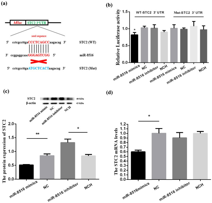
TargetScan database (targetscan.org) was used to select a target gene of miR-8516 in GMECs. We chose wild-type (WT) STC2-3′UTR or mutant (MUT)-STC2-3′UTR to set up a two-tier luciferase assay with psiCHECK-2 vector. The results showed that the relative luciferase activities of co-transfection with miR-8516 mimics and psC-STC2-3′UTR-WT were significantly attenuated compared with co-transfection with NC and psC-STC2-3′UTR-WT; by contrast, no change was observed in the psC-STC2-3′UTR-MUT (Figure 1a,b).
Figure 1.
MiR-8516 down-regulated the expression level of STC2 via the 3′UTR. (a) The seed sequence of miR-8516 could match with WT-STC2-3′UTR and could not match with Mut-STC2-3′UTR. (b) Luciferase reporter assay of 293T cells co-transfected with WT-STC2-3′UTR or Mut-STC2-3′UTR and miR-8516 mimic, NC (negative control), miR-8516 inhibitor or NCH (NC-inhibitor). (c) miR-8516 decreased STC2 protein level in GMECs. (d) miR-8516 decreased STC2 mRNA level in GMECs. ** p < 0.01, * p < 0.05.
Western blot and RT-PCR (Quantitative real-time polymerase chain reaction) were used to detect the influence of miR-8516 on mRNA and proteins of STC2. The expression levels of miR-8516 were significantly increased or decreased by transfecting miR-8516 mimic or miR-8516 inhibitor (Appendix A Figure A1). The results showed that overexpression of miR-8516 significantly decreased mRNA and proteins levels of STC2 down-regulation of the expression of miR-8516 increased the proteins levels of STC2 (Figure 1c,d). In summary, these results proved that miR-8516 acts as a demotivated regulator of STC2 by directly binding to 3′UTR in GMECs in vitro.
3.2. CircRNA8220 Acted as a Sponge for miR-8516
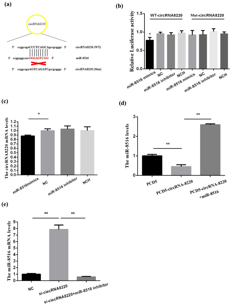
miR-8516 seed sequence could match with circRNA8220 (Figure 2a). To verify this prediction, dual-luciferase reporter vectors were structured the luciferase activity reduced after co-transfection WT-circRNA8220 and miRNA-8516 mimic. However, the luciferase activity did not change after co-transfection MT-circRNA8220 and miRNA-8516 mimic (Figure 2b). We found that up-regulation of miRNA-8516 could decrease the mRNA levels of circRNA8220 in GMECs (Figure 2c). Overexpression of circRNA8220 decreased the mRNA levels of miR-8516 (Figure 2d). The level of miRNA-8516 was recovered after co-transfection pCD5-circRNA8220 vector and miRNA-8516. miR-8516 increased after silencing the expression of circRNA8220 using small interfering RNAs (siRNAs) the result was recovered by co-transfection si-circRNA8220 and miR-8516 inhibitor (Figure 2e). In summary, these findings proved that circRNA8220 could serve as a sponge for miR-8516 a negative regulatory relationship existed between circRNA8220 and miR-8516 in GMECs.
Figure 2.
circRNA8220 acted as a sponge for miR-8516. (a) The seed sequence of miR-8516 could match with WT-circRNA8220 and could not match with Mut-circRNA8220. (b) Luciferase reporter assay of 293T cells co-transfected with wild type (WT)-circRNA8220 or Mut-circRNA8220 and miR-8516 mimic, NC (negative control), miR-8516 inhibitor or NCH (NC-inhibitor). (c) miRNA-8516 inhibited the mRNA expression of circRNA8220. (d) circRNA8220 decreased miR-8516 mRNA level in GMEC. (e) si-circRNA8220 promoted the mRNA expression of circRNA8220. ** p < 0.01, * p < 0.05.
3.3. CircRNA8220 Increased the Expression of STC2 in GMECs In Vitro
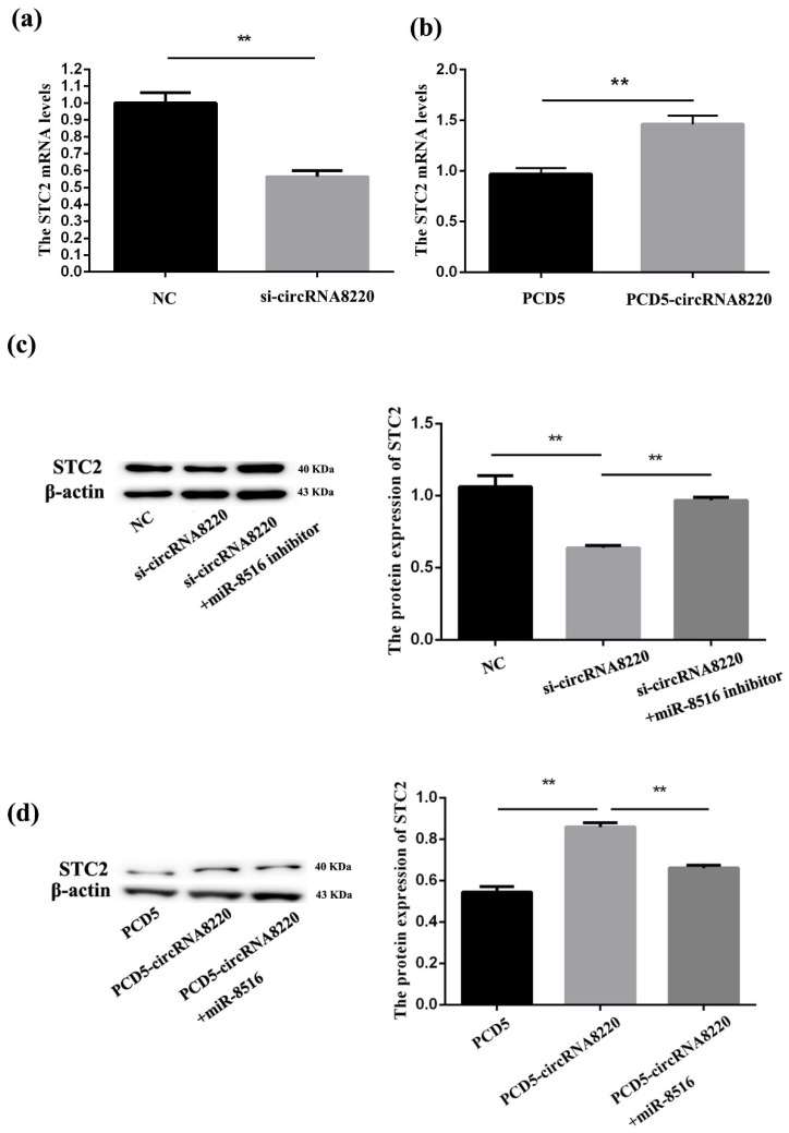
qPCR was used to explore the impact of circRNA8220 on STC2 in GMECs the results showed the mRNA level STC2 was significantly down- or up-regulated after transfection of si-circRNA8220 or pCD5-circRNA8220. Western blot was also used to explore the impact of circRNA8220 on STC2 in GMECs. We found that the protein level of STC2 was down- or up-regulated after transfection of si-circRNA8220 or pCD5-circRNA8220; the mRNA and protein level of STC2 were consistent with those of circRNA8220 (Figure 3). These results revealed that circRNA8220 could positively regulate mRNA and protein levels of STC2.
Figure 3.
circRNA8220 increased the expression of STC2 in GMEC in vitro. (a) si-circRNA8220 reduced the mRNA expression of STC2. (b) circRNA8220 increased the mRNA expression of STC2. (c) si-circRNA8220 blocked the protein expression of STC2. (d) circRNA8220 raised the protein expression of STC2. ** p < 0.01.
3.4. STC2 Inhibited Apoptosis and Promoted Proliferation of GMECs In Vitro
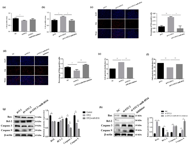
STC2-siRNA and pcDNA3.1-STC2 vector were synthesized to study the effect of STC2 on GMECs. The CCK8 assay was applied to explore the effect of viability of STC2 on GMECs after transfection for 24 h. The data revealed that overexpression or inhibition of STC2 enhanced or attenuated the viability of GMECs (Figure 4a,b). The Edu assay was conducted to verify the credibility of the proliferation assay results in GMECs; the result of the Edu assay was consistent with that of the CCK8 assay (Figure 4c,d).
Figure 4.
STC2 inhibited apoptosis and promoted proliferation of GMEC in vitro. (a,b) Cell proliferation was assessed using the cell counting kit-8 (CCK-8) assay after transfection with pc3.1-STC2 or si-STC2. (c,d) Cell proliferation indices were assessed after treatment with Edu after transfection with pc3.1-STC2 or si-STC2. (e,f) Apoptosis analysis of GMEC was detected with flow cytometry method after transfection with pc3.1-STC2 or si-STC2. (g,h) Protein levels of Bcl-2, Bax, caspase 3 and caspase 9 in GMEC after transfection with pc3.1-STC2 or si-STC2. Protein levels were measured by WB (western blot) densitometry was normalised to the β-actin density from the same lane. Data were expressed as the means ± SEM. ** p < 0.01, * p < 0.05.
Apoptosis ratios of GMECs after transfection with STC2-siRNA/pcDNA3.1-STC2 and co-transfection of STC2-siRNA with miR-8516 inhibitor/pcDNA3.1-STC2 with miR-8516 mimic were detected by Annexin V-FITC (fluoresceine isothiocyanate)/PI (propidium iodide) staining. The results showed that the rise or drop in the expression of STC2 could attenuate or enhance the apoptosis ratio of GMECs (Figure 4e,f). The protein levels of critical apoptotic genes, including Bcl-2, Bax, caspase 9 and caspase 3, were also researched. We found that Bax, caspase 9 and caspase 3 all decreased significantly Bcl-2 increased (p < 0.01) after transfection with pcDNA3.1-STC2 after 48 h; the result was opposite when transfected with STC2-siRNA compared with pcDNA3.1-STC2 (Figure 4g,h). As expected, Western blot showed that miR-8516 mimic or inhibitor co-transfection with pcDNA3.1-STC2 or STC2-siRNA could partially weaken the effect of pcDNA3.1-STC2 or STC2-siRNA on apoptosis and viability. CCK8 and Edu in GMECs proved that miR-8516 could negatively regulate STC2. These consequences suggested that STC2 could decrease GMEC apoptosis and induce proliferation in vitro.
3.5. Effect of circRNA8220 Was in Accordance with STC2 on Apoptosis and Proliferation of GMECs In Vitro
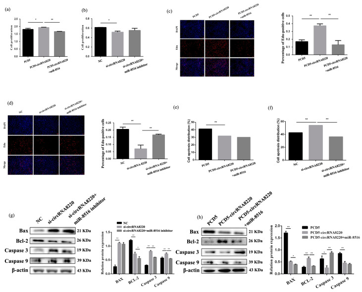
The effect of circRNA8220 on proliferation was checked by CCK8 and Edu assay in GMECs. The results of CCK8 showed that circRNA8220 promoted cell proliferation the result was recovered after co-transfection of pCD5-circRNA8220 with miR-8516 mimic (Figure 5a). Knockdown circRNA8220 inhibited cell proliferation (Figure 5b). The Edu results showed that circRNA8220 promoted cell proliferation, whereas si-circRNA8220 inhibited cell proliferation. The results were recovered after co-transfection of pCD5-circRNA8220 or si-circRNA8220 with miR-8516 mimic or miR-8516 inhibitor compared with transfection with pCD5-circRNA8220 or si-circRNA8220 (Figure 5c,d).
Figure 5.
circRNA8220 inhibited apoptosis and promoted proliferation of GMEC in vitro. (a,b) Cell proliferation was assessed using the cell counting kit-8 (CCK-8) assay after transfection with PCD5-circRNA8220 or si-circRNA8220. (c,d) Cell proliferation indices were assessed after treatment with Edu after transfection with PCD5-circRNA8220 or si-circRNA8220. (e,f) Apoptosis analysis of GMEC was detected with FCM after transfection with PCD5-circRNA8220 or si-circRNA8220. (g,h) Protein levels of Bcl-2, Bax, caspase 3 and caspase 9 in GMEC after transfection with PCD5-circRNA8220 or si-circRNA8220. Protein levels were measured by WB densitometry was normalised to the β-actin density from the same lane. Data were expressed as the means ± SEM. ** p < 0.01, * p < 0.05.
Apoptosis ratio was tested after transfection of pCD5-circRNA8220 or si-circRNA8220 by Annexin V-FITC/PI staining. The results showed that circRNA8220 inhibited the apoptosis of GMECs (Figure 5e), si-circRNA8220 induced the apoptosis of GMECs and the rate of apoptosis was decreased when si-circRNA8220 and miR-8516 inhibitors were co-transfected into GMECs (Figure 5f). The protein levels of Bax, caspase 3 and caspase 9 were up- or down-regulated after transfection with si-circRNA8220 or pCD5-circRNA8220 (Figure 5g,h).
3.6. STC2 Enhanced Synthesis of β-Casein and Triglycerides in GMECs
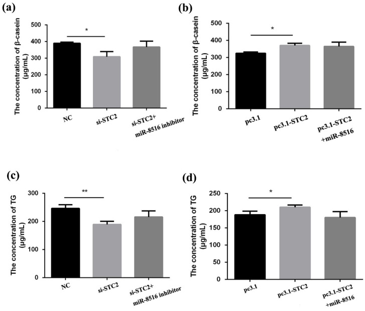
The concentration of β-casein and triglycerides were tested by ELISA KIT after 24 h transfection with STC2-siRNA or pcDNA3.1-STC2 to explore the effects of STC2 on milk production in GMECs. The results displayed that production of β-casein and triglycerides was restrained after transfection of STC2-siRNA in GMECs (Figure 6a,c). Overexpression of STC2 strengthened the concentration of β-casein and triglycerides (Figure 6b,d).
Figure 6.
STC2 enhanced the synthesis of β-casein and triglycerides in GMEC. (a,b,c,d) Secretion of β-casein and triglycerides (TG) after transfection with pc3.1-STC2 or si-STC2 was measured by enzyme-linked immunosorbent assay kit. ** p < 0.01, * p < 0.05.
3.7. Ability of circRNA8220 to Promote Cell Synthesis β-Casein and Triglycerides Was Consistent with That of STC2
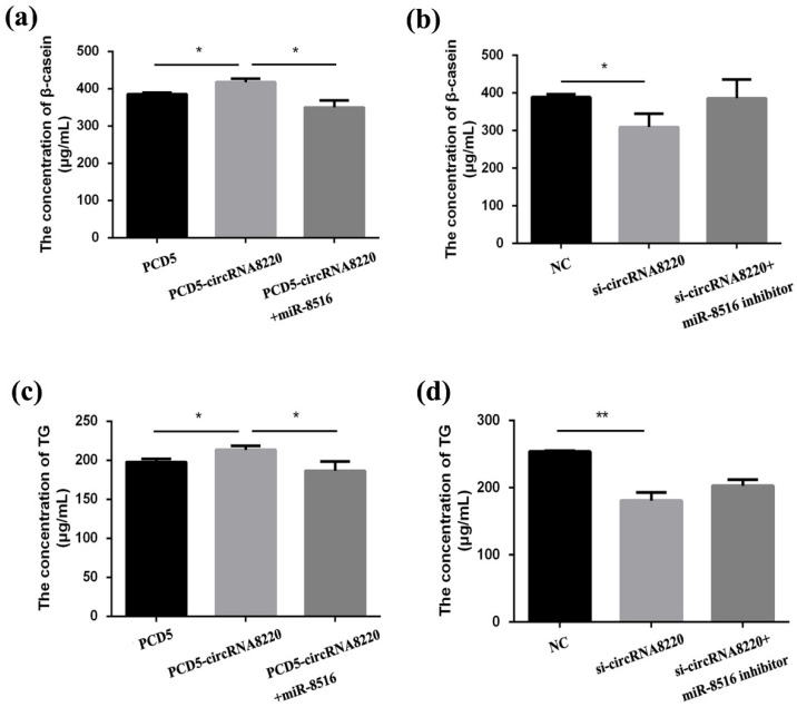
The concentrations of β-casein and triglycerides were also tested by ELISA KIT. Overexpression of circRNA8220 enhanced the synthesis ability of β-casein and triglycerides in GMECs (Figure 7a,c). Knockdown of circRNA8220 decreased the synthesis ability of β-casein and triglycerides in GMECs (Figure 7b,d). The result could be recovered after co-transfection of pcDNA2.1-circRNA8220 with miR-8516 mimic. Thus, circRNA8220 might facilitate the synthesis of β-casein and triglycerides through miR-8516-STC2 pathways.
Figure 7.
circRNA8220 enhanced the synthesis of β-casein and triglycerides in GMEC. (a–d) Secretion of β-casein and triglycerides (TG) after transfected with PCD5-circRNA8220 or si-circRNA8220 was measured by enzyme-linked immunosorbent assay kit. ** p < 0.01, * p < 0.05.
3.8. CircRNA8220 and STC2 Activated Ras/MEK/ERK Signaling Pathways in GMECs
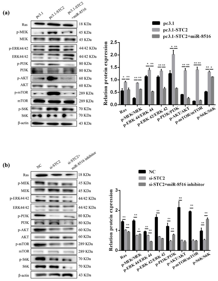
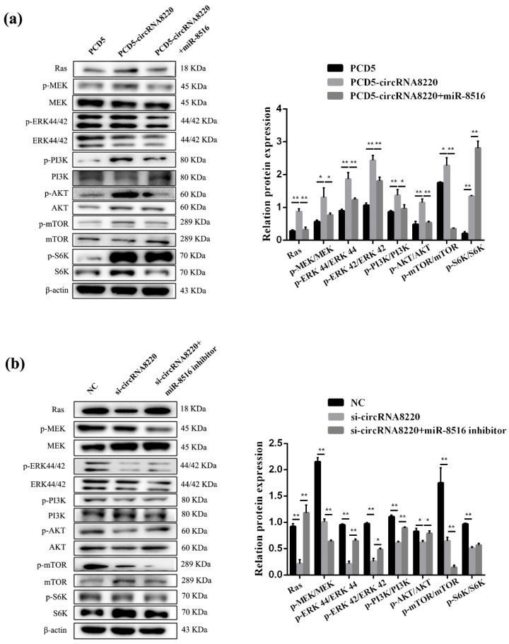
Ras/MEK/ERK signaling pathways were tested to explore the regulation mechanism of STC2 on apoptosis and proliferation in GMECs. Thus, the protein expression of Ras and the phosphorylation of MEK and ERK1/2 were evaluated by Western blot. The protein expression of Ras and the phosphorylation of MEK and ERK1/2 increased after transfection with pcDNA3.1-STC2 (Figure 8a). Compared with pcDNA3.1-STC2, STC2-siRNA had the reverse results (Figure 8b).
Figure 8.
STC2 activated the signaling pathways of Ras/MEK/ERK and PI3K/AKT/mTOR in GMEC. (a,b) Western blot analysis was performed to detect the protein expression of Ras, p-MEK, MEK, p-ERK44/42, ERK44/42, p-PI3K, PI3K, p-AKT, AKT, p-mTOR, mTOR, p-S6K, S6K and β-actin in GMEC after transfection with pc3.1-STC2 or si-STC2. Protein levels were measured by WB densitometry was normalised to the β-actin density from the same lane. Data were expressed as the means ± SEM. ** p < 0.01, * p < 0.05.
Similarly, Ras/MEK/ERK signaling pathways were tested after transfection with si-circRNA8220 or pCD5-circRNA8220. The results showed that circRNA8220 increased the protein expression of Ras and the phosphorylation of MEK and ERK1/2 (Figure 9a). Knockdown circRNA8220 decreased the protein expression of Ras and the phosphorylation of MEK and ERK1/2 (Figure 9b). The effects of circRNA8220 on Ras/MEK/ERK signaling pathways were consistent with those of STC2. Moreover, circRNA8220 could positively regulate STC2 in GMECs. These results showed that the protein levels of Bax, Bcl-2, caspase 3 and caspase 9 were consistent with those of STC2 (Figure 5g,h).
Figure 9.
circRNA8220 activated the signaling pathways of Ras/MEK/ERK and PI3K/AKT/mTOR in GMEC. (a,b) Western blot analysis was performed to detect the protein expression of Ras, p-MEK, MEK, p-ERK44/42, ERK44/42, p-PI3K, PI3K, p-AKT, AKT, p-mTOR, mTOR, p-S6K, S6K and β-actin in GMEC after transfection with PCD5-circRNA8220 or si-circRNA8220. Protein levels were measured by WB densitometry was normalised to the β-actin density from the same lane. Data were expressed as the means ± SEM. ** p < 0.01, * p < 0.05.
3.9. CircRNA8220 and STC2 Activated PI3K/AKT/mTOR Signaling Pathways in GMECs
The molecular mechanism of STC2 affecting milk synthesis was explored in GMECs. The phosphorylation levels of PI3K/AKT/mTOR signal pathways after transfection with pcDNA3.1-STC2 or si-STC2 were detected by Western blot. The results showed that high expression of STC2 increased the ratio of p-PI3K/PI3K, p-AKT/AKT, p-mTOR/mTOR and p-S6K/S6K (Figure 8a) the phosphorylation levels of PI3K/AKT/mTOR/S6K signal pathway decreased when the expression of STC2 was down-regulated in GMECs (Figure 8b). In the meantime, STC2-siRNA + miR-8516 inhibitor and pcDNA3.1-STC2 + miR-8516 mimic could more or less recover to the level of the control group.
As mentioned above, the ELISA result of circRNA8220 was consistent with that of STC2. The phosphorylation levels of PI3K/AKT/mTOR signal pathway were detected in GMECs after transfection with pCD5-circRNA8220 or si-circRNA8220 by Western blot to explore whether the molecular mechanism of circRNA8220 on lactation was consistent with that of STC2. The results showed that the effect of circRNA8220 on PI3K/AKT/mTOR signal pathways was consistent with that of STC2 miR-8516 mimic or miR-8516 inhibitor could weaken the effect of pCD5-circRNA8220 or si-circRNA8220 on the PI3K/AKT/mTOR signal pathways in GMECs (Figure 9a,b). The above-mentioned results suggested a positive regulatory relationship between circRNA8220 and STC2. In other words, circRNA8220 could control PI3K/AKT/mTOR signal pathways through miR-8516/STC2.
4. Discussion
Many studies have reported that circRNAs could function as miRNA sponges to adjust the expression of gene in many physiological processes, such as gastric cancer [19], development of endometrial receptivity [20]. Recent studies showed that miRNA, circRNA and lncRNAs could function as regulators in mammary epithelial cell and be helpful for lactation, such as BTAT017009.2-miR-21-3p-IGFBP5 play an important role in cells proliferation in bovine mammary gland epithelial cells [21]. circHIPK3 promotes proliferation of cow mammary epithelial cells [22]. However, studies on circRNA in the regulation of lactation in dairy goat are limited. In this study, circRNA8220 displayed a sponging effect for miR-8516 in GMECs there was a negative feedback relationship between circRNA8220 and miR-8516 in GMECs the molecular mechanism of the negative feedback relationship require further research.
The nutritional value of goat milk is related to the proportion of milk components [23]. Many studies have reported that the PI3K/AKT/mTOR/S6K pathway was related to the synthesis of β-casein and triglycerides [24,25,26], study showed that overexpression of menin caused significant suppression of factors involved in the mTOR pathway, as well as milk protein κ-casein [27]. In this study, the results from ELISA KIT showed that overexpression of circRNA8220 promoted the synthesis of β-casein and triglycerides in GMECs. The result was contrary when the level of circRNA8220 was knockdown. Interestingly, the effects of si-circRNA8220 or pCD5-circRNA8220 on lactation were partially weakened by miR-8516 inhibitor or miR-8516 mimic. The results of WB showed that overexpressed or knockdown circRNA8220 up- or down-regulated the phosphorylation levels of PI3K, AKT, mTOR and S6K. These results explained that circRNA8220 promoted synthesis of β-casein and triglycerides by sponging miR-8516 in GMECs via PI3K/AKT/mTOR/S6K pathway.
ERK pathway inhibited cell apoptosis in many kinds of cells. For example, SOCS-1 inhibited apoptosis in cardiac myocytes via ERK1/2 pathway activation [28] varicella–zoster virus ORF12 protein inhibited apoptosis by induced phosphorylation of ERK1/2 in melanoma cells [29]. Our results showed that circRNA8220 could activate Ras/MEK/ERK pathway by sponging miR-8516, these results were consistent with Edu, FCM and CCK8. It was known that the number and activity of mammary epithelial cells are closely related to lactation and play an important role in breast development [1]. The casepase family is the initiator and executor of cell apoptosis in mammals, among which, caspase-3 is the most crucial apoptotic protease in the downstream of the caspase cascade reaction [30]. Bcl-2 inhibits the activation of the upstream caspase protease by interfering with release of cytochrome c, result in inhibition cell apoptpsis [30]. As a composition of the ion channel on the mitochondrial membrane, Bax protein allows cytochrome c to pass through the mitochondrial membrane, activating caspase-9 further activating caspase-3, thus resulting in cell apoptosis [31]. Procaspase-9, an initiator caspase in the mitochondrial pathway, is recruited and activated by the apoptosome leading to downstream casepase-3 processing [32]. The protein levels of critical apoptotic genes, including Bcl-2 [33], Bax [34], caspase 9 [35] and caspase 3 [36], we researched to prove the effect of circRNA8220 on critical genes of apoptosis. Our results showed that circRNA8220 could promote cell proliferation and inhibit apoptosis by sponging miR-8516. This result was opposite that of knockdown circRNA8220 in GMECs. Recent studies have shown that the expression level of Bax or Bcl-2 was inhibited or promoted by ERK in GMECs [37,38]. Together we draw a conclusion that circRNA8220 could promote cell proliferation and inhibit apoptosis by sponging miR-8516 in GMECs via Ras/MEK/ERK pathway.
STC2 promoted proliferation in many kinds of cells, for example, STC2 promoted hepatocellular carcinoma proliferation in vitro [15] and hepatocellular carcinoma cells proliferation [39]. The results of dual luciferase analysis, RT-qPCR and WB further proved that STC2 was a target gene of miR-8516. We predicted that STC2 may be in the regulation network of circRNA8220/miR-8516. The results showed that circRNA8220 increased the mRNA and protein levels of STC2. On the contrary, miR-8516 mimics decreased the mRNA and protein levels of STC2, miR-8516 inhibitor increased the protein level of STC2. As expected, the effects of pCD5-circRNA8220 and si-circRNA8220 were weakened by miR-8516 mimic and inhibitor. These results explained that circRNA8220 regulated the STC2 by sponging miR-8516 as a ceRNA and proved the conjecture that STC2 is in the regulation network of circRNA8220/miR-8516.
We speculated that circRNA8220 functioned as a miR-8516 sponge to promote proliferation, inhibit apoptosis and enhance the synthesis of β-casein and triglycerides via regulating STC2 expression in vitro. The results showed that STC2 promoted proliferation and inhibited apoptosis in GMECs in vitro. We also found that STC2 enhanced the synthesis of β-casein and triglycerides in GMECs by the way of ELISA. All the results were consistent with those for circRNA8220. Therefore, STC2 is in the regulation network of circRNA8220/miR-8516. Previous studies have shown that STC2 could activate ERK in osteoblast [16,40]. ERK pathway inhibited cell apoptosis in many kinds of cells [28,29]. The results showed that overexpression of STC2 enhanced the protein level of Ras and the phosphorylation level of MEK and ERK. This result was opposite that of knockdown STC2 in GMECs. In addition, the protein levels of Bcl-2, Bax, caspase 9 and caspase 3 were explored in GMECs. As expected, the results showed that overexpression of STC2 enhanced the protein level of Bcl-2 and inhibited the protein level of Bax, caspase 9 and caspase 3. These results were consistent with effect of circRNA8220 on Bcl-2, Bax, caspase 9 and caspase 3. In summary, circRNA8220 functioned as a miR-8516 sponge to promote proliferation and inhibit apoptosis via regulating STC2 expression by Ras/MEK/ERK pathway in GMECs.
Similarly, we studied the regulatory mechanism of circRNA8220 and STC2 on the effect of milk synthesis. The results of WB showed that STC2 could activate PI3K/AKT/mTOR/S6K pathway. The results of STC2 were consistent with those of circRNA8220 in PI3K/AKT/mTOR pathway. Overall, these results suggested that circRNA8220 functioned as a miR-8516 sponge to promote the synthesis of β-casein and triglycerides by regulating STC2 expression via PI3K/AKT/mTOR pathway in GMECs.
5. Conclusions
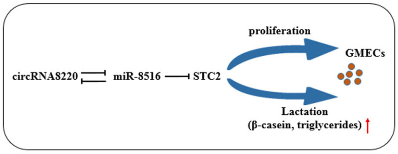
In summary, this study researched the effect and regulatory mechanism of circRNA8220 and STC2 on cell apoptosis, proliferation and lactation in GMECs in vitro. Therefore, a circRNA–miRNA–mRNA network is presented. We conclude that circRNA8220 as the sponge of miR-8516 can activate Ras/MEK/ERK pathway, promote proliferation and inhibit apoptosis by raising of STC2; it can also activate PI3K/AKT/mTOR pathway and enhance the synthesis of β-casein and triglycerides by up-regulating STC2 in GMECs in vitro (Appendix A Figure A2).
Acknowledgments
The authors would like to thank Yuxuan Song for excellent assistance in editing the manuscript.
Abbreviations
| circRNA | Circular RNA |
| GMECs | Goat mammary epithelial cells |
| STC2 | Stanniocalcin 2 |
| TG | Triglycerides |
| RNA | Ribonucleic |
| mRNA | Messenger RNA |
| miRNA | MicroRNA |
| UTR | Untranslated region |
| HEK293T | Human embryonic kidney cell line |
Appendix A
Figure A1.
Transfection efficiency of pcDNA3.1-STC2 and pCD5-circRNA8220 vectors and STC2-siRNA, circRNA8220-siRNA and miR-8516 mimics/inhibitors. (a,b) mRNA levels of circRNA8220. (c) mRNA levels of miR-8516. (d) The mRNA levels of STC2. (e) Protein levels of STC2. ** p < 0.01, * p < 0.05.
Figure A2.
CircRNA8220 promote cell proliferation and enhance the synthesis of β-casein and triglycerides in GMEC in vitro.
Table A1.
RT-qPCR primers used in this study.
| Gene | Genbank Accession No. | Primer Sequence (5′→3′) |
|---|---|---|
| β-actin | XM_018039831.1 | F: GATCTGGCACCACACCTTCT |
| R: GGGTCATCTTCTCACGGTTG | ||
| STC2 (qPCR) | XM_005694539.3 | F: CGGAAGTGTCCAGCCATCAAGG |
| R: CACAGGTCAGCAGCAGGTTCAC | ||
| STC2 (Check2) | / | F: CCCTCGAGTTGCCACCAGAGCAAAGCC |
| R: TAAAGCGGCCGCTCTTGTCCCCCAGTGACGTG | ||
| Circ-8220 (qPCR) | / | F: GCCACAGCCTGGACATGAA |
| Circ-8220 (Check2) | R: CCTCTTGGTCACAGGGATGG | |
| U6 | / | F: cgCTCGAGTTCAAGATGCCCTGACCCCC |
| R: atGCGGCCGCAGGTGAACTTCATGTCCAGGCT | ||
| miR-8516-Loop | / | F: CTCGCTTCGGCAGCACA |
| miR-449a | R: AACGCTTCACGAATTTGCGT | |
| Reverse Primer | / | gtcgtatccagtgcagggtccgaggtattcgcactggatacgacGGCCTCCG |
| gcgcgcGGCTGAGGGCAACGGAGGCC | ||
| / | GTGCAGGGTCCGAGGT |
Note: The characters with underscore were restriction enzyme cutting site of Xho I and Not I for constructing psiCHECK2.
Author Contributions
X.A. and C.Z. conceived of and designed the experiments. C.Z., H.Y., J.Z., Y.H., Y.J. and Q.D. performed the experiments. C.Z. analyzed the data. C.Z. and X.A. wrote the paper. L.Z. and B.C. helped perform the analysis, with constructive discussions. All authors have read and agreed to the published version of the manuscript.
Funding
This work was supported by the National Natural Science Foundation of China [31601925], the Nature Science Foundation of Shaanxi Province [2018JM3006 and 2015JM3087], the China Postdoctoral Science Foundation [2016T90954 and 2014M552498] and the Shaanxi Science and Technology Innovation Project Plan [2015KTCQ03-08, 2016KTZDNY02-04, 2017ZDXM-NY-081 and 2018ZDCXL-NY-01-04].
Conflicts of Interest
The authors declare no competing financial interest.
Ethics Statement
All procedures in our animal study were approved by the Animal Care and Use Committee of the Northwest A&F University (Yangling, China) (permit number: 17-347, data:2017-10-13).
References
- 1.Boutinaud M., Guinardflament J., HélèneJammes The number and activity of mammary epithelial cells, determining factors for milk production. Reprod. Nutr. Dev. 2004;44:499–508. doi: 10.1051/rnd:2004054. [DOI] [PubMed] [Google Scholar]
- 2.Stefanon B., Colitti M., Gabai G., Knight C.H., Wilde C.J. Mammary apoptosis and lactation persistency in dairy animals. J. Dairy Res. 2002;69:37–52. doi: 10.1017/S0022029901005246. [DOI] [PubMed] [Google Scholar]
- 3.Zhang H.D., Jiang L.H., Sun D.W., Hou J.C., Ji Z.L. CircRNA: A novel type of biomarker for cancer. Breast Cancer (Tokyo Jpn.) 2018;25:1–7. doi: 10.1007/s12282-017-0793-9. [DOI] [PubMed] [Google Scholar]
- 4.Chen L., Zhang S., Wu J., Cui J., Zhong L., Zeng L., Ge S. circRNA_100290 plays a role in oral cancer by functioning as a sponge of the miR-29 family. Oncogene. 2017;36:4551–4561. doi: 10.1038/onc.2017.89. [DOI] [PMC free article] [PubMed] [Google Scholar] [Retracted]
- 5.Liu W., Ma W., Yuan Y., Zhang Y., Sun S. Circular RNA hsa_circRNA_103809 promotes lung cancer progression via facilitating ZNF121-dependent MYC expression by sequestering miR-4302. Biochem. Biophys. Res. Commun. 2018;500:846–851. doi: 10.1016/j.bbrc.2018.04.172. [DOI] [PubMed] [Google Scholar]
- 6.Chen G., Shi Y., Zhang Y., Sun J. CircRNA_100782 regulates pancreatic carcinoma proliferation through the IL6-STAT3 pathway. Oncotargets Ther. 2017;10:5783–5794. doi: 10.2147/OTT.S150678. [DOI] [PMC free article] [PubMed] [Google Scholar]
- 7.Huang X.Y., Huang Z.L., Xu Y.H., Zheng Q., Chen Z., Song W., Zhou J., Tang Z.Y., Huang X.Y. Comprehensive circular RNA profiling reveals the regulatory role of the circRNA-100338/miR-141-3p pathway in hepatitis B-related hepatocellular carcinoma. Sci. Rep. 2017;7:5428. doi: 10.1038/s41598-017-05432-8. [DOI] [PMC free article] [PubMed] [Google Scholar]
- 8.Fang H., Xie J., Zhang M., Zhao Z., Wan Y., Yao Y. miRNA-21 promotes proliferation and invasion of triple-negative breast cancer cells through targeting PTEN. Am. J. Transl. Res. 2017;9:953–961. [PMC free article] [PubMed] [Google Scholar]
- 9.Lopes-Ramos C.M., Barros B.P., Koyama F.C., Carpinetti P.A., Pezuk J., Doimo N.T.S., Habr-Gama A., Perez R.O., Parmigiani R.B. E2F1 somatic mutation within miRNA target site impairs gene regulation in colorectal cancer. PLoS ONE. 2017;12:e0181153. doi: 10.1371/journal.pone.0181153. [DOI] [PMC free article] [PubMed] [Google Scholar]
- 10.Maller Schulman B.R., Liang X., Stahlhut C., DelConte C., Stefani G., Slack F.J. The let-7 microRNA target gene, Mlin41/Trim71 is required for mouse embryonic survival and neural tube closure. Cell Cycle (Georget. Tex.) 2008;7:3935–3942. doi: 10.4161/cc.7.24.7397. [DOI] [PMC free article] [PubMed] [Google Scholar]
- 11.Yang Y., Fang X., Yang R., Yu H., Jiang P., Sun B., Zhao Z. MiR-152 Regulates Apoptosis and Triglyceride Production in MECs via Targeting ACAA2 and HSD17B12 Genes. Sci. Rep. 2018;8:417. doi: 10.1038/s41598-017-18804-x. [DOI] [PMC free article] [PubMed] [Google Scholar]
- 12.Wang H., Luo J., He Q., Yao D., Wu J., Loor J.J. miR-26b promoter analysis reveals regulatory mechanisms by lipid-related transcription factors in goat mammary epithelial cells. J. Dairy Sci. 2017;100:5837–5849. doi: 10.3168/jds.2016-12440. [DOI] [PubMed] [Google Scholar]
- 13.Hou J., An X., Song Y., Cao B., Yang H., Zhang Z., Shen W., Li Y. Detection and comparison of microRNAs in the caprine mammary gland tissues of colostrum and common milk stages. BMC Genet. 2017;18:38. doi: 10.1186/s12863-017-0498-2. [DOI] [PMC free article] [PubMed] [Google Scholar]
- 14.Li Q., Zhou X., Fang Z., Pan Z. Effect of STC2 gene silencing on colorectal cancer cells. Mol. Med. Rep. 2019;20:977–984. doi: 10.3892/mmr.2019.10332. [DOI] [PMC free article] [PubMed] [Google Scholar]
- 15.Wang H., Wu K., Sun Y., Li Y., Wu M., Qiao Q., Wei Y., Han Z.G., Cai B. STC2 is upregulated in hepatocellular carcinoma and promotes cell proliferation and migration in vitro. BMB Rep. 2012;45:629–634. doi: 10.5483/BMBRep.2012.45.11.086. [DOI] [PMC free article] [PubMed] [Google Scholar]
- 16.Chen B., Zeng X., He Y., Wang X., Liang Z., Liu J., Zhang P., Zhu H., Xu N., Liang S. STC2 promotes the epithelial-mesenchymal transition of colorectal cancer cells through AKT-ERK signaling pathways. Oncotarget. 2016;7:71400–71416. doi: 10.18632/oncotarget.12147. [DOI] [PMC free article] [PubMed] [Google Scholar]
- 17.Steffensen L.B., Conover C.A., Bjørklund M.M., Ledet T., Bentzon J.F., Oxvig C. Stanniocalcin-2 overexpression reduces atherosclerosis in hypercholesterolemic mice. Atherosclerosis. 2016;248:36–43. doi: 10.1016/j.atherosclerosis.2016.02.026. [DOI] [PubMed] [Google Scholar]
- 18.Chen K., Hou J., Song Y., Zhang X., Liu Y., Zhang G., Wen K., Ma H., Li G., Cao B., et al. Chi-miR-3031 regulates beta-casein via the PI3K/AKT-mTOR signaling pathway in goat mammary epithelial cells (GMECs) BMC Vet. Res. 2018;14:369. doi: 10.1186/s12917-018-1695-6. [DOI] [PMC free article] [PubMed] [Google Scholar]
- 19.Cheng J., Zhuo H., Xu M., Wang L., Xu H., Peng J., Hou J., Lin L., Cai J. Regulatory network of circRNA-miRNA-mRNA contributes to the histological classification and disease progression in gastric cancer. J. Transl. Med. 2018;16:216. doi: 10.1186/s12967-018-1582-8. [DOI] [PMC free article] [PubMed] [Google Scholar]
- 20.Zhang L., Liu X., Che S., Cui J., Liu Y., An X., Cao B., Song Y. CircRNA-9119 regulates the expression of prostaglandin-endoperoxide synthase 2 (PTGS2) by sponging miR-26a in the endometrial epithelial cells of dairy goat. Reprod. Fertil. Dev. 2018;30:1759–1769. doi: 10.1071/RD18074. [DOI] [PubMed] [Google Scholar]
- 21.Zhang X., Cheng Z., Wang L., Jiao B., Yang H., Wang X. MiR-21-3p Centric Regulatory Network in Dairy Cow Mammary Epithelial Cell Proliferation. J. Agric. Food Chem. 2019;67:11137–11147. doi: 10.1021/acs.jafc.9b04059. [DOI] [PubMed] [Google Scholar]
- 22.Liu J., Zhang M., Li D., Li M., Kong L., Cao M., Wang Y., Song C., Fang X., Chen H., et al. Prolactin-Responsive Circular RNA circHIPK3 Promotes Proliferation of Mammary Epithelial Cells from Dairy Cow. Genes. 2020;11:5109. doi: 10.3390/genes11030336. [DOI] [PMC free article] [PubMed] [Google Scholar]
- 23.Rafiee-Tari N., Fan M.Z., Archbold T., Arranz E., Corredig M. Effect of milk protein composition and amount of β-casein on growth performance, gut hormones, inflammatory cytokines in an in vivo piglet model. J. Dairy Sci. 2019;102:8604–8613. doi: 10.3168/jds.2018-15786. [DOI] [PubMed] [Google Scholar]
- 24.Samant G.V., Sylvester P.W. gamma-Tocotrienol inhibits ErbB3-dependent PI3K/Akt mitogenic signalling in neoplastic mammary epithelial cells. Cell Prolif. 2006;39:563–574. doi: 10.1111/j.1365-2184.2006.00412.x. [DOI] [PMC free article] [PubMed] [Google Scholar]
- 25.Laplante M., Sabatini D.M. An emerging role of mTOR in lipid biosynthesis. Curr. Biol. CB. 2009;19:R1046–R1052. doi: 10.1016/j.cub.2009.09.058. [DOI] [PMC free article] [PubMed] [Google Scholar]
- 26.Burgos S.A., Cant J.P. IGF-1 stimulates protein synthesis by enhanced signaling through mTORC1 in bovine mammary epithelial cells. Domest. Anim. Endocrinol. 2010;38:211–221. doi: 10.1016/j.domaniend.2009.10.005. [DOI] [PubMed] [Google Scholar]
- 27.Li H., Liu X., Wang Z., Lin X., Yan Z., Cao Q., Zhao M., Shi K. MEN1/Menin regulates milk protein synthesis through mTOR signaling in mammary epithelial cells. Sci. Rep. 2017;7:5479. doi: 10.1038/s41598-017-06054-w. [DOI] [PMC free article] [PubMed] [Google Scholar]
- 28.Yan L., Tang Q., Shen D., Peng S., Zheng Q., Guo H., Jiang M., Deng W. SOCS-1 inhibits TNF-alpha-induced cardiomyocyte apoptosis via ERK1/2 pathway activation. Inflammation. 2008;31:180–188. doi: 10.1007/s10753-008-9063-5. [DOI] [PubMed] [Google Scholar]
- 29.Liu X., Li Q., Dowdell K., Fischer E.R., Cohen J.I. Varicella-Zoster virus ORF12 protein triggers phosphorylation of ERK1/2 and inhibits apoptosis. J. Virol. 2012;86:3143–3151. doi: 10.1128/JVI.06923-11. [DOI] [PMC free article] [PubMed] [Google Scholar]
- 30.Li Y., Jia Z., Zhang L., Wang J., Yin G. Caspase-2 and microRNA34a/c regulate lidocaine-induced dorsal root ganglia apoptosis in vitro. Eur. J. Pharmacol. 2015;767:61–66. doi: 10.1016/j.ejphar.2015.10.008. [DOI] [PubMed] [Google Scholar]
- 31.Zhang C., Shi J., Qian L., Zhang C., Wu K., Yang C., Yan D., Wu X., Liu X. Nucleostemin exerts anti-apoptotic function via p53 signaling pathway in cardiomyocytes. In Vitr. Cell. Dev. Biol. Anim. 2015;51:1064–1071. doi: 10.1007/s11626-015-9934-7. [DOI] [PubMed] [Google Scholar]
- 32.Shakeri R., Kheirollahi A., Davoodi J. Apaf-1: Regulation and function in cell death. Biochimie. 2017;135:111–125. doi: 10.1016/j.biochi.2017.02.001. [DOI] [PubMed] [Google Scholar]
- 33.Adams J.M., Cory S. The Bcl-2 protein family: Arbiters of cell survival. Science. 1998;281:1322–1326. doi: 10.1126/science.281.5381.1322. [DOI] [PubMed] [Google Scholar]
- 34.Yin X.M., Oltvai Z.N., Veis-Novack D.J., Linette G.P., Korsmeyer S.J. Bcl-2 gene family and the regulation of programmed cell death. Cold Spring Harb. Symp. Quant. Biol. 1994;59:387–393. doi: 10.1101/SQB.1994.059.01.043. [DOI] [PubMed] [Google Scholar]
- 35.Kitazawa M., Hida S., Fujii C., Taniguchi S., Ito K., Matsumura T., Okada N., Sakaizawa T., Kobayashi A., Takeoka M., et al. ASC Induces Apoptosis via Activation of Caspase-9 by Enhancing Gap Junction-Mediated Intercellular Communication. PLoS ONE. 2017;12:e0169340. doi: 10.1371/journal.pone.0169340. [DOI] [PMC free article] [PubMed] [Google Scholar]
- 36.Kivinen K., Kallajoki M., Taimen P. Caspase-3 is required in the apoptotic disintegration of the nuclear matrix. Exp. Cell Res. 2005;311:62–73. doi: 10.1016/j.yexcr.2005.08.006. [DOI] [PubMed] [Google Scholar]
- 37.Liu Y., Hou J., Zhang M., Seleh-Zo E., Wang J., Cao B., An X. circ-016910 sponges miR-574-5p to regulate cell physiology and milk synthesis via MAPK and PI3K/AKT-mTOR pathways in GMECs. J. Cell Physiol. 2020;235:4198–4216. doi: 10.1002/jcp.29370. [DOI] [PMC free article] [PubMed] [Google Scholar]
- 38.Xu Y., Liu L., Qiu X., Liu Z., Li H., Li Z., Luo W., Wang E. CCL21/CCR7 prevents apoptosis via the ERK pathway in human non-small cell lung cancer cells. PLoS ONE. 2012;7:e33262. doi: 10.1371/journal.pone.0033262. [DOI] [PMC free article] [PubMed] [Google Scholar]
- 39.Wu F., Li T.Y., Su S.C., Yu J.S., Zhang H.L., Tan G.Q., Liu J.W., Wang B.L. STC2 as a novel mediator for Mus81-dependent proliferation and survival in hepatocellular carcinoma. Cancer Lett. 2017;388:177–186. doi: 10.1016/j.canlet.2016.11.039. [DOI] [PubMed] [Google Scholar]
- 40.Zhou J., Li Y., Yang L., Wu Y., Zhou Y., Cui Y., Yang G., Hong Y. Stanniocalcin 2 improved osteoblast differentiation via phosphorylation of ERK. Mol. Med. Rep. 2016;14:5653–5659. doi: 10.3892/mmr.2016.5951. [DOI] [PubMed] [Google Scholar]


