Abstract
Individuals infected with the novel coronavirus (Severe Acute Respiratory Syndrome Coronavirus 2 [SARS‐CoV‐2]) who develop coronavirus disease 2019 (COVID‐19) experience many symptoms; however, cutaneous manifestations are relatively rare. The authors encountered three patients with COVID‐19 who presented with erythema and suspected viral rash. In all cases, erythema appeared after the onset of the initial symptoms of COVID‐19. Erythema was considered to be caused by COVID‐19 and not a drug‐induced eruption because, in all cases, erythema was relieved merely by external medicine and oral antihistamines, without discontinuing the original medication. The authors’ hospital accepted 69 COVID‐19 patients between 22 February 2020 and 31 May 2020 and, of these, three (4.3%) exhibited eruptions, and all cases presented erythema. Except for seven patients who exhibited positive nasopharyngeal swab tests for SARS‐CoV‐2 RNA but no symptoms, three (4.8%) of the remaining 62 patients exhibited erythema. Although various types of eruptions have been reported in patients with COVID‐19, erythema was the only type in our patients. Erythema in the three patients exhibited many similarities to that previously reported in COVID‐19 patients, particularly in the manner it appeared and disappeared. For these reasons, these three cases were considered typical examples of erythema in patients with COVID‐19. Considering previous studies and the three cases reported here, there is a high probability that SARS‐CoV‐2 can cause erythema.
Keywords: COVID‐19, cutaneous manifestations, eruption, erythema, rash
Introduction
Infections with the novel coronavirus (Severe Acute Respiratory Syndrome Coronavirus 2 [SARS‐CoV‐2]) resulting in coronavirus disease 2019 (COVID‐19) have become widespread around the world. Between 22 February and 31 May 2020, 69 patients diagnosed with COVID‐19 required hospitalization at our hospital. COVID‐19 has a wide range of primary symptoms, including fever, cough, malaise and digestive symptoms; however, the frequency of cutaneous manifestations is comparatively lower. 1 , 2 Various types of cutaneous manifestations have been reported, including erythema, urticaria, purpura, reticular and frostbite. 3 There are, however, few reports of COVID‐19 patients with erythema that include detailed information such as symptom progression. Herein, we report three patients with COVID‐19 who presented with erythema suspicious for viral exanthema.
Case Report
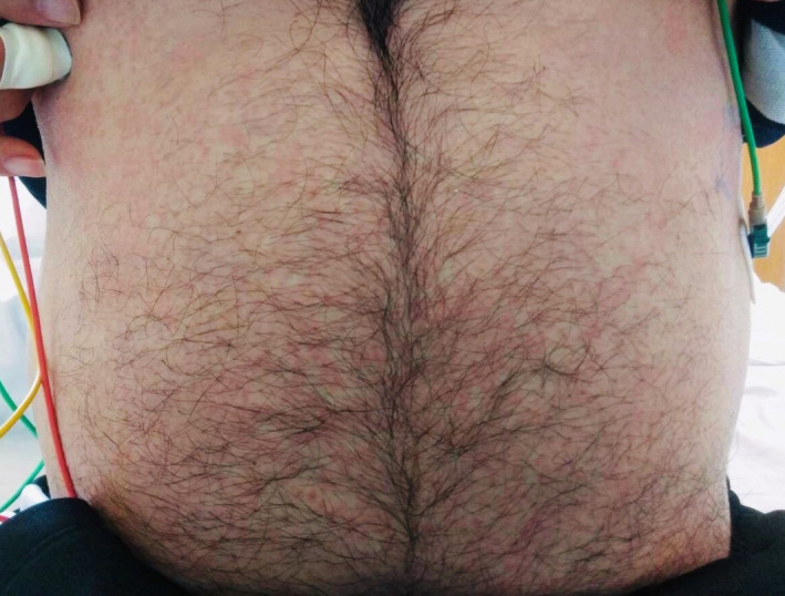
A 54‐year‐old man experiencing cough and slight fever (37°C) tested positive for SARS‐CoV‐2 RNA on nasopharyngeal swab 4 days after symptom onset, and was admitted to the authors’ hospital on day 5. Laboratory tests revealed normal white blood cell and platelet counts (6000 and 171 000/mm3, respectively), slightly impaired liver function (aspartate aminotransferase [AST], 31 IU/L; alanine aminotransferase [ALT], 45 IU/L) and an increased C‐reactive protein level (5.6 mg/L). Chest radiography revealed a pattern suggestive of pneumonia. The patient’s comorbidities included bronchial asthma and hypertension, for which he used antihypertensive drugs and rescue bronchodilators. Inhaled ciclesonide and internal use of hydroxychloroquine and favipiravir was initiated on day 5. On day 11, the patient noticed a small itchy rash. On examination, multiple small erythematous papules with slight infiltrate on the extremities and trunk were observed (Fig. 1). No erosion, blistering, purpura or mucosal lesions were observed. Suspecting COVID‐19‐induced viral rash, topical steroids and oral antihistamines were started. On day 14, his fever resolved and erythema disappeared without pigmentation, and two consecutive negative polymerase chain reaction (PCR) tests were confirmed; the patient was discharged 26 days after onset.
Figure 1.

Multiple small erythematous papules with slight infiltrates on the abdomen.
A 24‐year‐old man with no comorbidity including allergic diseases, presented with fever (38°C) and, on day 8, a nasopharyngeal swab test was positive for SARS‐CoV‐2 RNA; he was admitted to hospital and inhaled ciclesonide was started. Laboratory investigations revealed a low white blood cell count (3100/mm3), normal platelet count (146 000/mm3), slightly impaired liver function (AST, 40 IU/L; ALT, 32 IU/L) and increased C‐reactive protein level (47.5 mg/L). Chest computed tomography revealed a pattern suggestive of pneumonia. On day 10, the patient noticed an itchy rash. Physical examination revealed multiple papular erythemas with mild infiltration of the extremities and trunk, some of which coalesced with adjacent lesions, and his face was mildly flushed with edema. No erosions, blistering, purpura or mucosal lesions were observed. Suspecting COVID‐19‐induced viral rash, topical steroids and oral antihistamines were started. p.o. administration of hydroxychloroquine was started on day 10; thereafter, fever resolved on day 15 and erythema disappeared without pigmentation on day 17. After two consecutive negative PCR tests were confirmed, the patient was discharged on day 19.
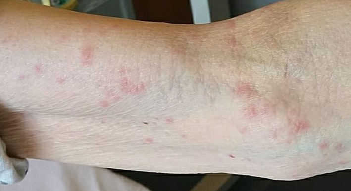
A 81‐year‐old woman with no comorbidity including allergic diseases, experiencing cough and slight fever (37°C), tested positive for SARS‐CoV‐2 RNA on nasopharyngeal swab on day 14, and was admitted to the authors’ hospital on day 16. After admission, inhaled ciclesonide was started. Laboratory investigations revealed a high white blood cell count (9700/mm3), normal platelet count (267 000/mm3), slightly impaired liver function (AST, 44 IU/L; ALT, 31 IU/L) and increased C‐reactive protein level (87.3 mg/L). Chest computed tomography revealed a pattern suggestive of pneumonia. On day 22, the patient noticed an itchy rash. Physical examination revealed multiple papular erythemas with mild infiltration of the extremities and trunk (Fig. 2). No erosions, blistering, purpura or mucosal lesions were observed. Suspecting COVID‐19‐induced viral rash, topical crotamiton and oral antihistamines were started, and erythema almost disappeared without pigmentation on day 26. After two consecutive negative PCR tests were confirmed, the patient was discharged on day 28.
Figure 2.

Multiple papular erythemas with mild infiltration of the upper limb.
Discussion
The primary symptoms of COVID‐19 include fever and dry cough; however, skin rashes are relatively rare. In a study by Recalcati, 4 , 5 skin rash was recognized in 20.8% (18/88) of COVID‐19 patients. In the only recent retrospective study, Hedou et al. 4 , 5 reported that cutaneous manifestations of COVID‐19 occurred in 4.9% (5/103) of patients. 4 , 5 According to a review by Sachdeva et al., 3 COVID‐19 patients exhibit various types of eruptions, including erythema, urticaria, purpura, reticular ecchymosis and frostbite, and erythema accounted for 36.1% at most among them. Galván et al. 6 also reported that erythema was the most common, accounting for 47% of the total. In addition, there are reports of the erythema multiforme type. 7 A search of the PubMed database retrieved only seven case reports describing COVID‐19 patients with an erythema that included basic patient information such as sex, age and time course of rash; 8 , 9 , 10 , 11 , 12 , 13 , 14 these reports are summarized in Table 1. The mean patient age was 47.1 years, with a range of 6–67 years. The number of days from the onset of COVID‐19 symptoms to the appearance of erythema were between 0 and 30 days, and all cases exhibited erythema after the appearance of fever.
Table 1.
Summary of seven case reports describing COVID‐19 patients with an erythema type eruption
| Age, years | Sex | Features of erythema | Body parts where erythema appeared | Days after onset until erythema appears | No. of days that erythema appeared | Drugs used against COVID‐19 |
|---|---|---|---|---|---|---|
| 64 | F | Erythematous rash | Trunk, antecubital fossa | 4 days | 5 days | Paracetamol |
| 67 | F | Erythematous confluent rash | Neck, trunk, back, proximal portions of upper and lower limbs | 30 days (originally stated as 1 month) | 7 days (originally stated as 1 month) | Hydroxychloroquine omeprazole piperacillin/tazobactam remdesivir |
| 57 | F | Erythematous confluent rash, with undefined margins, bleaching | Limbs and trunk, palms | 2 days | 9 days | Paracetamol |
| 60 | M | Scattered erythematous macules coalescing into papules | Back, bilateral flanks, groin and proximal lower extremities | 3 days | 7 days (originally stated that it turned into small round purpuric macules) | None |
| 37 | F | Erythematous maculopapular lesions | Trunk, neck and face | 3 days | 8 days | Acetaminophen |
| 6 | M | Maculopapular rash | Cheeks and upper and lower extremities, reaching the palms of the hands | 2 days | 5 days | No information |
| 39 | M | Erythematous and edematous non‐pruritic annular fixed plaques | Upper limbs, chest, neck, abdomen and palms, sparing the face and mucous membranes | 0 days (the same day) | 7 days | None |
Our hospital accepted 69 COVID‐19 patients between 22 February 2020 and 31 May 2020, of whom three (4.3%) exhibited eruptions, all of which were erythema. We excluded a patient who had a skin eruption approximately 2 weeks before the onset of COVID‐19 symptoms because the eruption was unrelated. Except for seven patients with positive PCR for SARS‐CoV‐2, but no symptoms, three of 62 patients (4.8%) exhibited erythema. In all cases, we did not alter the drug regimen before and after the erythema appeared, and the erythema improved with oral antihistamines and topical medicine; therefore, we considered that the possibility of drug‐induced eruption was low. COVID‐19 is a viral infection caused by SARS‐CoV‐2, and it has been reported that SARS‐CoV‐2 glycoproteins are present along with complement in skin blood vessels, which may induce viral rash. 15 For this reason, we conclude that the erythema observed in our three cases was a viral rash and not a drug‐induced eruption.
In all cases, erythema appeared after the onset of the initial symptoms of COVID‐19 and improved with the alleviation of other symptoms, as described in other case reports. From this perspective, our cases can be considered to be typical examples of erythema that appear in COVID‐19 patients.
One limitation of our study was that more patients in our hospital might have experienced cutaneous manifestation than we report. This is because it was difficult for us to perform a detailed examination of the patients’ skin (including biopsy) due to infection control measures against COVID‐19, and we had to determine cutaneous manifestation depending only on the patient’s self‐report. Some patients may fail to recognize eruption, or even if a patient is aware of eruption, more serious symptoms, such as dyspnea, may cause them to devote less attention to any eruption. Second, the number of COVID‐19 patients in our hospital was limited to 69, and our hospital specializes only in those with mild to moderate COVID‐19 (i.e. those who do not require artificial respiration). As such, the patients at our hospital cannot be regarded as truly representative of the entire COVID‐19 population. Third, there were 69 cases in our hospital, of whom four were transferred to another hospital and seven were still under treatment; therefore, we did not observe the entire disease course. It is possible that a skin rash might have been present in some of these cases.
Based on our experience and those described in other reports, it is highly possible that SARS‐CoV‐2 can cause erythema. COVID‐19 is an emerging infectious disease, and it is hoped that further analysis of cutaneous manifestations will be performed and reported as the number of cases accumulate.
Conflict of Interest
None declared.
Acknowledgments
We thank all of our hospital staff who contribute to the treatment of COVID‐19 patients.
References
- 1. Guan W, Ni Z, Hu Y et al. Clinical characteristics of coronavirus disease 2019 in China. N Engl J Med 2020; 382(18): 1708–1720. [DOI] [PMC free article] [PubMed] [Google Scholar]
- 2. Zhang JJ, Dong X, Cao YY. Clinical characteristics of 140 patients infected by SARS‐CoV‐2 in Wuhan, China. Allergy 2020; 75(7): 1730–1741. [DOI] [PubMed] [Google Scholar]
- 3. Sachdeva M, Gianotti R, Shah M etal. Cutaneous manifestations of COVID‐19: report of three cases and a review of literature. J Dermatol Sci 2020; 98(2): 75–81. [DOI] [PMC free article] [PubMed] [Google Scholar]
- 4. Recalcati S. Cutaneous manifestations in COVID‐ 19: a first perspective. J Eur Acad Dermatol Venereol 2020; 34(5): e212–e213. [DOI] [PubMed] [Google Scholar]
- 5. Hedou M, Carsuzaa F, Chary E, Hainaut E, Cazenave‐Roblot F, Masson Regnault M. Comment on “Cutaneous manifestations in COVID‐19: a first perspective ” by Recalcati S. J Eur Acad Dermatol Venereol 2020; 4(7): e299–e300. [DOI] [PMC free article] [PubMed] [Google Scholar]
- 6. Galván Casas C, Català A, Carretero Hernández G etal. Classification of the cutaneous manifestations of COVID‐19: a rapid prospective nationwide consensus study in Spain with 375 cases. Br J Dermatol 2020; 183(1): 71–77. [DOI] [PMC free article] [PubMed] [Google Scholar]
- 7. Jimenez‐Cauhe J, Ortega‐Quijano D, Carretero‐Barrio I etal. Erythema multiforme‐like eruption in patients with COVID‐19 infection: clinical and histological findings. Clin Exp Dermatol 2020; 10.1111/ced.14281 [DOI] [PMC free article] [PubMed] [Google Scholar]
- 8. Mahé A, Birckel E, Krieger S, Merklen C, Bottlaender L. A distinctive skin rash associated with Coronavirus Disease 2019? J Eur Acad Dermatol Venereol 2020; 34(6): e246–e247. [DOI] [PMC free article] [PubMed] [Google Scholar]
- 9. Zengarini C, Orioni G, Cascavilla A etal. Histological pattern in COVID‐19‐induced viral rash. J Eur Acad Dermatol Venereol 2020; 10.1111/jdv.16569 [DOI] [PMC free article] [PubMed] [Google Scholar]
- 10. Ahouach B, Harant S, Ullmer A etal. Cutaneous lesions in a patient with COVID‐19: are they related? Br J Dermatol 2020; 10.1111/bjd.19168 [DOI] [PMC free article] [PubMed] [Google Scholar]
- 11. Rivera‐Oyola R, Koschitzky M, Printy R et al. Dermatologic findings in 2 patients with COVID‐19. JAAD Case Rep 2020; 6(6): 537–539. [DOI] [PMC free article] [PubMed] [Google Scholar]
- 12. Paolino G, Canti V, Raffaele Mercuri S, Rovere Querini P, Candiani M, Pasi F. Diffuse cutaneous manifestation in a new mother with COVID‐19 (SARS‐Cov‐2). Int J Dermatol 2020; 59(7): 874–875. [DOI] [PMC free article] [PubMed] [Google Scholar]
- 13. Morey‐Olivé M, Espiau M, Mercadal‐Hally M, Lera‐Carballo E, García‐Patos V. Cutaneous manifestations in the current pandemic of coronavirus infection disease (COVID 2019). An Pediatr (Engl Ed) 2020; 92(6): 374–375. [DOI] [PMC free article] [PubMed] [Google Scholar]
- 14. Amatore F, Macagno N, Mailhe M etal. SARS‐CoV‐2 infection presenting as a febrile rash. J Eur Acad Dermatol Venereol 2020; 34(7): e304–e306. [DOI] [PMC free article] [PubMed] [Google Scholar]
- 15. Magro C, Mulvey JJ, Berlin D etal. Complement associated microvascular injury and thrombosis in the pathogenesis of severe COVID‐19 infection: a report of five cases. Transl Res 2020; 220: 1–13. [DOI] [PMC free article] [PubMed] [Google Scholar]


